
Archetypomys erlianensi, MENG & LI & NI & WANG & BEARD, 2007
|
publication ID |
https://doi.org/ 10.1206/0003-0082(2007)536[1:ANERFT]2.0.CO;2 |
|
persistent identifier |
https://treatment.plazi.org/id/03A9141B-FF83-FFCD-FF5E-FB159891E65C |
|
treatment provided by |
Carolina |
|
scientific name |
Archetypomys erlianensi |
| status |
sp. nov. |
Archetypomys erlianensi , sp. nov.
HOLOTYPE: IVPP V14623.1 , an upper M1.
INCLUDED SPECIMENS: V 14623.2, left P4 (or dP4); V 14622.1, right M1 with metacone broken; V 14622.2, left M1; V 14622.3, right M1; V 14622.4, left M1; V 14622.5, right M1; V 14622.6, right M2; V 14622.7, right M2; V 14622.8, left M2; V 14622.9, left M2; V 14623.3, left M2; V 14623.4, left M2; V 14623.5, left M2; V 14623.6, right M3; V 14622.10, left dp4; V 14622.11, right dp4; V 14622.12, right dp4; V 14622.13, left dp4; V 14622.14, right dp4; V 14622.15, right dp4; V 14622.16, right dp4; V 14622.17, left dp4; V 14623.7, left dp4; V 14623.8, left dp4; V 14623.9, right dp4; V 14622.18, left m1; V 14622.19, right m1; V 14622.20, left m1; V 14622.21, right m1; V 14622.22, right m1; V 14623.10, right m1; V 14623.11, right m1; V 14623.12, left m1; V 14623.13, right m1; V 14622.23, right m2; V 14622.24, left m2; V 14622.25, left m2; V 14622.26, left m2; V 14622.27, left m2; V 14622.28, right m2; V 14623.14, right m2; V 14623.15, left m2; V 14623.16, left m2; V 14623.17, left m2; V 14622.29, right m3; V 14622.30, right m3; V 14622.31, left m3; V 14622.32, right m3; V 14622.33, left m3.
ETYMOLOGY: Archetypus (L.), original; species name from Erlian, Nei Mongol (Inner Mongolia), the nearest city to the outcrops where the specimens were collected.
DIAGNOSIS: Same as for the family.
TYPE LOCALITY AND HORIZON: Nuhetingboerhe (Camp Margetts area), Erlian Basin, Nei Mongol (Inner Mongolia); lower part of the Arshanto Formation; late Early Eocene. Specimens were collected from two sites that are geographically separated by about 200 m, with the ‘‘chalicothere pit’’ being slightly lower stratigraphically than the other, unnamed site. Those from the chalicothere pit were cataloged with the number V 14622 View Materials , and the others from the unnamed site bear the number V 14623 View Materials .
DESCRIPTION: Because the current sample consists only of isolated teeth showing unique morphology unknown previously, the orientation of the teeth is problematic. Based on the similar occlusal pattern in Tribosphenomys and Alagomys , we interpret upper cheek teeth of Archetypomys as having a protocone that tapers anterobuccally and is separated from a large metaconule by a talon basin ( Meng and Wyss, 2001) or hypocone basin ( Lopatin and Averianov, 2004a), while the protoconule (paraconule) joins the preprotocrista. Without better material to show the upper dentition in serial association, we cannot rule out the possibility that the anteroposterior tooth orientation we currently recognize is reversed.
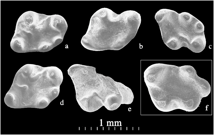
All upper cheek teeth have one major lingual and two minor buccal roots. Upper and lower teeth have distinct cusps but weak crests; they are small and low-crowned. P4 (or DP4) is identified as such because of its relatively simple occlusal pattern ( fig. 1a View Fig ). It is basically triangular in shape, with the protocone forming the lingual apex. The protocone is tilted anterobuccally and is connected with the large paracone by a crest. A weak crest is anterior to the paracone. There is no paraconule. An oval, transversely oriented trigon basin occupies the central part of the tooth and opens buccally. The metacone occurs at the posterobuccal corner of the tooth and is much smaller than the paracone. The metaconule is subequal to the metacone in size and is separated from the latter by a small basin. A more distinct basin separates the metaconule from the protocone.
Six teeth are identified as M1 ( fig. 1b–g View Fig ) because of their relatively narrow anterior margins. These teeth are somewhat triangular in occlusal view, with all apexes rounded, and are anteroposteriorly longer than P4. The protocone is somewhat comma-shaped with the preprotocrista being its tail. The anterolingual surface of the protocone bears an inclined wear facet in V14622.1 ( fig. 1b View Fig ), on which there are microstriations indicating a primarily transverse movement of mastication. A neomorphic cusp occurs on the preprotocrista between the protocone and protoconule (paraconule). Because this cusp is unique among early rodents with which we are familiar, we designate it as the preprotoconule (see below). Buccal to the preprotoconule is a distinct protoconule, and between them is a short, weak ridge, which we consider to be the buccal part of the preprotocrista. A wear facet occurs on the anterolingual side of the preprotoconule in V14622.1, parallel to that on the protocone. The paracone is conical and connects to the paraconule by a short and low postparaconule crista. The buccal surface of the paracone forms the anterobuccal margin of the tooth, but a vestigial buccal cingulum occurs in some specimens. Anterior to the paracone and protoconule is a transverse crest that projects anteriorly and is similar in position to the precingulum of rodents. Because of its relationship with the preprotoconule and its similarity with those of alagomyids, we regard it as the preprotoconule crista, which may be homologous with the precingulum of rodents (see below). Posterobuccal to the paracone is a small cusp that may be called the mesostyle. The central basin of the tooth is long and curved, starting from the region between the protocone and preprotoconule and ending at the buccal edge of the tooth between the mesostyle and the metacone. Wear striations within the basin are roughly parallel to the long axis of the basin, indicating primarily transverse movements of the lower jaw during mastication. The metaconule, which is as large as the metacone, is connected to the protocone by a low postprotocrista lingually, and to the metacone by a low premetaconule crista buccally. The posterior part of the metaconule merges with the postcingulum, which is low but more distinctive between the metacone and metaconule than it is farther lingually. Between the protocone and metaconule is a conspicuous basin, which was homologized with the talon basin of a tribosphenic tooth ( Meng and Wyss, 2001). A smaller basin is present between the metaconule and metacone. The metacone is smaller than the paracone and occupies the posterobuccal corner of the tooth; there is no cingulum buccal to the metacone.
Seven teeth are identified as M2 ( fig. 2a–g View Fig ). M2 is basically similar to M 1 in general morphology. They are, however, relatively larger and have a more transversely extended preprotoconule crista. These teeth show that a narrow cingulum usually exists buccal to the paracone but not the metacone; the metaconule is subequal to the metacone in occlusal view, but is lower than the latter ( fig. 2f View Fig 1 View Fig ); the preprotoconule crista is low, along the anterior edge of the tooth ( fig. 2d View Fig 1 View Fig ). The buccal margin of M2 can be quite uneven, depending on the development of the buccal cingulum and mesostyle.
A single M3 (V14623.6, fig. 2h View Fig ) is identified, and its metacone is broken. It is smaller than the other molars and is narrower posteriorly than anteriorly; its metaconule is not so pronounced as in M1–2, and the central basin is shorter than in the other upper cheek teeth. The preprotoconule is relatively indistinct, and there is no connection between it and the paraconule.
In identifying the lower cheek teeth, we consider those bearing an anteroconid (we do not assume homology between this cusp and those in, for instance, myodont rodents) as terminal teeth because the anteroconid usually occurs on the anteriormost cheek tooth in rodents, such as the m 1 in early myodont rodents ( Emry and Korth, 1989; Emry et al., 1998; Tong, 1992, 1997; Wang and Dawson, 1994; Dawson and Tong, 1998) and because there is no contact facet on the anterior surface of any of these teeth. We interpret these teeth as dp4 ( fig. 3 View Fig ), although other possibilities, such as their being p4 or m1, cannot be absolutely ruled out. However, if they are p4s, then these teeth are quite molariform, which is inconsistent with the general trend in mammal tooth replacement for a successive tooth to be simpler than its deciduous precursor. In particular, it has been shown that p4 is much simpler than dp4 of Tribosphenomys ( Meng and Wyss, 2001; Lopatin and Averianov, 2004a, b). Similar p4–dp4 morphologies are also present in rodents of modern aspect, such as Paramys adamus ( Dawson and Beard, 1996) . Given the similarity between the molars of Archetypomys and Tribosphenomys , it seems that similar p4– dp4 morphologies can be inferred for Archetypomys . Although a molariform p4 was recognized in Alagomys ( Tong and Dawson, 1995; Dawson and Beard, 1996), Meng and Wyss (2001) have pointed out that these molariform teeth probably represent delayed or nonreplaced deciduous teeth, a view that is supported by additional evidence ( Lopatin and Averianov, 2004a).
These teeth are unlikely to be m1s, because this would imply that the lower dentition consists of only three molars, which is inconsistent with both the primitive structure of these teeth and the identification of the P4. A myodont rodent from the same quarries retains a p4 or dp4 (unpubl. data); suppression of the last lower premolar therefore seems unlikely for Archetypomys . Moreover, m1 and m2 seem to be recognizable within the current sample, inasmuch as the m1 trigonid is relatively narrower than that of m2, although the difference is not dramatic.
Among the 11 dp4s, three specimens are poorly preserved, with the enamel layer largely gone ( fig. 3a–c View Fig ). Because they are considerably worn, owing to postmortem preservation, these specimens look relatively small and lack an anteroconid. The remaining specimens are quite molariform in having the major cusps of a lower molar. The dp4 is significantly narrower anteriorly than posteriorly. It has a small anteroconid at its anterior tip, situated near the longitudinal axis of the tooth, as is the case on m 1 in some Eocene myodonts, such as Pappocricetodon ( Tong, 1992, 1997; Wang and Dawson, 1994), but there is no anterior cingulid. There is no contact facet on the anterior surface of the tooth. The protoconid and metaconid are conical and closely spaced; they are separated by a longitudinal groove. The metaconid is larger than the protoconid and extends posteriorly as a crest that forms the lingual border of the tooth. The cristid obliqua is distinct and quite diagonally oriented. The anterior end of the cristid obliqua extends to the posterolingual base of the protoconid. A mesoconid is present on the cristid obliqua. Because of the orientation of the cristid obliqua, there is a deep sinusid. The hypoconid is the largest cusp on the tooth and is more posterobuccally extended than the hypoconulid; the latter is transversely extend- ed. The entoconid occupies the posterolingual corner of the tooth. There is no crest developed from the main cusps of the tooth.
The differences between what we regard as m1 and m2 are comparatively trivial. The trigonid of m1 ( fig. 4 View Fig ) is slightly narrower than that of m2 ( fig. 5 View Fig ), as is the case in Tribosphenomys ( Meng and Wyss, 2001; Lopatin and Averianov, 2004 a, 2004b) and Alagomys ( Tong and Dawson, 1995; Dawson and Beard, 1996). Each of the specimens identified as m2 has a contact facet on its posterior surface, excluding it from being an m3. Because of their similarity, we describe m1 and m2 collectively. The crown tapers anteriorly, but to a lesser degree than that of dp4, and it lacks the anteroconid. It is doublerooted, with the posterior root being more robust ( fig. 4b View Fig 1 View Fig ). The metaconid extends more anteriorly than the protoconid and is the highest cusp of the tooth. The anterior margin of the tooth is raised to form a low ridge; otherwise, there is no crest between the protoconid and metaconid. The posterior wall of the metaconid is a large sloping surface that descends to the central basin of the tooth. A ridge from the metaconid is developed to a variable degree among the specimens at hand and extends posteriorly to form the lingual edge of the tooth. In some specimens, a small cuspid at the posterior end of the ridge may be called the mesostylid. The cristid obliqua is diagonally oriented. Its anterior end terminates at the posterolingual base of the protoconid, either merging with the base or being separated from the base by a narrow notch. Posteriorly, it develops from the anterobuccal side of the hypoconid. The anterior part of the cristid obliqua inflates to form a sizable, elongated mesoconid. Because of the orientation of the cristid obliqua, the sinusid is narrow and lingually deep. Similar to the metaconid, the entoconid is more anterior than the hypoconid so that the tooth is more or less diamond-shaped in occlusal view. The hypoconid projects posterobuccally and is the most robust cuspid of the tooth. The hypoconulid is prominent and transversely oriented. The postcingulid or postlophid is weak between these cuspids. No other crest is developed from these talonid cuspids. V14623.13 ( fig. 4i View Fig ) is a tooth that had not fully erupted in life; its cuspids are not fully developed and bear no wear; it could also be an m2.
The m3 is slightly wider anteriorly than posteriorly and has a hypoconulid that is more conical than transverse, although in one tooth ( fig. 6b View Fig ) the hypoconulid is not distinct. In addition, there is no contact fact on the posterior surface of the tooth and the posterior root is more posteriorly flared than those of m1–2. The metaconid of m3 is relatively higher and even more anteriorly extended than that of m1–2. A peculiar cuspulid is developed between the protoconid and metaconid in two specimens ( fig. 6a, d View Fig ); it sends a weak ridge to the anterior edge of the tooth. As in m1–2, the m3 hypoconid is the largest talonid cusp and is slightly more posterior than the hypoconulid.


| IVPP |
Institute of Vertebrate Paleontology and Paleoanthropology |
| V |
Royal British Columbia Museum - Herbarium |
No known copyright restrictions apply. See Agosti, D., Egloff, W., 2009. Taxonomic information exchange and copyright: the Plazi approach. BMC Research Notes 2009, 2:53 for further explanation.
|
Kingdom |
|
|
Phylum |
|
|
Class |
|
|
Order |
|
|
Family |
|
|
Genus |