
Xerobiotus reductus, Vincenzi & Cesari & Kaczmarek & Roszkowska & Mioduchowska & Rebecchi & Guideưi, 2024
|
publication ID |
https://doi.org/10.1093/zoolinnean/zlad129 |
|
DOI |
https://doi.org/10.5281/zenodo.10470591 |
|
persistent identifier |
https://treatment.plazi.org/id/03E04041-6E18-FFB8-FEBE-28AA68D0F864 |
|
treatment provided by |
Plazi (2024-01-08 13:24:01, last updated 2024-11-27 14:18:03) |
|
scientific name |
Xerobiotus reductus |
| status |
sp. nov. |
Xerobiotus reductus sp.nov.
( Figs 6 View Figure 6 , 7 View Figure 7 ; measurements and statistics are in Tables 2 View Table 2 and 3 View Table 3 ; Supporting Information, Table S1 View Table 1 ).
ZooBank: urn:lsid:zoobank.org:act:0116DF21-ACDA-436F-806C-A4E1DE09E979
Type locality: Burgenland, near Rust , Austria (47°47ʹ59″N, 16°40ʹ42″E, 118 m a.s.l.), moss from sandy soil, September 2019, coll. Johenn Sholl. GoogleMaps
Additional locality: Notecka Forest , Wiełkopolska Province ( Poland) (52°48ʹ48.32″N, 16°14ʹ59.49″E), in a clump of grass GoogleMaps .
Material examined: Eighty-four adults and 28 eggs (slides AU1–AU8).
Type repositories: One hundred and six specimens deposited at the Department of Animal Taxonomy and Ecology , Institute of Environmental Biology , Adam Mickiewicz University in Poznań, Poznań, Poland ; five specimens [slides: M8/17 and M8/27 (four adults and one egg)] deposited at the Institute of Systematics and Evolution of Animals , Polish Academy of Sciences, Kraków, Poland .
Etymology: The name of the species refers to the reduced claws on all pairs of legs.
Description: Body whitish, eye-spots present (in ≥ 60% of fixed and measured specimens). Very small, scaưered pores (~0.5 µm in diameter; visible only with SEM; Fig. 7C, E, F View Figure 7 ) in the dorsolateral cuticle of the body and legs. Very small single granules (visible only with SEM), distributed almost regularly, present on the entire cuticle ( Fig. 7E View Figure 7 ). Legs of the first pair clearly smaller than those of the second and third pairs. The area of the leg cuticle surrounding the claws with a swelling (forming a furbelow-like structure; Figs 6A View Figure 6 , 7C, F View Figure 7 ) covered with spheroidal microdigitations visible with SEM ( Fig. 7D View Figure 7 ).
Buccopharyngeal apparatus of the Macrobiotus type, with anteroventral mouth and 10 small peribuccal lamellae ( Fig. 7A, B View Figure 7 ). Oral cavity armature of the maculatus type (with LM), with only third band of teeth (transversal crests) formed by one continuous dorsal tooth (ridge) and ventral transversal ridges as two-to-three granular teeth ( Fig. 6 View Figure 6 D-F); with SEM, first (with one or two rows of teeth) and second (with two or three rows of a bit larger teeth) bands visible ( Fig. 7B View Figure 7 ). Typically shaped stylet furcae, with oval condyles supported by short branches provided with rounded apophyses. In the pharynx ( Fig. 6C View Figure 6 ): large and triangular pharyngeal apophyses overlapping the first macroplacoid; two rod-shaped macroplacoids (in lateral view), length sequence 2 <1, and evident triangular microplacoid. In frontal view, the first macroplacoid in the shape of a drop with a median slight constriction, the second rectangular with rounded corners and with a subterminal slight constriction, at least in some specimens.
Claws I–III of Xerobiotus type, small and compact, with the common tract similar in length to the main branch; main branch with accessory points; lunules absent ( Figs 6G, H View Figure 6 , 7C View Figure 7 ). Claw IV with a longer common tract, smaller and shorter claw branches, without lunules or thickening under the claws ( Figs 6I, J View Figure 6 , 7F View Figure 7 ). Primary branches of all claws with small accessory points.
Eggs spherical, white, ornamented and laid freely ( Figs 6K View Figure 6 , 7G View Figure 7 ). Surface between processes of hufelandi type, i.e. covered with a reticulum formed by a mesh of small, densely distributed pores, uniform in size and evenly distributed ( Figs 6L, M View Figure 6 , 7H View Figure 7 ). Processes of the hufelandi type, with a straight trunk and a relatively small and concave terminal disc. The terminal disc greatly indented on the disc margin, forming evident but irregular teeth covered by microgranules (visible only with SEM; Fig. 7H, I View Figure 7 ).
Reproduction: Gonochoristic amphimictic species; females and male with spermatozoa present ( Fig. 6B View Figure 6 ). The diploid karyotype resulted in six chromosomes (2 n) ( Fig. 6N View Figure 6 ).
Molecular characterization: One haplotype for cox1, two haplotypes for ITS2 (p-distance 0.5%), one haplotype for 18S, and one haplotype for 28S genes (GenBank accession numbers in Supporting Information, Table S1 View Table 1 ; p-distances in Supporting Information,Table S8). The more similar sequences of X.reductus belong: for cox1, to X. naginae and to specimens from Skwierzyna population with p-distances of 1.3% and 1.9%, respectively; for ITS2, to X. naginae and P. degenerans with p-distances of 0.0%- 0.3% and 0.0%-0.8%, respectively (Supporting Information, Table S8).
Differential diagnosis: Xerobiotus reductus differs from: X. euxinus by the absence of semilunar thickening at the base of the claws I–III and absence of lunules in claw IV; X. gretae by shorter branches of claws I–III and absence of lunules and cuticular thickening at the base of claw IV; X. naginae by the third band of teeth (transversal crests) visible with LM and formed by one continuous dorsal tooth (ridge); X. xerophilus by the absence (visible with SEM) of cuticular plates at the base of all claws (see Dastych and Alberti 1990) and the shape of egg processes (without a funnel-like depression).
Dastych H, Alberti G. Redescription of Macrobiotus xerophilus (Dastych, 1978) comb. nov., with some phylogenetic notes (Tardigrada, Macrobiotidae). Mitteilungen aus dem Hamburgischen zoologische Museum und Institut. Hamburgisches Zoologisches Museum und Institut: Hamburg 1990; 87: 157 - 69.
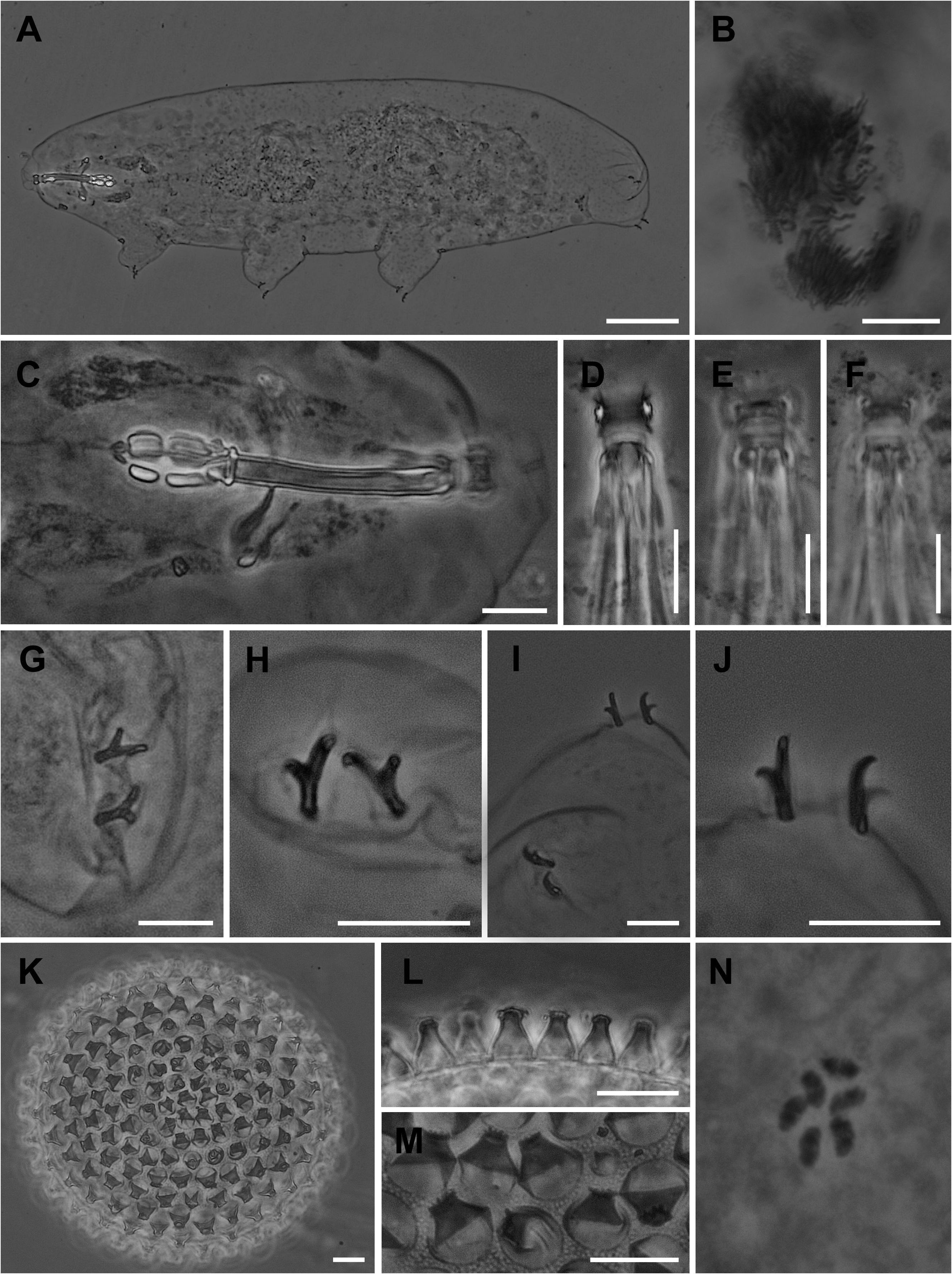
Figure 6. Xerobiotus reductus (type locality).A, animal in toto. B, gonad with sperm (stained with Orcein). C, buccopharyngeal apparatus. D, buccal armature (dorsal view). E, buccal armature (ventral view). F, buccal armature (ventral view). G, claw II. H, claw I. I, claw IV. J, claw IV. K, egg. L, egg processes.M, egg surface and processes. N, chromosomes (six bivalents, stained with Orcein).A, C–N, PhC; B, DIC. Scale bars: 50 µm in A; 10 µm in B–M.
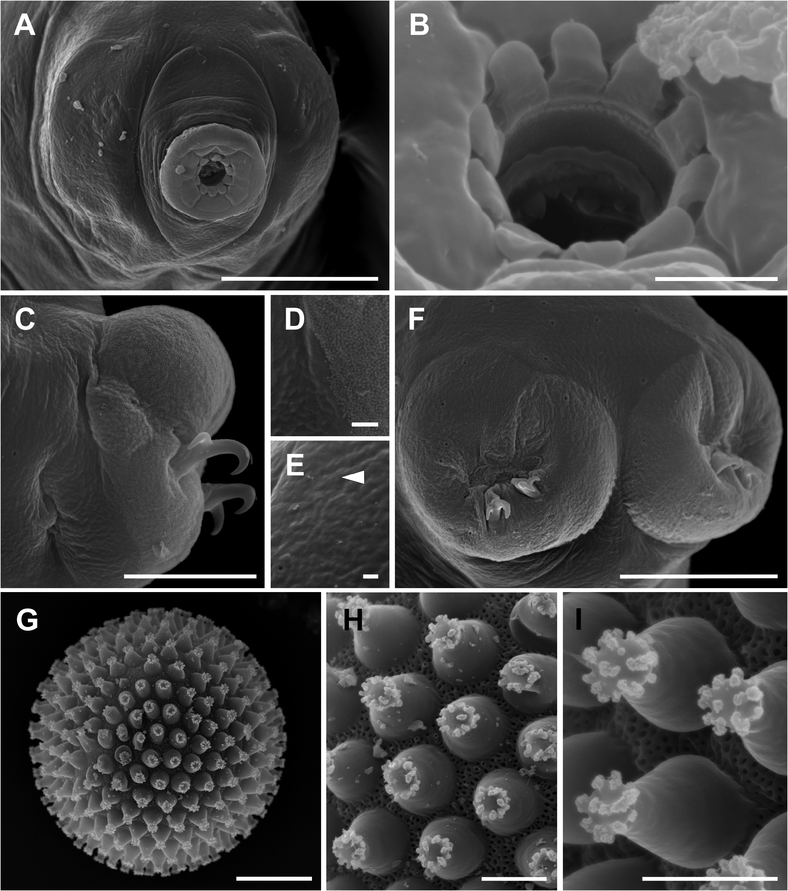
Figure 7. Xerobiotus reductus (type locality).A, head in frontal view.B, mouth opening.C, leg I, with the swelling of the furbelow-like structure. D, detail of image C, showing the boundary between the swelling with microgranulations and the remaining cuticle.E, cuticle, with a pore and single granules (arrowhead).F, legs IV, visible pores on dorsal cuticle. G, egg. H, egg processes. I, egg surface and processes.A–I, SEM. Scale bars:20 µm in A, F, G; 3 µm in B; 10 µm in C; 5 µm in H, I; 1 µm in D, E.
No known copyright restrictions apply. See Agosti, D., Egloff, W., 2009. Taxonomic information exchange and copyright: the Plazi approach. BMC Research Notes 2009, 2:53 for further explanation.
|
Kingdom |
|
|
Phylum |
|
|
Class |
|
|
Order |
|
|
Family |
|
|
Genus |