
Johnmannia Irwin & Lyneborg, 1989
|
publication ID |
https://doi.org/ 10.5281/zenodo.170862 |
|
DOI |
https://doi.org/10.5281/zenodo.6264989 |
|
persistent identifier |
https://treatment.plazi.org/id/F6528787-FFBE-842A-1526-D1410FDBFCBB |
|
treatment provided by |
Plazi |
|
scientific name |
Johnmannia Irwin & Lyneborg, 1989 |
| status |
|
Johnmannia Irwin & Lyneborg, 1989 View in CoL View at ENA
( Figs 1–7 View FIGURE 1 View FIGURE 2 View FIGURE 3 View FIGURE 4 View FIGURE 6 View FIGURE 7 )
Mannia Paramonov, 1950: 525 ; Liepa, 1969: 12. [Preoccupied Davidson, 1874: 156 (Fossil Brachiopoda); Prout, 1915: 382 (Lepidoptera)].
Johnmannia Irwin & Lyneborg, 1989: 357 View in CoL [new name for Mannia ]; Winterton et al., 1999c: 276.
Type species. Mannia tasmanica Paramonov, 1950 , by original designation
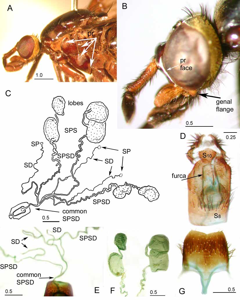
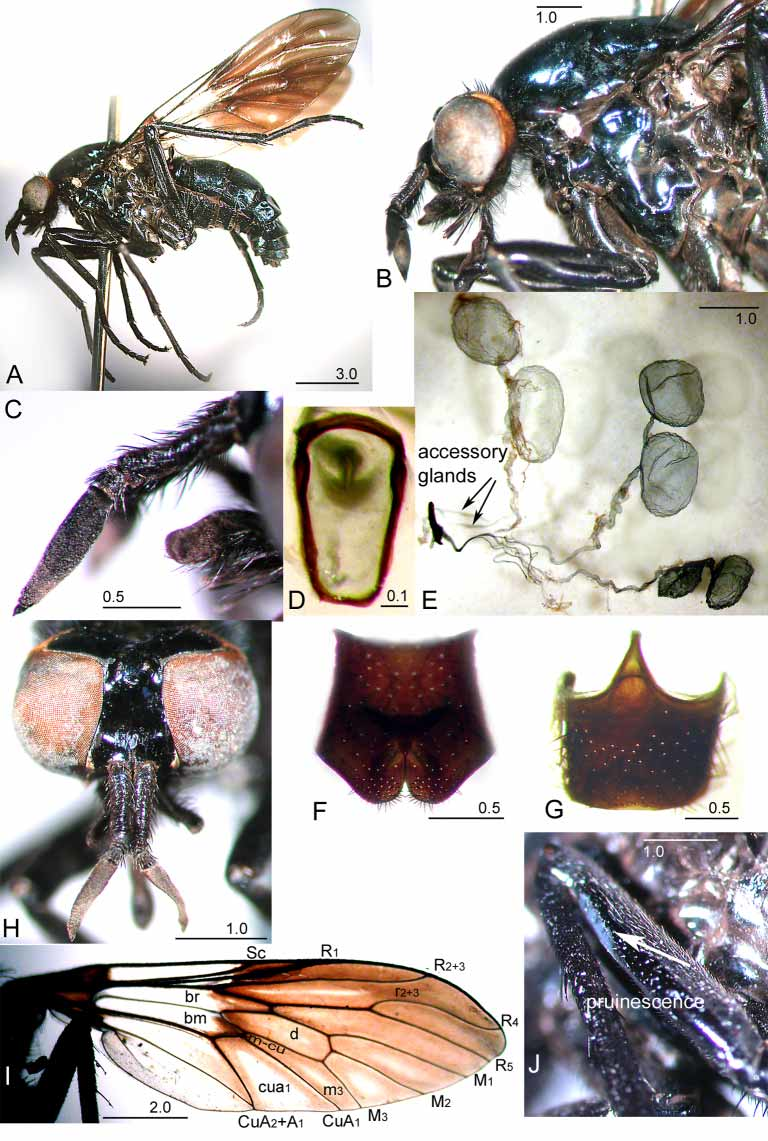
Diagnosis. Colouration black ( Fig. 6 View FIGURE 6 A) with metallic blue or green reflectivity; scape elongate, with uniform long dark setae ( Fig. 3 View FIGURE 3 F–G); face with pruinescence laterally from antennae along compound eye, narrowed medially ( Fig. 3 View FIGURE 3 F), covering anteriorly directed flange of gena at base of compound eye ( Fig. 4 View FIGURE 4 B); occiput with gold pruinescence, indistinct row of dark postocular setae almost length of ocellar triangle from compound eye; 4– 6 short black postspiracular setae (Fig. 5B) (metepisternal ( Yeates 1994) or hypopleural pile (Lyneborg 2001)); wing cell m3 open (Fig. 5C); seta present anteroventrally subapically on hind femur ( Fig. 1 View FIGURE 1 ); without velutum patches on fore and hind femora and ventral surface of gonocoxites; wing with black infuscation distally and medially (Fig. 5C) or entire blade ( Fig. 7 View FIGURE 7 F), without orange infuscation; two scutellar setae ( Fig. 3 View FIGURE 3 C) (rarely 4, with outer setae weak); abdomen strongly recurved apically ( Fig. 1 View FIGURE 1 ), appears globular dorsally ( Fig. 3 View FIGURE 3 E); spermathecal sac arrangement trilobate with each spermathecal sac with narrow tube to outer lobe ( Fig. 4 View FIGURE 4 C); spermathecal duct broadening basally before joining individual spermathecal sac duct ( Fig. 4 View FIGURE 4 E); subepandrial sclerite well developed sclerotised plate ( Fig. 7 View FIGURE 7 K–M); outer gonocoxal process well developed, pointed (Fig. 5G–I); inner gonocoxal process long, narrow, apically acuminate (Fig. 5H); gonostylus longer than inner gonocoxal process, dorsal recurved triangulate hook 1/3 length of gonostylus from apex ( Fig. 7 View FIGURE 7 I–J); hypandrium triangular, joined laterally to gonocoxites (Fig. 5I); distiphallus directed ventrally (Fig. 5L).
Generic Redescription. Female. Head shape laterally oval ( Fig. 6 View FIGURE 6 B). Frons glossy black; upper frons width greater than width of ocellar triangle ( Fig. 3 View FIGURE 3 C), short dark setae on ocellar triangle, deep broad medial concavity; lower frons rounded dark glossy callus or antennal tubercule ( Fig. 4 View FIGURE 4 B); short, dark, setae laterally above antennae; face with dense, silver pruinescence. Occiput rounded ( Fig. 3 View FIGURE 3 B), indistinct rows of dark postocular setae distant from compound eye; dense, gold pruinescence at least ventrally; very long fine dark setae ventrally. Gena covered with fine long, dark setae, overlain with dense, gold pruinescence; distinct protrusion of gena at base of compound eye ( Fig. 4 View FIGURE 4 B). Palp twosegmented, apical segment spatulate, fine long dark setae basally, less dense medially ( Fig. 4 View FIGURE 4 B); labellum dark, long dark setae medially, short dark setae apically. Antenna slightly longer than head ( Fig. 3 View FIGURE 3 F–G); scape as long or longer than postpedicel, wider than pedicel;
scape and pedicel covered with uniformly long dark setae on all surfaces; postpedicel (1st antennal flagellomere) long (postpedicel/pedicel = 4.4–6.3); laterally flattened, broadest subbasally, tapering apically, indentation on lateromedial surface; basal stylomere (2nd antennal flagellomere) short broad cylinder, antennal apical stylomere (3rd antennal flagellomere) conical with pale brown, short pointed style. Thorax: Scutum glossy dark, reflective metallic dark blue to green, numerous fine, short, dark setae; gold pruinescence anteriorly. Scutellum black, reflective, metallic dark blue to green. Pleuron glossy, dark; overlain with areas of reddishgold pruinescence, denser ventrally ( Fig. 4 View FIGURE 4 A); pale, short, dense setae around anterior spiracle. Scutal chaetotaxy: np 4–5; sa 2–3; pa 1; dc 1–2; sc 1 (2 rarely: outer finer, shorter, not equal). Legs dark reddishbrown to black, with short dark setae; 12 large black anteroventral subapical macrosetae on hind femur; tarsomeres sometimes with orange bands, ventral surface of 4th and 5th tarsomeres lacking setae, partial loss on 3rd. Hind legs long. Wing covered with microtrichia, variably infuscated, with darker opaque triangular basal area. Triangulate darker opaque area between apex of Sc and R1, extending to join of CuA2 and A1, over mcu crossvein and CuA2; across base of r2+3, d, m3 and cua1; apex of bm and br, indistinct posteriorly ( Fig. 3 View FIGURE 3 D). Apical half of wing darker, paler posteriorly ( Fig. 3 View FIGURE 3 D). Halter stem and knob dark. Abdomen: Conical, apically recurved thus appears globular in dorsal view ( Fig. 3 View FIGURE 3 E). Dark, glossy, reflective, metallic dark blue to green; setae sparse, fine short, T12 with longer setae laterally; reddishgold pruinescence on T1 basally, over entire S1–4, densest laterally at ST sutures.
Genitalia: T8 ( Fig. 4 View FIGURE 4 G) subquadrate, anterior process long narrowed medially, 0.38 length of T8; furca ( Fig. 6 View FIGURE 6 D) heavily sclerotised, long (1.7 x as long as wide), narrowed posteriorly, closed; T8–10 joined through sclerotised band; acanthophorites with 6–8 broad dark brown to black A1 setae and 10–15 long fine, black A2 setae; S8 ( Fig. 6 View FIGURE 6 F) with posterior margin indented medially, forming blunt rounded lobes; three spermathecae ( Fig. 4 View FIGURE 4 C); spermathecal duct threadlike, twice length of furca to sclerotised cuticular valve, broadening basally ( Fig. 4 View FIGURE 4 E) for the length of furca before joining individual spermathecal sac duct, less than length of furca from spermathecal sac duct join; spermathecae indistinct, unpigmented, unsclerotised; spermathecal sac arrangement trilobate ( Fig. 4 View FIGURE 4 C), with spermathecal sac joining common spermathecal sac duct less than length of furca from furca; long ovoid median spermathecal sac with narrow tube to longer oval lobe ( Fig. 4 View FIGURE 4 F); pair of ovoid outer spermathecal sacs with narrow tube to large round lobe ( Fig. 4 View FIGURE 4 C).
Male. Similar to female except: larger and darker. Upper frons narrower dorsally ( Fig. 7 View FIGURE 7 C). Apical half of wing darker (Fig. 5C). Genitalia: Gonocoxites with dense tufts of posteriorly directed setae along ventral ridge on basomedial margin (Fig. 5I), without medial atrium ( Winterton et al. 1999a). Epandrium (Fig. 5D) narrowed posteriorly, with posterolateral extensions, indented medially on posterior surface. Subepandrial sclerite ( Fig. 7 View FIGURE 7 K– M) with very well developed, sclerotised, broad plate strongly attached to basiphallus. T8 ( Fig. 7 View FIGURE 7 O–P) emarginate, broadly narrowed medially, setae along posterolateral margin, with lateral spiracular pore (Fig. 5J); S8 ( Fig. 7 View FIGURE 7 N) trapezoid, narrowed basally, posterior margin longer than basal margin, black setae along posterior margin. Outer gonocoxal process (Fig. 5G–I) well developed, pointed; ventral lobe ( Fig. 7 View FIGURE 7 I) broad, flattened, sclerotised; gonocoxites distinctly separate, not fused ventromedially ( Fig. 7 View FIGURE 7 I); gonocoxal apodeme (Fig. 5G–I) as long as ventral lobe, expanded apically to form broad gonocoxal apodeme plate. Inner gonocoxal process ( Fig. 7 View FIGURE 7 I) long, narrow, apically acuminate, cupshaped on ventroapical surface, glabrous. Gonostylus (Fig. 5G–I) longer than inner gonocoxal process, basally broad and twisted, some setae on basolateral surface, many setae on medial surface; dorsal recurved triangulate subapical hook, 1/3 length of gonostylus from apex; apical portion of gonostylus narrowing, recurved dorsomedially, cupshaped on dorsal surface. Hypandrium (Fig. 5G) triangular, separate from gonocoxites but joined laterally. Distiphallus ( Fig. 7 View FIGURE 7 K–M) smoothly recurved from base, directed ventrally, longer than ejaculatory apodeme, spinulose on dorsal surface; dorsal apodeme ( Fig. 7 View FIGURE 7 K–M) of parameral sheath with anteriorly directed lateral arm; ventral apodeme of parameral sheath short; ejaculatory apodeme (Fig. 5L) broad anteriorly; lateral ejaculatory apodeme ( Fig. View FIGURE 7
7K–M) narrow, ribbonlike, separation less than base of distiphallus; basal ejaculatory apodeme broad, spinulose on ventral surface.
Comments. Very rare. In this genus, female specimens have more often been collected, described, and given holotype status. We will continue this trend by basing our descriptions upon the female, and then comparing the male to that description.
Irwin & Lyneborg (1989) erected the name Johnmannia citing preoccupation of the name Mannia by Dewalque (1868). However, the Dewalque 1868: 432 name for a fossil had been declared a nomen nudum ( Neave 1940). The name Mannia was preoccupied by Davidson (1874) for a fossil brachiopod, and again by Prout (1915) for a moth.
Distribution. SE Queensland; Lansdowne (31°47'S 152°32'E), Kosciuszko National Park, NSW; Tasmania.
Included species: Johnmannia tasmanica (Paramonov) , Johnmannia powerae Lambkin & Recsei , Johnmannia kosciuszkoensis Lambkin & Recsei.


No known copyright restrictions apply. See Agosti, D., Egloff, W., 2009. Taxonomic information exchange and copyright: the Plazi approach. BMC Research Notes 2009, 2:53 for further explanation.
|
Kingdom |
|
|
Phylum |
|
|
Class |
|
|
Order |
|
|
Family |
Johnmannia Irwin & Lyneborg, 1989
| Lambkin, Christine L., Recsei, Jacqueline M. & Yeates, David K. 2005 |
Johnmannia
| Winterton 1999: 276 |
| Irwin 1989: 357 |
Mannia
| Liepa 1969: 12 |
| Paramonov 1950: 525 |
| Prout 1915: 382 |
| Davidson 1874: 156 |