
Helicopsyche (Feropsyche) shaamunensu, Dumas & Nessimian, 2019
|
publication ID |
https://doi.org/ 10.11646/zootaxa.4619.2.2 |
|
publication LSID |
lsid:zoobank.org:pub:E9CFFBFF-E437-4919-9E59-730E87875B62 |
|
DOI |
https://doi.org/10.5281/zenodo.5945279 |
|
persistent identifier |
https://treatment.plazi.org/id/D30B23D9-C044-4B3C-A913-649AB8FF2CA8 |
|
taxon LSID |
lsid:zoobank.org:act:D30B23D9-C044-4B3C-A913-649AB8FF2CA8 |
|
treatment provided by |
Plazi |
|
scientific name |
Helicopsyche (Feropsyche) shaamunensu |
| status |
sp. nov. |
Helicopsyche (Feropsyche) shaamunensu , new species
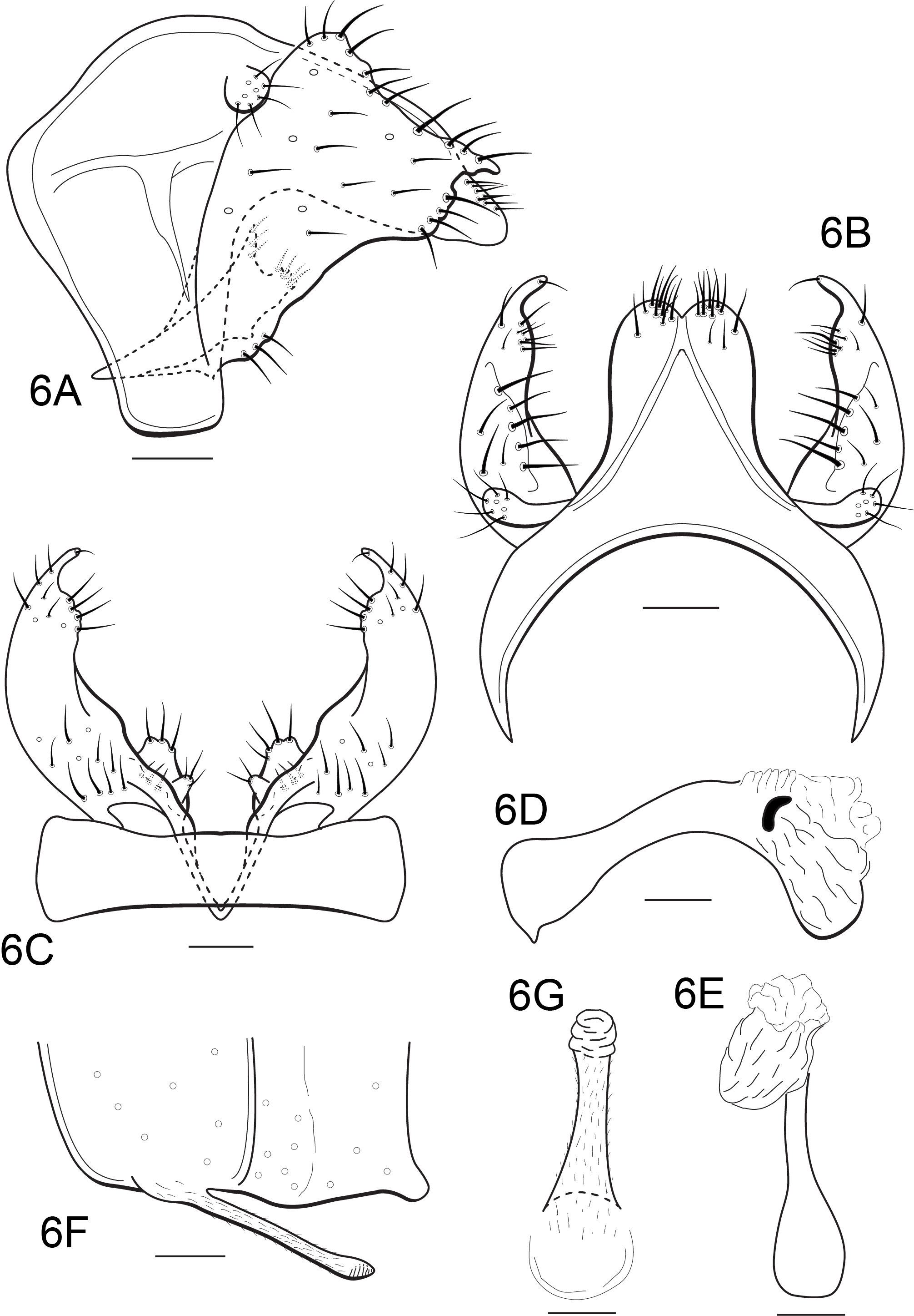
( Figures 6 View FIGURE 6 A-G)
urn:lsid:zoobank.org:act:D30B23D9-C044-4B3C-A913-649AB8FF2CA8
Diagnosis. Helicopsyche (F.) shaamunensu sp. nov. resembles Helicopsyche (F.) lazzariae Holzenthal, Bahnik & Calor 2016 by the overall deltoid shape of the primary branches of inferior appendages. The new species can be easily distinguished from H. (F.) lazzariae by the presence of basomesal lobes of inferior appendages, which are absent in the second species, and by the shape and setation of tergum X in dorsal view.
Description. Adult. Similar to H. (F.) bendego sp. nov. except as follows: Overall color (in alcohol) uniformly golden-brown; antennae, palps, and legs pale yellow. Maxillary palps with proximal segment longer than distal one. Each foreleg anterior apical tibial spur about 2x longer than posterior spur. Male forewings each 3.2 mm long (holotype male). Abdominal sternum VI ventral process long, about as long as segment VI, slender, tubular along its length, oriented posteroventrad, slightly wider at apex, nearly straight in lateral view with apex subacute, with lamellae apically ( Figs. 6F, 6G View FIGURE 6 ).
Male genitalia. Segment IX short ventrally; in lateral view with anteromesal margin well-developed, anterodorsal margin strongly convex near dorsum, and anteroventral margin slightly convex; lateral apodeme well-developed, located midlaterally in the segment ( Fig. 6A View FIGURE 6 ); in dorsal view with anterior margin strongly concave, lateral margins concave ( Fig. 6B View FIGURE 6 ); in ventral view subrectangular, with anterior and posterior margins slightly concave ( Fig. 6C View FIGURE 6 ). Segment X, in lateral view, subtriangular, strongly tapering to apex from apical 2/3 ( Fig. 6A View FIGURE 6 ); in dorsal view slightly narrowed near base, about as long as inferior appendages, mesodorsal borders inverted V-shaped, bearing 1–3 short, stout setae subapically and pair of clusters of short, stout setae apically, apex rounded on either side of slight mesal notch ( Fig. 6B View FIGURE 6 ). Superior appendages originate dorsolaterally, setose, rounded in lateral view, club-like and laterally directed in dorsal view ( Figs. 6A, 6B View FIGURE 6 ). Basal plate of inferior appendages triangular in lateral and ventral views, tapering to apex, slightly surpassing anteroventral margin of segment IX ( Figs. 6A, 6C View FIGURE 6 ); primary branches of inferior appendages large, covered by long, stout setae; in lateral view each deltoid, with distal portion about 2.5x broader than its proximal portion, dorsal margin slightly convex, ventral margin slightly convex near base, with 3 stout setae, and strongly convex near apex, with 5–6 stout setae, posterodorsal margin concave, slightly undulated, with sparse long, stout setae along apical margin, apex pointed ( Fig. 6A View FIGURE 6 ); in dorsal view curved mesad, tapering apically, dorsal margin projecting at midlength, bearing several stout setae along apical margin, apex subacute, with single short, stout seta ( Fig. 6B View FIGURE 6 ); in ventral view, with inner margin sinuous, with 3 stout setae basally and row of 4–5 long, stout setae subapically ( Fig. 6C View FIGURE 6 ); basomesal lobes of inferior appendages well-developed, narrow basally, widening apically, bifid at apex and protruding beyond primary branches in lateral view ( Fig. 6A View FIGURE 6 ); in ventral view each of these lobes bearing 3–4 stout setae on upper apical surface and with small lateral protuberance bearing 2–3 short, stout setae at inner margin ( Fig. 6C View FIGURE 6 ). Phallus tubular, strongly down-curved along its length; phallobase narrow, narrowest in middle, inflated basodorsally, with apicoventral margin elongate, strongly sclerotized, rounded apically, semi-membranous apically in lateral view; endotheca large, long, membranous lobe extending dorsally over endophallus; endophallic membranes not inflated; phallotremal sclerite of moderate size, slightly C-shaped ( Figs. 6D, 6E View FIGURE 6 ).
Holotype male: BRAZIL: Rio de Janeiro: Nova Iguaçu, Tinguá, Reserva Biológica do Tinguá , afluente do Rio Tinguá , 22°35’02.1” S, 43°26’43.5” W, 13.ix. 2016, 233 m, LL Dumas, JL Nessimian & JF Barbosa leg. ( DZRJ). GoogleMaps
Distribution. Brazil (Rio de Janeiro).
Etymology. This species is named for Sha-Amun-En-Su, an Egyptian priestess and singer who lived in Thebes during the first half of the 8th century B.C., responsible for ceremonial duties at the Temple of Karnak, dedicated to the god Amun. The wonderful wooden sarcophagus and its mummy were given as presents to the Brazilian emperor Dom Pedro II during his second travel to Egypt, in 1876, by the Khedive Ismail Pasha. In 1889 they became part of the great Egyptian artifacts collection of the Museu Nacional, one of the specialties of the museum.
No known copyright restrictions apply. See Agosti, D., Egloff, W., 2009. Taxonomic information exchange and copyright: the Plazi approach. BMC Research Notes 2009, 2:53 for further explanation.
|
Kingdom |
|
|
Phylum |
|
|
Class |
|
|
Order |
|
|
Family |
|
|
Genus |