
Cetopsorhamdia, Bockmann & Reis, 2021
|
publication ID |
https://doi.org/10.11606/1807-0205/2021.61.56 |
|
publication LSID |
lsid:zoobank.org:pub:C5CF39C6-4841-41A7-AACB-A41CC95994B7 |
|
persistent identifier |
https://treatment.plazi.org/id/F5338795-FF9A-7979-FF50-048BA3A5F4A2 |
|
treatment provided by |
Felipe |
|
scientific name |
Cetopsorhamdia |
| status |
|
Cetopsorhamdia View in CoL View at ENA ’s internal relationships
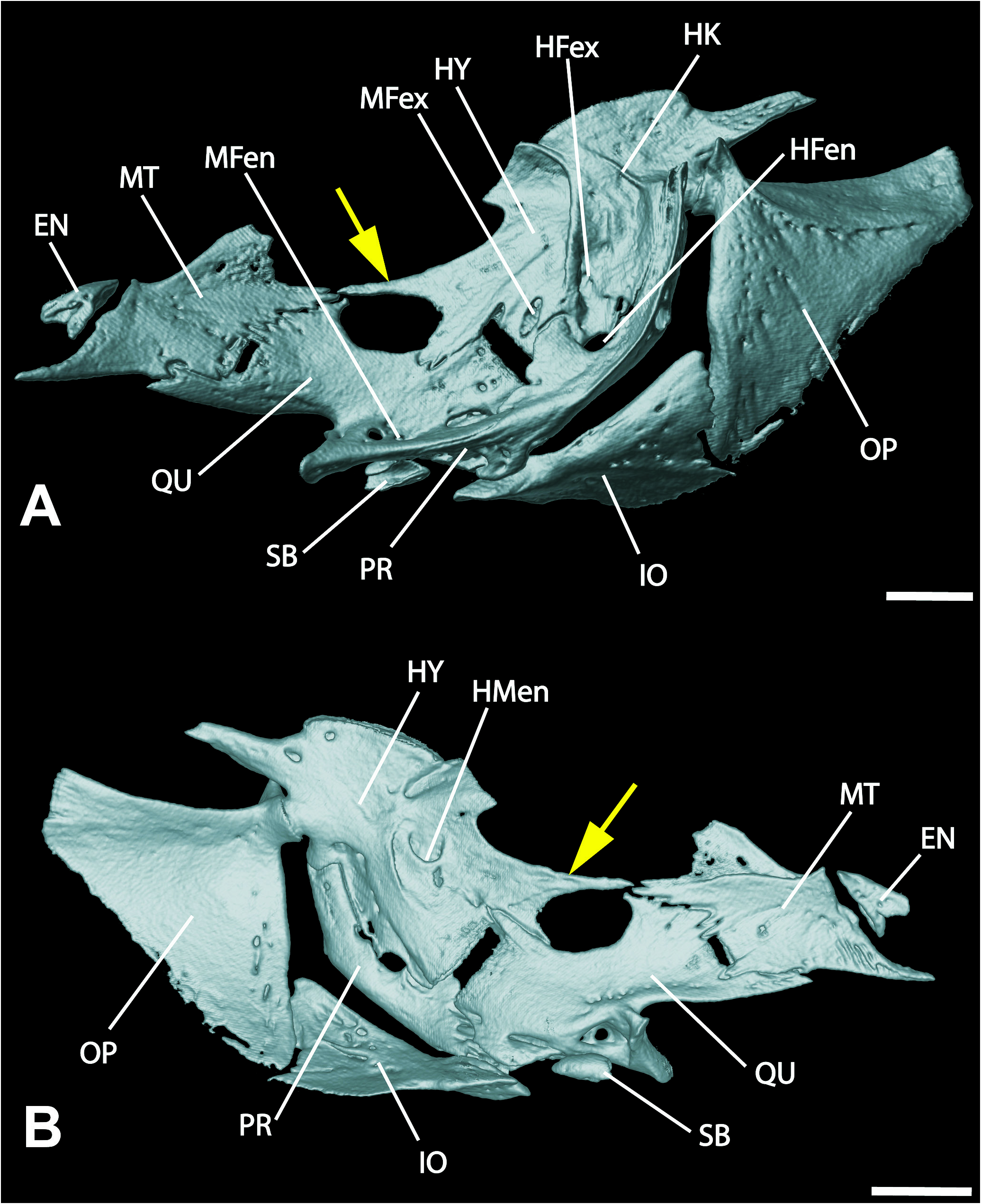
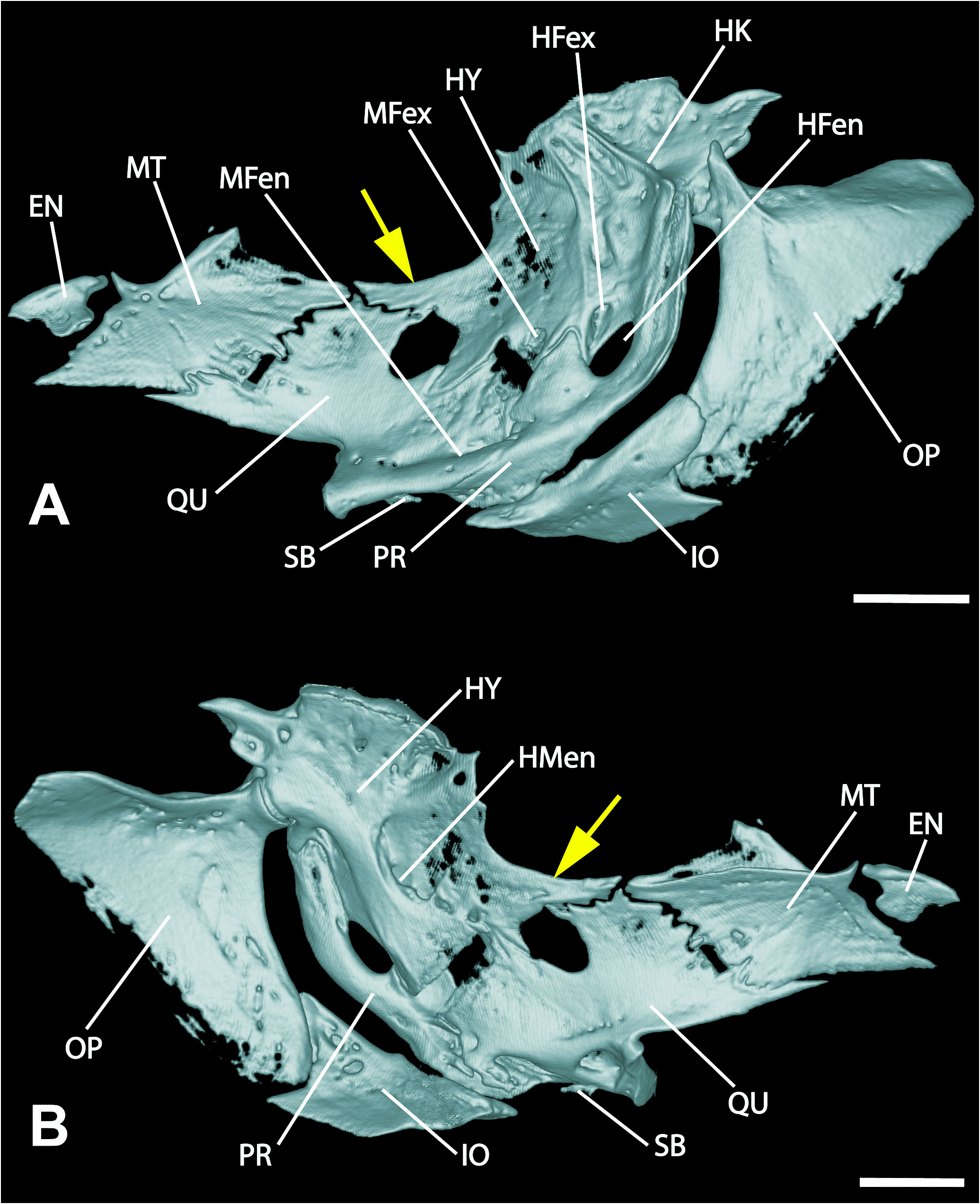
Cetopsorhamdia clathrata and C. spilopleura share with C. iheringi , C. insidiosa , C. nasus , and C. picklei two putatively apomorphic characteristics that are absent in C. boquillae . One is the presence of a conspicuous pointed process on the anterior border of hyomandibula ( Figs. 21-22 View Figure 21 View Figure 22 , see arrows), as illustrated and described for C.nasus by Ortega-Lara (2012:fig. 10). On the other hand, C.boquillae has the generalized heptapterid hyomandibula, lacking a pointed process at its anterior margin.
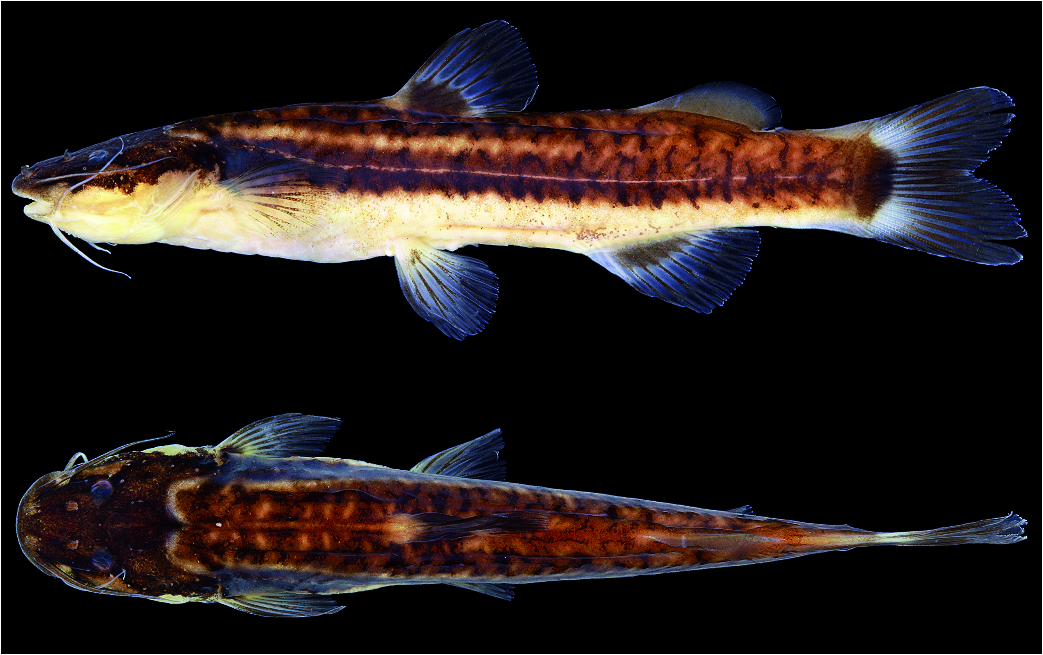
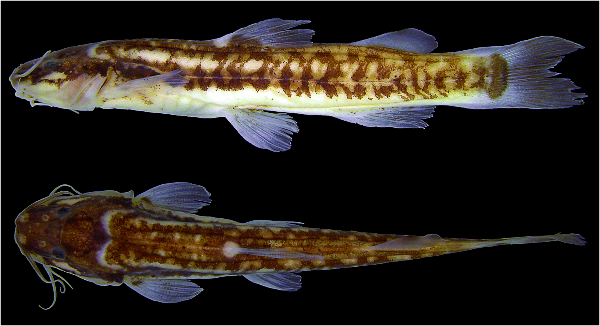
In addition, in all Cetopsorhamdia species other than C. boquillae the ventral lobe of the caudal fin is longer than the dorsal lobe ( cf. Eigenmann, 1916, 1922: pl. 4, fig. 1; Steindachner, 1915: pl. 12, fig. 7; Schultz, 1944: pl. 2, fig. d; Schubart & Gomes, 1959: fig. 1; Ortega-Lara, 2012: fig. 19). Such a condition, presumably derived, is exhibited by C. clathrata ( Figs. 1-3 View Figure 1 View Figure 2 View Figure 3 ) and C. spilopleura ( Figs. 12-14 View Figure 12 View Figure 13 View Figure 14 ), although the condition is more discrete in the latter species. Eigenmann (1922) imprecisely characterized the condition of ventral caudal-fin lobe of C. boquillae as “probably somewhat the longer” (likely due to the poor state of conservation of its type series). Indeed, the tips of the caudal-fin rays of the holotype and paratypes of C. boquillae are heavily damaged so that it is not possible to ascertain its state. However, the examination of a photograph of a live specimen of C. boquillae by Armando Ortega allowed us to determine the state of its caudal fin as having a dorsal lobe with the same length of the ventral lobe, or slightly longer.The caudal-fin lobes of approximately the same length or the dorsal lobe slightly longer than the ventral one is plesiomorphic for catfishes ( Bockmann & Miquelarena, 2008). A long ventral caudal-fin lobe also occurs homoplastically in the genus Phenacorhamdia ( cf. Britski, 1993; Bockmann, 1998; DoNascimiento & Milani, 2008).
Despite the striking morphological differences distinguishing Cetopsorhamdia clathrata from C. spilopleura , they share at least seven presumable apomorphies observed in the hyomandibula, in the dorsal and pectoral fins, and in the body coloration, which are suggestive of a sister group relationship between them.
As above commented, Cetopsorhamdia clathrata ( Fig. 21 View Figure 21 , see arrow) and C. spilopleura ( Fig. 22 View Figure 22 , see arrow) share with most of species of Cetopsorhamdia the presence of a conspicuous pointed process on the anterior border of hyomandibula. These two new species exhibit a further elongation in this process that reaches the metapterygoid so that these bones, together with quadrate, forming a large, rounded fenestra in the suspensorium ( Figs. 21-22 View Figure 21 View Figure 22 ).
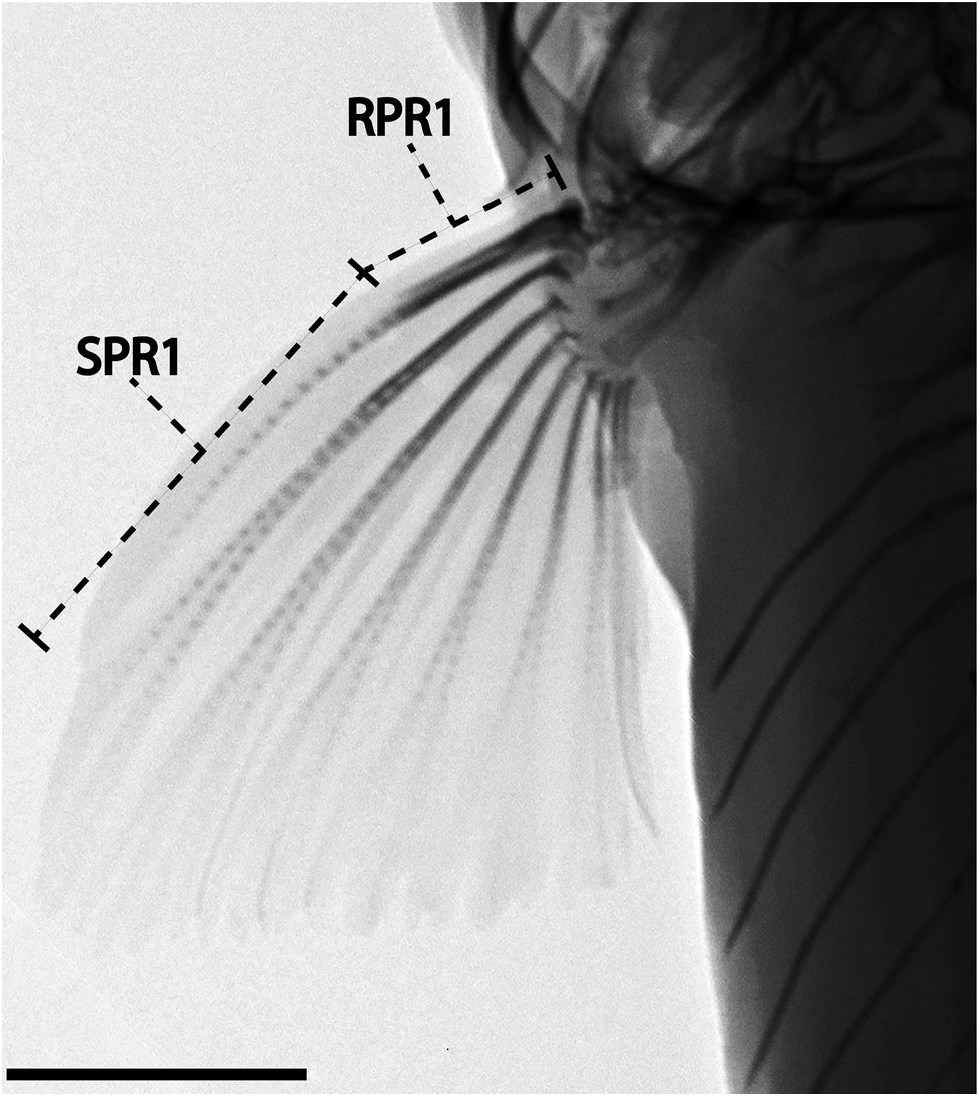
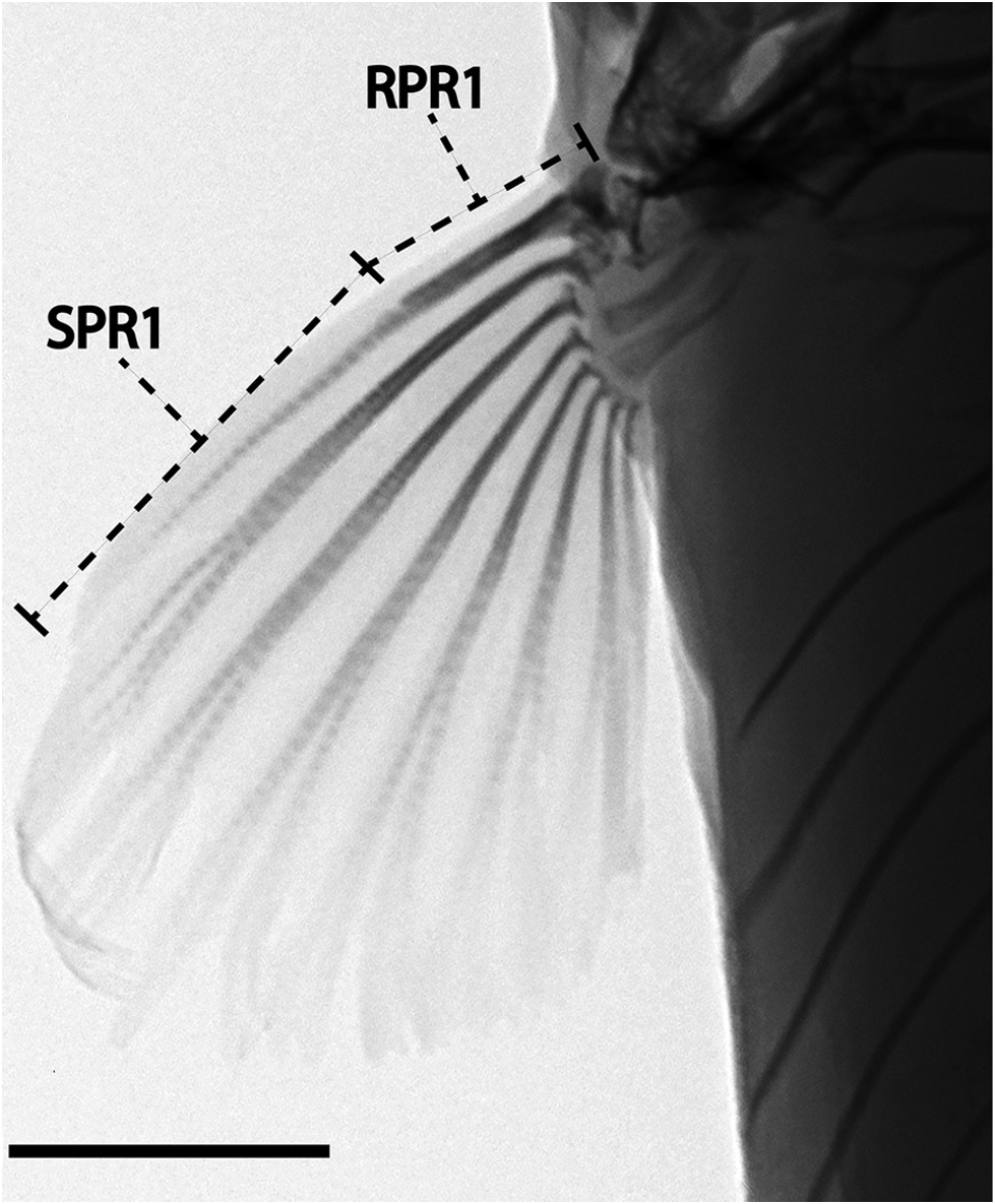
In Cetopsorhamdia clathrata and C. spilopleura the degree of ossification of the first (unbranched) dorsal- and pectoral-fin rays is quite distinct from the remaining Cetopsorhamdia species. In these species the first rays of the dorsal and pectoral fins are weakly ossified and stiffened only at their basal portions at most. In C. clathrata ( Fig. 9 View Figure 9 ) the range of variation of the length of the rigid part of the first dorsal-fin ray is 4.0-6.9% SL (x = 5.7, SD = 0.7) and of the first pectoral-fin ray is 3.4-6.2% SL (x = 4.6, SD = 0.7) ( Table 1), while in C. spilopleura ( Fig. 19 View Figure 19 ) is 5.3-6.4% SL (x = 5.8, SD = 0.4) and 4.1-5.1% SL (x = 4.4, SD = 0.3) ( Table 2). In opposition, the basal portion of first, undivided dorsal- and pectoral-fin rays of C. boquillae View in CoL , C. iheringi View in CoL ( Fig. 23A View Figure 23 ), C. insidiosa View in CoL ( Fig. 23B View Figure 23 ), C. nasus View in CoL , and C. picklei View in CoL is densely ossified in comparison to its distal segment, forming a somewhat rigid strut of about ⅓ and ½ size of the total length of those elements, respectively. This condition exhibited by most species of Cetopsorhamdia View in CoL is closer to that present in several successive basal lineages of the family Heptapteridae View in CoL in which more than half of the proximal portion of the first ray of the dorsal and the pectoral fins is heavily ossified and stiffened, sometimes forming a spine ( cf. Bockmann, 1998). Among these last-mentioned species of Cetopsorhamdia View in CoL , the proportions of the rigid part of the first dorsal-fin ray in SL are 8.2-12.9% SL whereas the proportions of the rigid part of the first pectoral-fin ray are 10.4-13.5% SL. No material of C. nasus View in CoL was available to be measured, but examination of the photographs and radiographs of its holotype and data provided by Ortega-Lara (2012) indicates that the first rays of the dorsal and pectoral fins are unquestionably rigid, at least for their ⅓ and ½ proximal parts, respectively, resembling the conditions exhibited by C. boquillae View in CoL , C. iheringi View in CoL , C. insidiosa View in CoL , and C. picklei View in CoL .
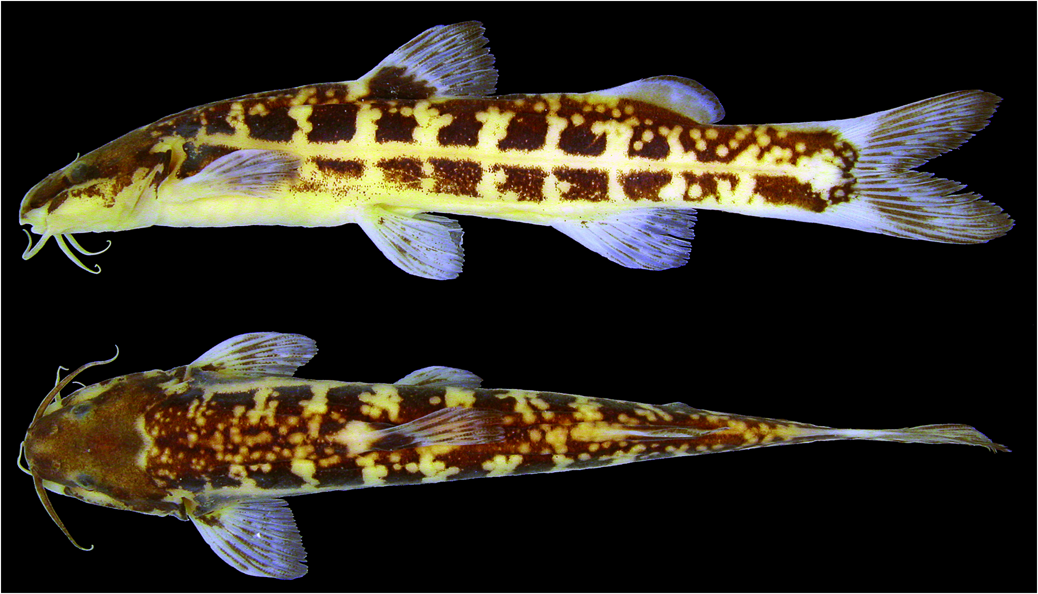
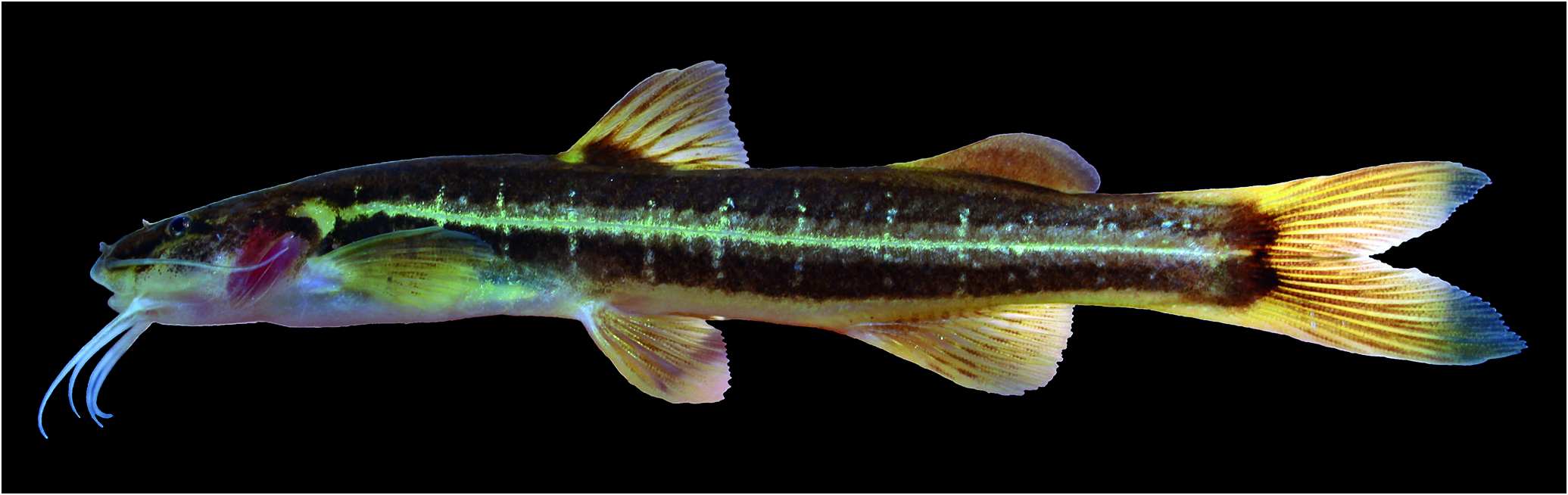
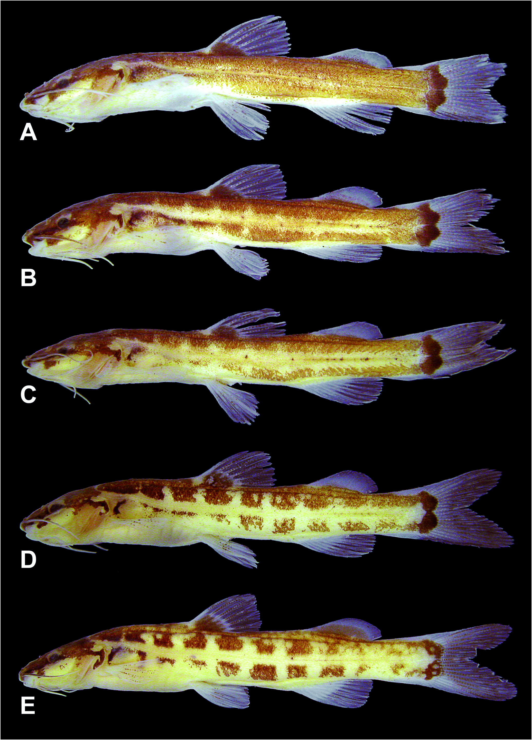
At last, most putative synapomorphies clustering together C. clathrata and C. spilopleura into a clade are observed in their color patterns, namely: (1) a dark stripe across the base of maxillary barbel to the region just posterior to the eye ( Figs. 1-4 View Figure 1 View Figure 2 View Figure 3 View Figure 4 , 12-14 View Figure 12 View Figure 13 View Figure 14 ); (2) a roughly rectangular unpigmented region just ventral to the eye ( Figs. 1-4 View Figure 1 View Figure 2 View Figure 3 View Figure 4 , 12-14 View Figure 12 View Figure 13 View Figure 14 ); (3) a dark, vertical bar at the posterior portion of opercle and branchiostegal membrane ( Figs. 1-4 View Figure 1 View Figure 2 View Figure 3 View Figure 4 , 12-14 View Figure 12 View Figure 13 View Figure 14 ); (4) a dark, vertical bar at the anterior portion of the trunk, dorsal to the pectoral fin ( Figs. 1-4 View Figure 1 View Figure 2 View Figure 3 View Figure 4 , 12-14 View Figure 12 View Figure 13 View Figure 14 ); and (5) each lobe of caudal fin with one ovoid, unpigmented area, of milky-looking in life, immediately posterior to the dark mark ( Figs. 1-3 View Figure 1 View Figure 2 View Figure 3 , 12-14 View Figure 12 View Figure 13 View Figure 14 ). None of the other species of Cetopsorhamdia View in CoL has any of these characteristics,alternatively possessing a mostly uniform dark coloration in the referred regions, without bands or unpigmented areas.
Four putative autapomorphies have been identified for Cetopsorhamdia clathrata : (1) unpigmented area in the posterodorsal part of the head, dorsally at the end of the opercular cleft ( Figs. 1-4 View Figure 1 View Figure 2 View Figure 3 View Figure 4 ); (2) trunk flanks with 10-12 quadrangular marks, separated by unpigmented vertical lines or bars ( Figs. 1-4 View Figure 1 View Figure 2 View Figure 3 View Figure 4 ); (3) midlateral region of the trunk, along the lateral line, devoid of pigmentation, forming a white stripe ( Figs. 1-3 View Figure 1 View Figure 2 View Figure 3 ); and (4) trunk with scattered unpigmented, rounded spots, mostly concentrated on its dorsal half ( Figs. 1 View Figure 1 , 3-4 View Figure 3 View Figure 4 ).
For Cetopsorhamdia spilopleura , in turn, three putative autapomorphies have been recognized: (1) anterodorsal region of the quadrate, outlining part of the suspensorium fenestra, expanded and turned backwards, broadly articulating with the ventral part of the anterior process of the hyomandibula ( Fig. 22 View Figure 22 ); (2) trunk flanks with 18-22 irregular, vertical brown bars, sometimes resembling inverted “v”,“y” or “x” ( Figs. 12-14 View Figure 12 View Figure 13 View Figure 14 ); and (3) trunk with laterodorsal unpigmented stripe, demarking a broad midlateral dark band along the lateral line ( Figs. 12-14 View Figure 12 View Figure 13 View Figure 14 ).


No known copyright restrictions apply. See Agosti, D., Egloff, W., 2009. Taxonomic information exchange and copyright: the Plazi approach. BMC Research Notes 2009, 2:53 for further explanation.
|
Kingdom |
|
|
Phylum |
|
|
Class |
|
|
Order |
|
|
Family |
|
|
Genus |
Cetopsorhamdia
| Bockmann, Flávio A. & Reis, Roberto E. 2021 |
Cetopsorhamdia
| Bockmann & Reis 2021 |
Cetopsorhamdia
| Bockmann & Reis 2021 |
Cetopsorhamdia
| Bockmann & Reis 2021 |
C. iheringi
| Schubart & Gomes 1959 |
C. iheringi
| Schubart & Gomes 1959 |
C. picklei
| Schultz 1944 |
C. picklei
| Schultz 1944 |
C. nasus
| Eigenmann & Fisher 1916 |
C. nasus
| Eigenmann & Fisher 1916 |
Heptapteridae
| T.N.Gill 1861 |