
Enterobius (Colobenterobius) emodensis, 2018
|
publication ID |
https://doi.org/ 10.11646/zootaxa.4514.1.5 |
|
publication LSID |
lsid:zoobank.org:pub:C9C5FC4C-4402-4FA0-BBE7-0814172BE2C0 |
|
DOI |
https://doi.org/10.5281/zenodo.5950814 |
|
persistent identifier |
https://treatment.plazi.org/id/BB1C842F-5D15-42D4-8941-7C4C109CF446 |
|
taxon LSID |
lsid:zoobank.org:act:BB1C842F-5D15-42D4-8941-7C4C109CF446 |
|
treatment provided by |
Plazi |
|
scientific name |
Enterobius (Colobenterobius) emodensis |
| status |
sp. nov. |
Enterobius (Colobenterobius) emodensis sp. n.
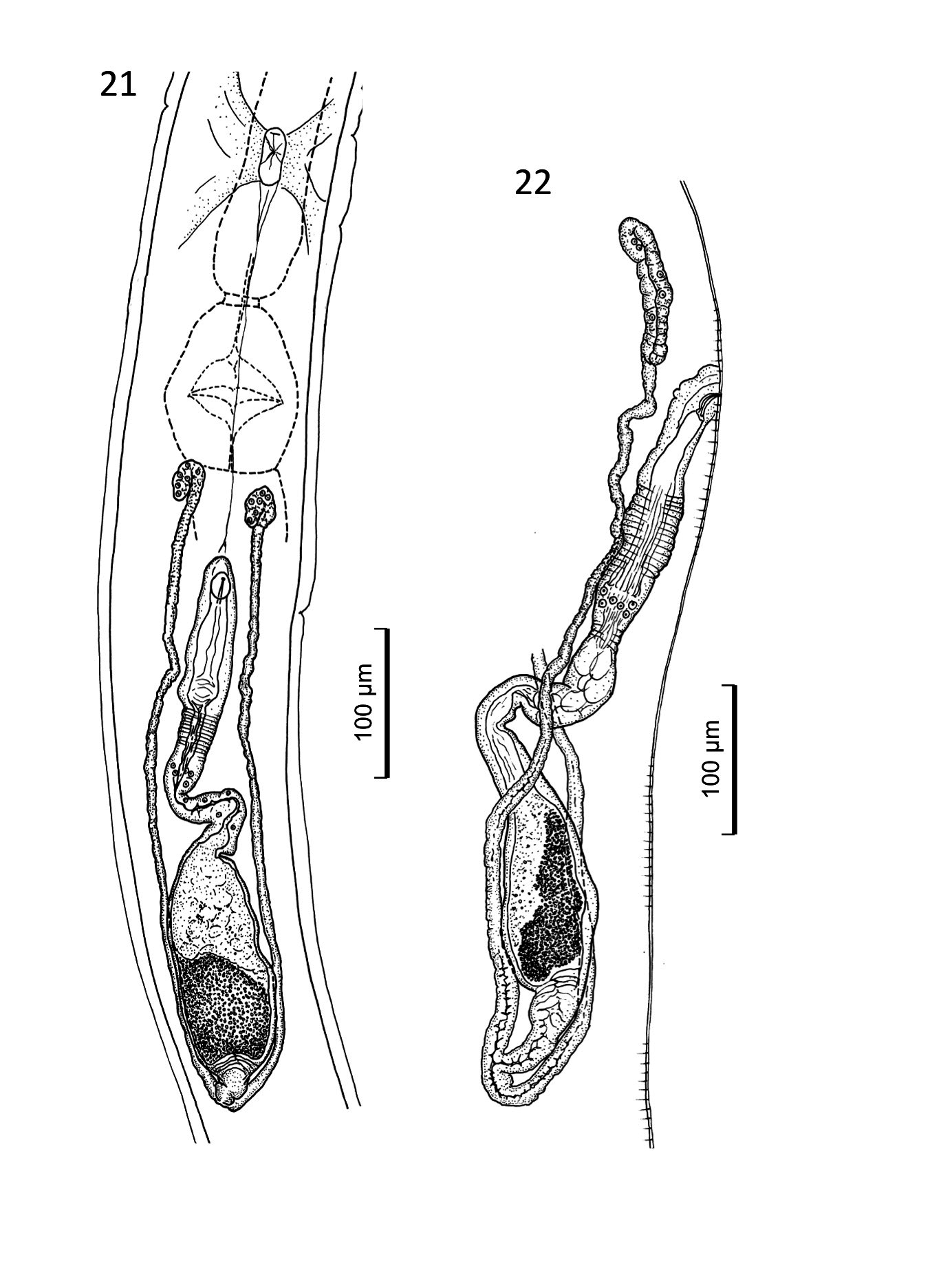
( Figs. 1–22 View FIGURES 1–7 View FIGURES 8–14 View FIGURES 15–20 View FIGURES 21, 22 )
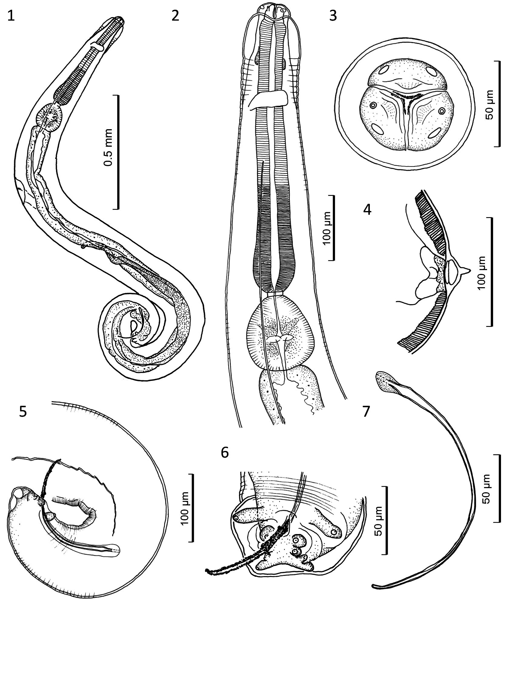
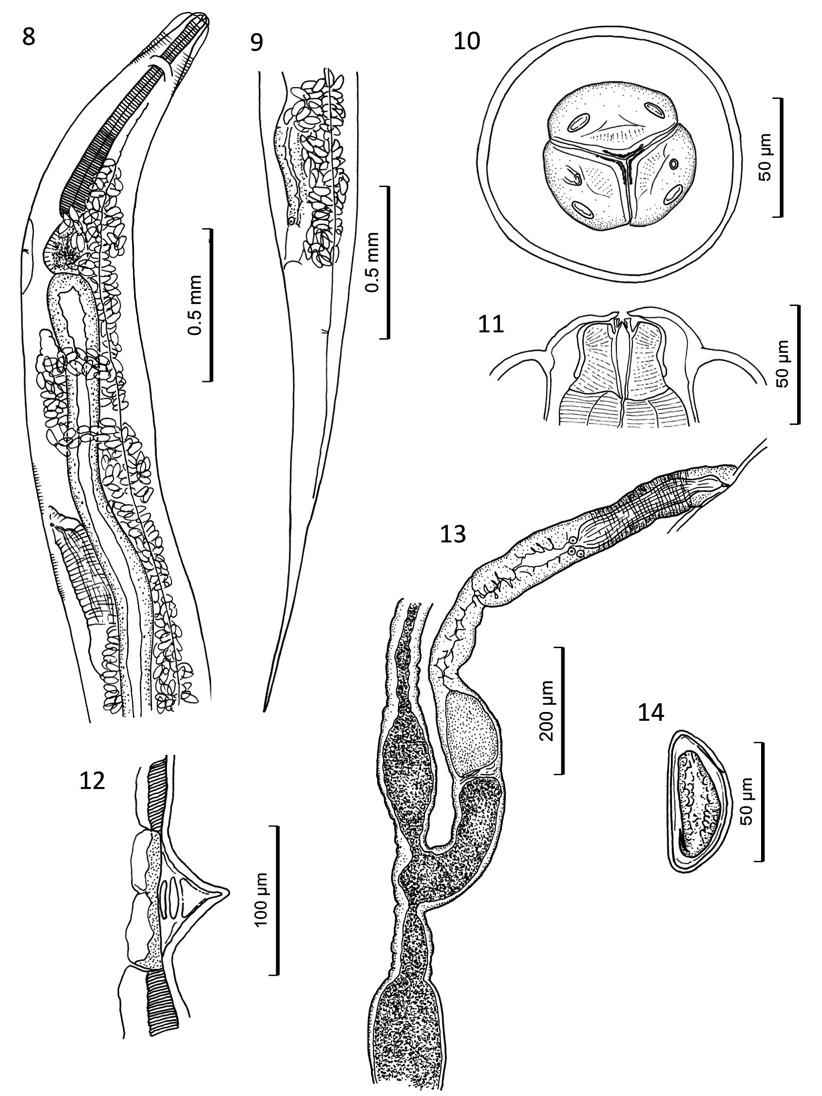
General: Body light brown in color. Cephalic expansion well developed, with internal septa in posterior half ( Figs. 1, 2 View FIGURES 1–7 , 8 View FIGURES 8–14 ). Cuticle with transverse striations. Lateral alae single crested, triangular in cross section ( Figs. 4 View FIGURES 1–7 , 12 View FIGURES 8–14 ). Cephalic end with 3 developed lips; dorsal lip with 2 cephalic papillae, subventral lips each with cephalic papilla and amphidial pore ( Figs. 3 View FIGURES 1–7 , 10 View FIGURES 8–14 ). Pharyngeal teeth with lamellated superstructures, corresponding to one-third of lip width in apical view (Figs, 3, 10); slots with cuticular wall extending to the posterior one-fourth of pharynx (Fig, 11). Esophageal corpus long club-shaped, with posterior one-third darker containing granules in wall ( Figs. 1, 2 View FIGURES 1–7 , 8 View FIGURES 8–14 ); short esophageal isthmus present but often hardly discernible in fully gravid females; esophageal bulb valved.
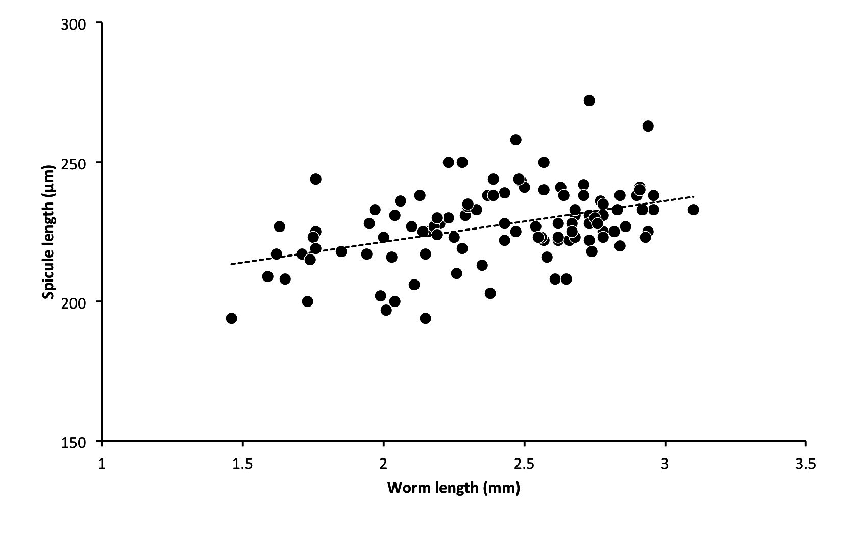
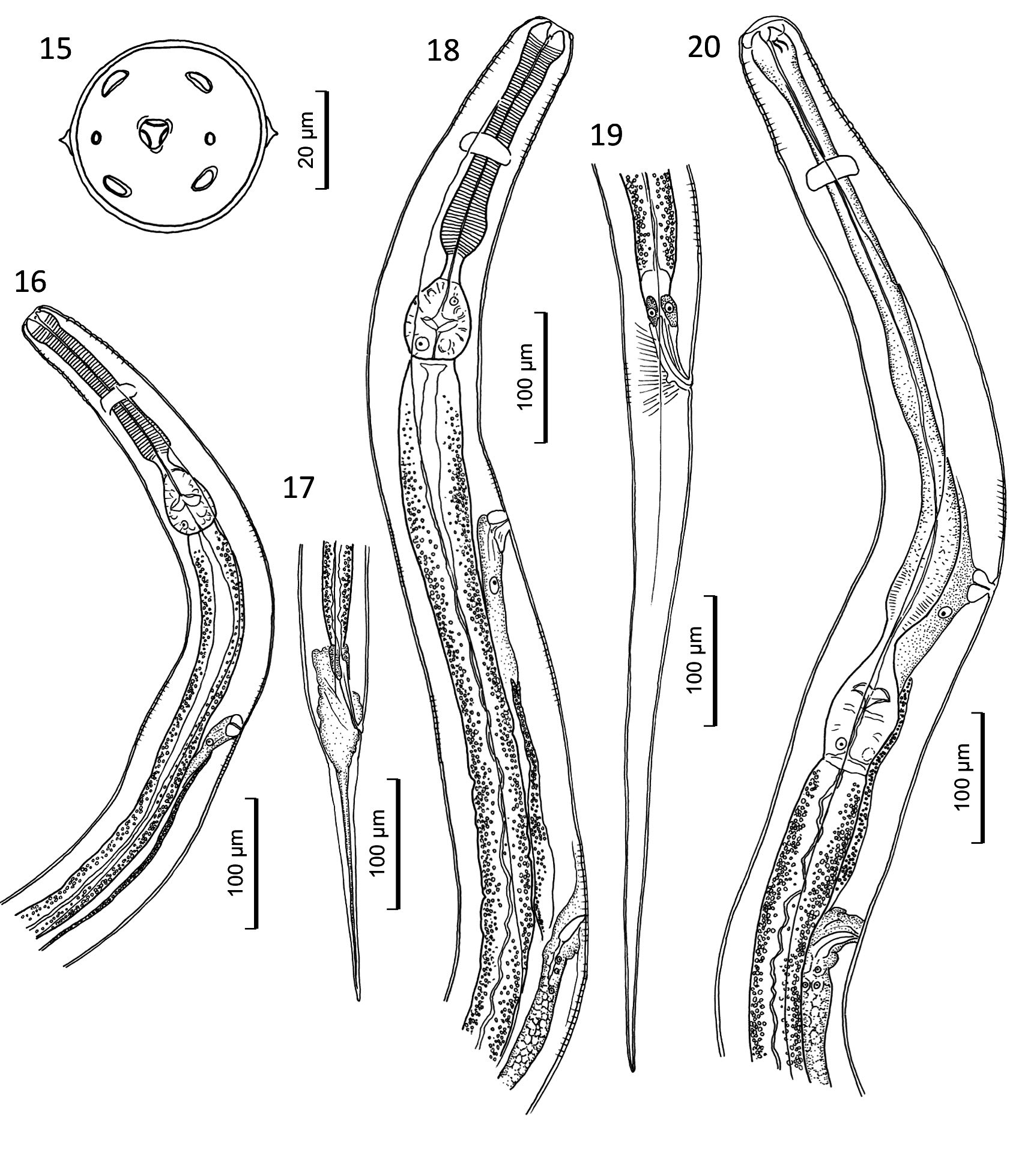
Male (Holotype and 10 paratypes): Body tapered to anterior end; posterior body bent ventrally, forming coils ( Fig. 1 View FIGURES 1–7 ). Length 2.61 (2.01–2.90) mm; maximum width 183 (143–216). Distance between amphidial pores 40. Lateral alae commencing at level slightly posterior to nerve ring, terminating at level anterior to caudal alae ( Figs. 2, 5 View FIGURES 1–7 ). Cephalic expansion 161 (123–186) long by 89 (75–97) wide. Pharynx 29 (28–32) long; esophageal corpus 431 (355–464) long by 66 (51–79) wide; isthmus 5 (3–6) long by 21 (18–24) wide; bulb 123 (91–135) long by 113 (95–125) wide. Total esophagus length including pharynx, corpus, isthmus and bulb 587 (480–620), corresponding to 22.5 (21.1–24.0) % of worm length. Nerve ring 157 (141–167) and excretory pore 795 (657–976) from cephalic apex. Caudal alae supported by 4 pairs of papillae: 1 st pair precloacal, pedunculate, large, directing laterally; 2 nd and 3 rd pairs near cloacal aperture, sessile, small, directing ventrally; 4 th pair pedunculate, directing posterolaterally; phasmidial pore near base of 4 th pair ( Figs. 5, 6 View FIGURES 1–7 ). Testis directing anteriorly ( Fig. 1 View FIGURES 1–7 ). Distal portion of testis filled with round spermatozoa each with ca. 2.5 diameter. Spicule slender, 223 (197–238) long, arched, bending ventrally distally, proximal end with light-refractive mass; portion protruding from the cloacal aperture often covered with dark material ( Figs. 5–7 View FIGURES 1–7 ). Relationship between worm length and spicule length shown in Fig. 23 View FIGURE 23 .
Female (Allotype and 11 paratypes): Body tapered to both extremities. Length 7.51 (7.15–8.10) mm, maximum width 428 (360–488). Distance between amphidial pores 42. Lateral alae commencing at level slightly posterior to nerve ring, terminating at middle of tail. Cephalic expansion 243 (204–257) long by 129 (106–143) wide. Pharynx 35 (32–36) long; esophageal corpus 739 (680–776) long by 103 (90–114) wide; isthmus 3 (2–4) long by 26 (24–28) wide (n=5); bulb 168 (162–178) long by 160 (147–172) wide. Total esophagus length 926 (796–992), corresponding to 12.4 (10.7–13.7) % of worm length. Nerve ring 210 (184–249), excretory pore 1.15 (0.94–1.32) mm (n=5) from cephalic apex. Distance from cephalic apex to vulva 1.88 (1.60–2.14) mm, corresponding to 25.0 (21.5–28.7) % of worm length ( Fig. 8 View FIGURES 8–14 ). Muscular vagina thick, directing posteriorly; vagina uterina with thick epithelium, about twice as long as muscular vagina; cellular wall present dividing length from vagina and opening to uterus ca. 3:1; amphidelphic; uterus of young gravid females filled with dark material, vagina uterina just before cellular wall containing mass of minute round structures each with ca. 2.5 diameter ( Fig. 13 View FIGURES 8–14 ). Tail slender, gradually tapering to sharp point, 1.49 (1.25–1.73) mm long, occupying 19.8 (16.8–21.6) % of worm length ( Fig. 9 View FIGURES 8–14 ). Phasmidial pore opening on lateral ala at level slightly posterior to anus. Eggs with one side flattened, 60.6 ± SD 1.2 (59–63) by 27.1 ± SD 0.6 (26–28) (n=25), convex side with operculum-like structure near pole ( Fig. 14 View FIGURES 8–14 ).
No known copyright restrictions apply. See Agosti, D., Egloff, W., 2009. Taxonomic information exchange and copyright: the Plazi approach. BMC Research Notes 2009, 2:53 for further explanation.