
Betaburmesebuthus fuscus, XUAN & CAI & HUANG, 2023
|
publication ID |
https://doi.org/ 10.11646/palaeoentomology.6.1.10 |
|
publication LSID |
lsid:zoobank.org:pub:B822ED53-D559-4DD8-8803-3F2850A00445 |
|
DOI |
https://doi.org/10.5281/zenodo.7757601 |
|
persistent identifier |
https://treatment.plazi.org/id/4788832D-992B-4A9A-BD43-E94C48BFC7DA |
|
taxon LSID |
lsid:zoobank.org:act:4788832D-992B-4A9A-BD43-E94C48BFC7DA |
|
treatment provided by |
Plazi |
|
scientific name |
Betaburmesebuthus fuscus |
| status |
sp. nov. |
Betaburmesebuthus fuscus sp. nov.
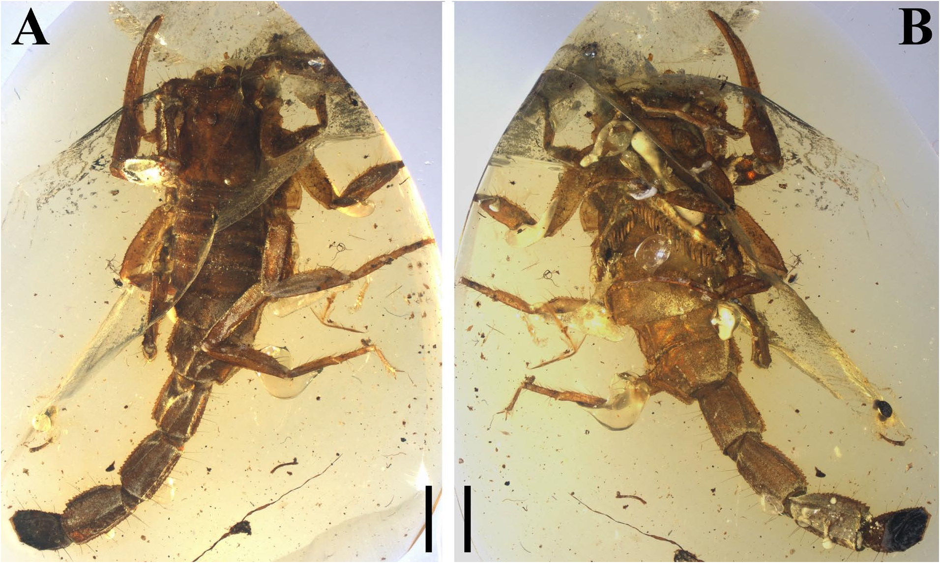
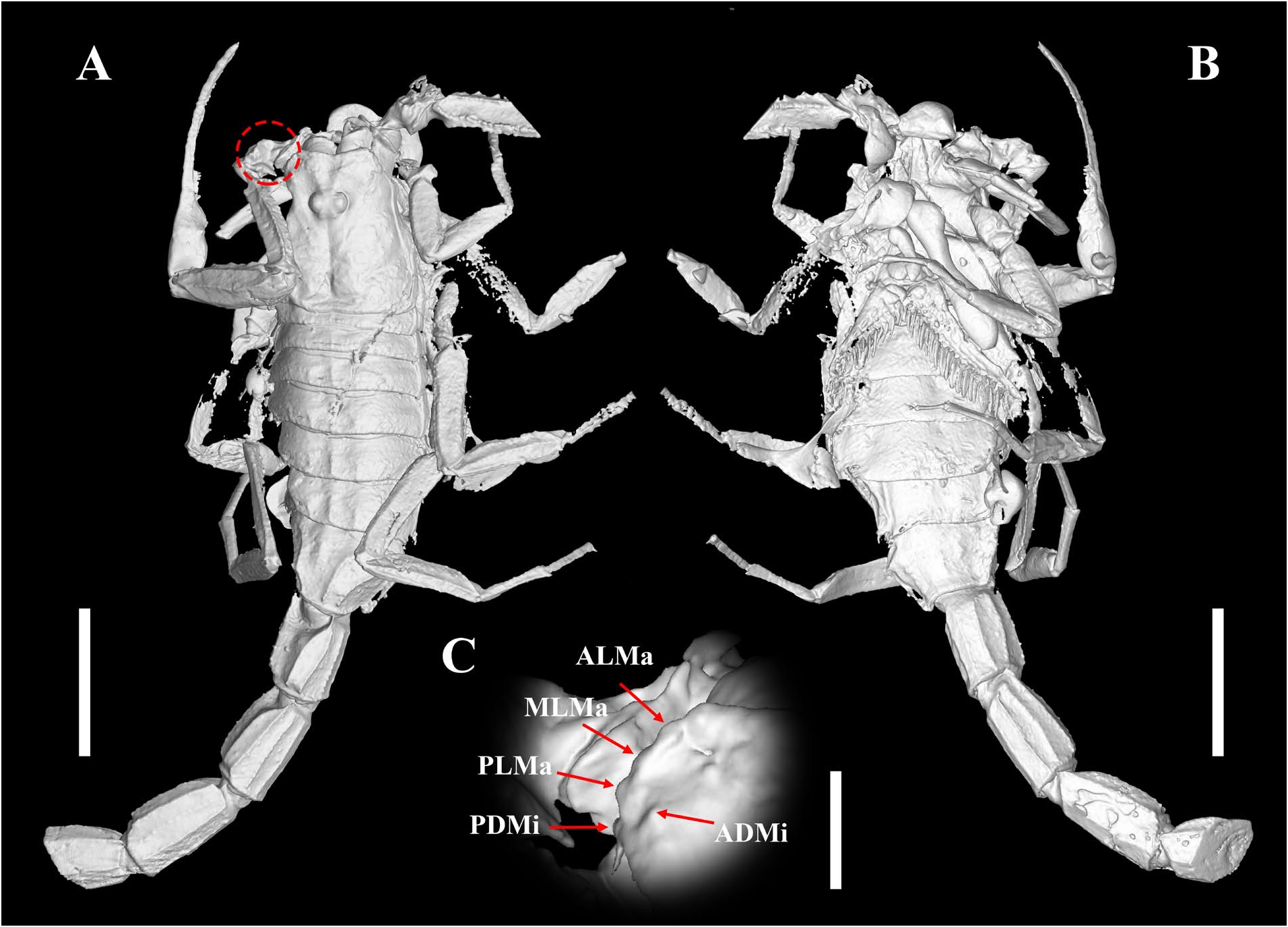
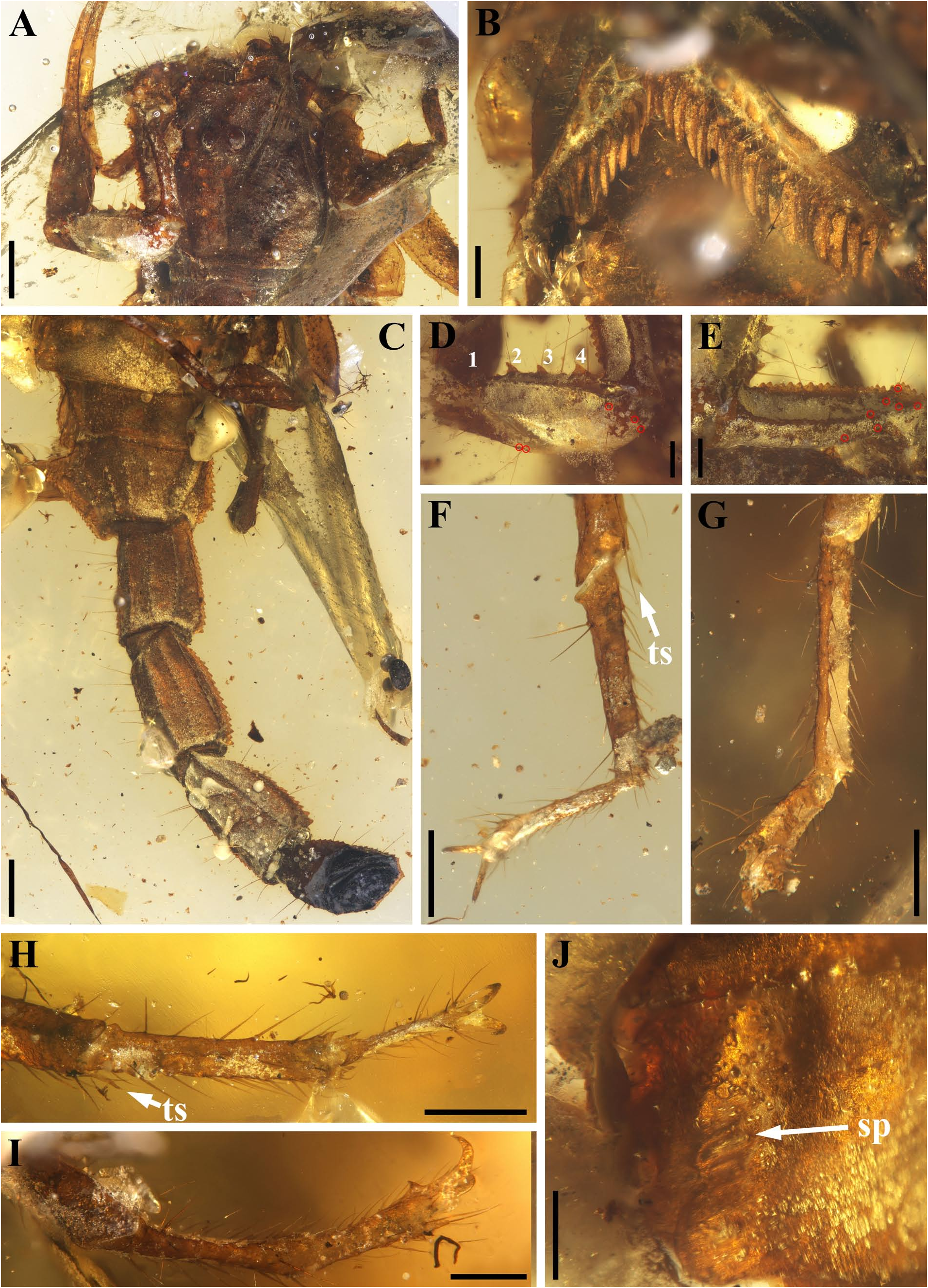
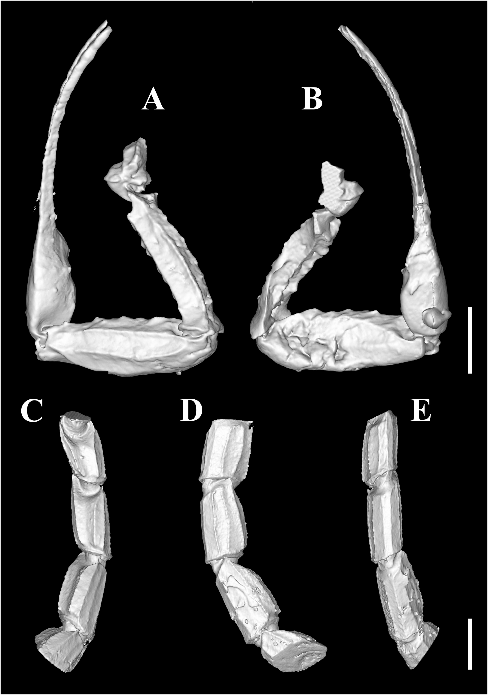
( Figs 27–30 View FIGURE 27 View FIGURE 28 View FIGURE 29 View FIGURE 30 )
Zoobank LSID urn:lsid:zoobank.org:act:4788832D-992B-4A9A-BD43-E94C48BFC7DA
Material. NIGP201154 , one probable adult male, metasoma IV and V, telson and part of right pedipalp missing.
Etymology. The species is named after the color of scorpion body; the Latin ‘ fuscus ’, meaning dark brown. The name masculine in gender.
Diagnosis(emended). Thisnewspeciescanbeclearly distinguished from other Betaburmesebuthus species by the following set of characters:1) general integument color dark brown 2) anterior margin of carapace with a single moderate marked median concavity ( Figs 28A View FIGURE 28 , 29A View FIGURE 29 ); 3) spiracles small, oblique and slit ( Fig. 29J View FIGURE 29 ); 4) pectines with 20 teeth ( Fig. 29B View FIGURE 29 ); 5) dorsal patellar spur carina with four strong tubercles ( Fig. 29D View FIGURE 29 ); 6) Chela manus with one small tubercle on internal surface ( Fig. 30 View FIGURE 30 ); and 7) telotarsus covered by numerous ventrosubmedian setae ( Fig. 29F–I View FIGURE 29 ).
Locality and horizon. Noije Bum near Tanai, Hukawng Valley, Kachin State of northern Myanmar; upper Albian to lower Cenomanian (mid-Cretaceous).
Description. Carapace. Covered by coarse granules without distinct carinae, posterior and posterior median furrows evident ( Figs 28A View FIGURE 28 , 29A View FIGURE 29 ); median eyes oval, large sized and separated by one ocular diameter ( Fig. 29A View FIGURE 29 ); PDMi posterodorsal to PLMa, and ADMi posterior to PLMa ( Fig. 28C View FIGURE 28 ).
Coxosternal region. A deep groove observed between coxapophysis I and coxa I ( Fig. 28B View FIGURE 28 ); genital operculum longitudinally divided and composed of two oval valves ( Fig. 28B View FIGURE 28 ).
Chelicerae. With setae on internal surface ( Fig. 29A View FIGURE 29 ); cheliceral dentition not visible except a long dorsal distal (dd) denticle ( Fig. 29A View FIGURE 29 ).
Pedipalps.Femur with five carinae( Figs29E View FIGURE 29 , 30A, B View FIGURE 30 ): internomedian carina well-marked and serratocrenulate; dorsointernal carina costate; dorsoexternal, ventroexternal and ventrointernal carinae crenulate. Patella with seven carinae ( Figs 29D View FIGURE 29 , 30A, B View FIGURE 30 ): Dorsal Patellar Spur carina (DPSc) and Ventral Patellar Spur carina (VPSc) welldeveloped; dorsointernal, dorsomedian and ventrointernal carinae costate with several granules; ventroexternal and dorsoexternal carinae smooth and costate; two evident macrosetae present on distal. Chela relatively slender (Cl/ Cw = 6.74, Table 1 View TABLE 1 ); finger denticle rows not visible.
Trichobothrial pattern ( Fig. 29D, E View FIGURE 29 ). Trichobothria not clear. Femur with 7 trichobothria observed, 5 dorsal and 2 external trichobothria, trichobothrium e 1 proximal to trichobothrium d 5. Patella with 4 trichobothria observed, including 1 dorsal and 3 external trichobothria.
Legs. Trochanter bearing a lateral apophysis ( Fig. 28B View FIGURE 28 ); femur with serrated internal and external carinae evident ( Fig. 27B View FIGURE 27 ); patella incrassate with serrated internal carina ( Fig. 27B View FIGURE 27 ). Prolateral and retrolateral pedal spurs present on all legs ( Fig. 29F–I View FIGURE 29 ). Ungues very long, dactyl pointed ( Fig. 29F–I View FIGURE 29 ).
Pectines ( Fig. 29B View FIGURE 29 ). Basal piece with an anterior median furrow evident ( Fig. 28B View FIGURE 28 ). Pectines with 3 marginal lamellae and 10 median lamellae; fulcra present, small. Numerous sensory hairs extending from surface of lamellae. Peg sensillae very short and sensory area fully occupied distal end of teeth.
Mesosoma. Tergites covered by coarse granules ( Fig. 27A View FIGURE 27 ), median carina evident on posterior half of all tergites ( Fig. 28A View FIGURE 28 ); dorsolateral carinae evident on tergites III and VI ( Fig. 28A View FIGURE 28 ); tergite VII with five costate carinae ( Fig. 28A View FIGURE 28 ): paired dorsolateral and lateral carinae, and one axial carina on anterior half. Sternites covered by coarse granules; Sternite VII with five carinae ( Fig. 29C View FIGURE 29 ).
Metasoma ( Figs 29C View FIGURE 29 , 30C–E View FIGURE 30 ). All segments longer than width and covered by macrosetae ventrally. Segments I–III with distinct depression dorsally; segments I–II with ten carinae (paired ventromedian, ventrolateral, lateral, dorsolateral, and dorsal carinae); segments III with eight well marked carinae (paired ventromedian, ventrolateral, dorsolateral, and dorsal carinae) and two feebly marked lateral carinae; all ventral and dorsal carinae obviously serrated.
Remarks. The most distinguishing character differentiating B. fuscus from others is the general body coloration and other species all exhibit yellowish integument. In addition, the slit-like spiracle is shared by B. larafleissnerae and B. villosus sp. nov. but not oblique in them.
No known copyright restrictions apply. See Agosti, D., Egloff, W., 2009. Taxonomic information exchange and copyright: the Plazi approach. BMC Research Notes 2009, 2:53 for further explanation.