
Betaburmesebuthus bellus Lourenço, 2016
|
publication ID |
https://doi.org/ 10.11646/palaeoentomology.6.1.10 |
|
publication LSID |
lsid:zoobank.org:pub:B822ED53-D559-4DD8-8803-3F2850A00445 |
|
DOI |
https://doi.org/10.5281/zenodo.7754824 |
|
persistent identifier |
https://treatment.plazi.org/id/F308E17F-4358-FFEA-FCCD-4B45FA701CC2 |
|
treatment provided by |
Plazi |
|
scientific name |
Betaburmesebuthus bellus Lourenço, 2016 |
| status |
|
Betaburmesebuthus bellus Lourenço, 2016
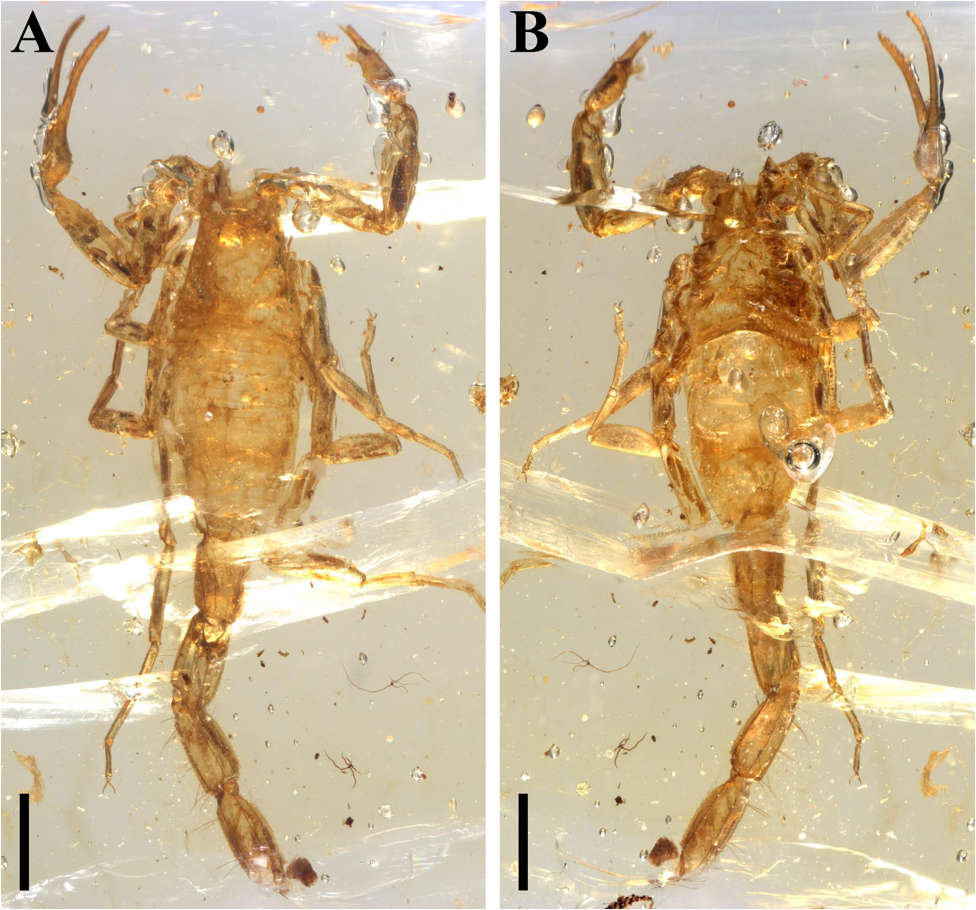
( Figs 1–7 View FIGURE 1 View FIGURE 2 View FIGURE 3 View FIGURE 4 View FIGURE 5 View FIGURE 6 View FIGURE 7 )
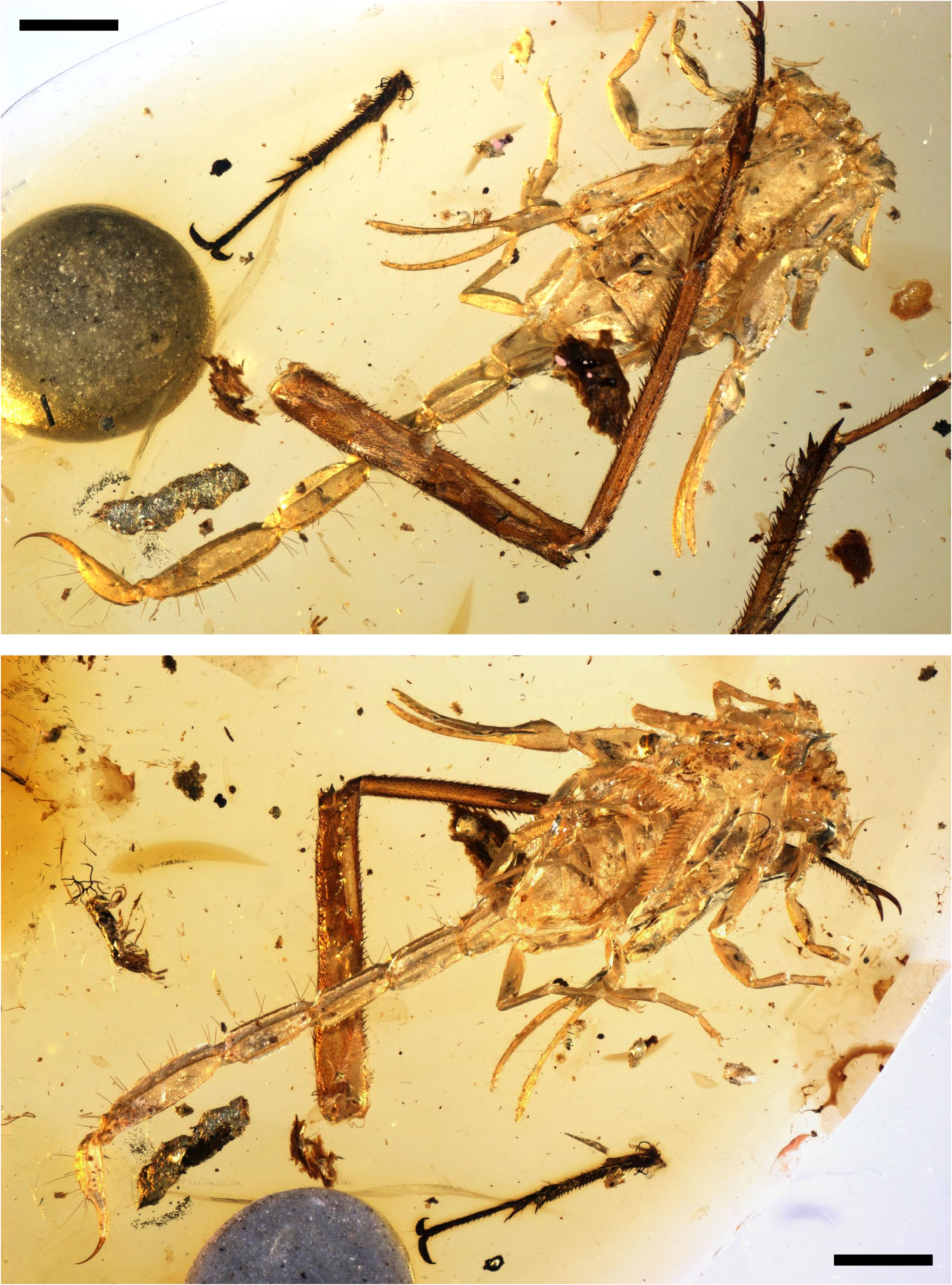
Material. NIGP 200648, one probable juvenile male, the part of right pedipalp finger and metasoma damaged. NIGP 200649, one probable juvenile female, a complete scorpion.
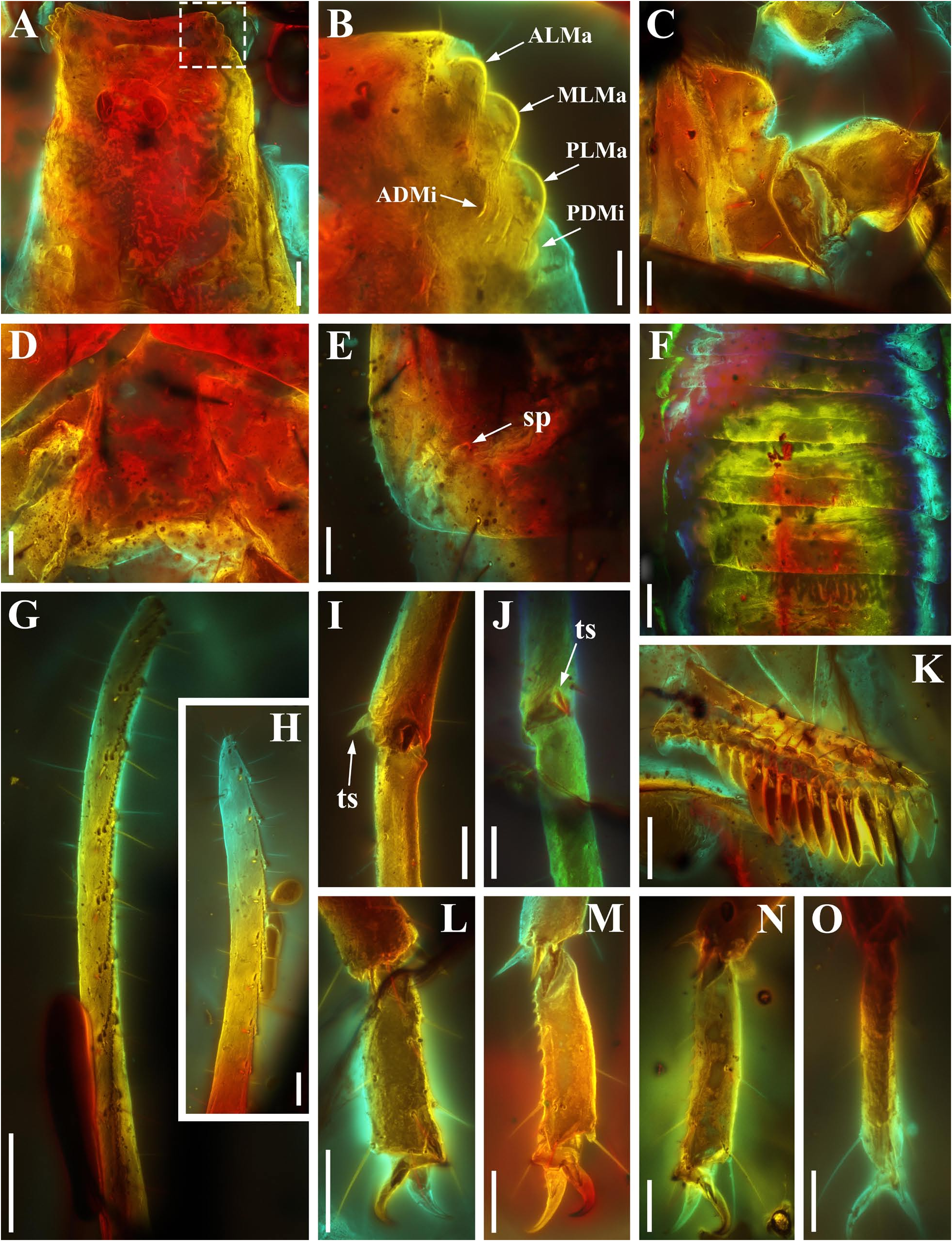
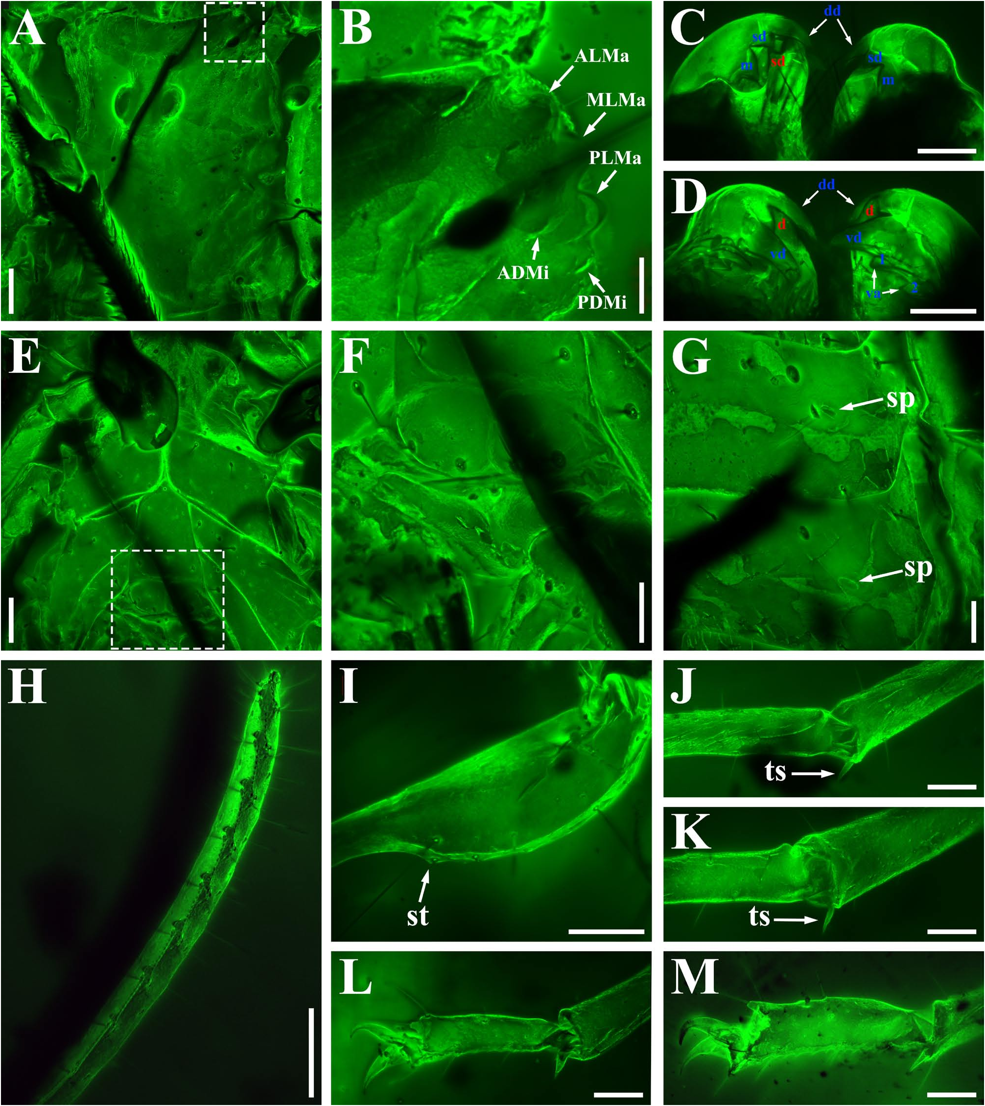
Diagnosis (emended). This species can be clearly distinguished from other congeners by the following set of characters: 1) anterior margin of carapace with a single moderately marked median concavity ( Figs 2A View FIGURE 2 , 6A View FIGURE 6 ); 2) spiracles small, oblique and oval to slit-like ( Figs 2E View FIGURE 2 , 6G View FIGURE 6 ; Lourenço, 2016: fig. 7); 3) pectines with 17–18 teeth ( Figs 2K View FIGURE 2 , 5B View FIGURE 5 ; Lourenço, 2016: fig. 7); 4) vesicle pear-shaped and very long, and aculeus very long and moderately curved ( Figs 5E View FIGURE 5 , 6I View FIGURE 6 ; Lourenço, 2016: fig. 4); 5) dorsal patellar spur carina well-developed with a strong tubercle and three relatively small spinous tubercles ( Figs 3D, E, F View FIGURE 3 , 7C, D View FIGURE 7 ); 6) Chela manus with two small spinous tubercles on internal surface ( Figs 3G, I, J, K View FIGURE 3 , 7F, G View FIGURE 7 ; Lourenço, 2016: figs 5, 9); 7) telotarsus with few ventrosubmedian setae and a ventromedian spinules row ( Figs 2M, N View FIGURE 2 , 6L, M View FIGURE 6 ).
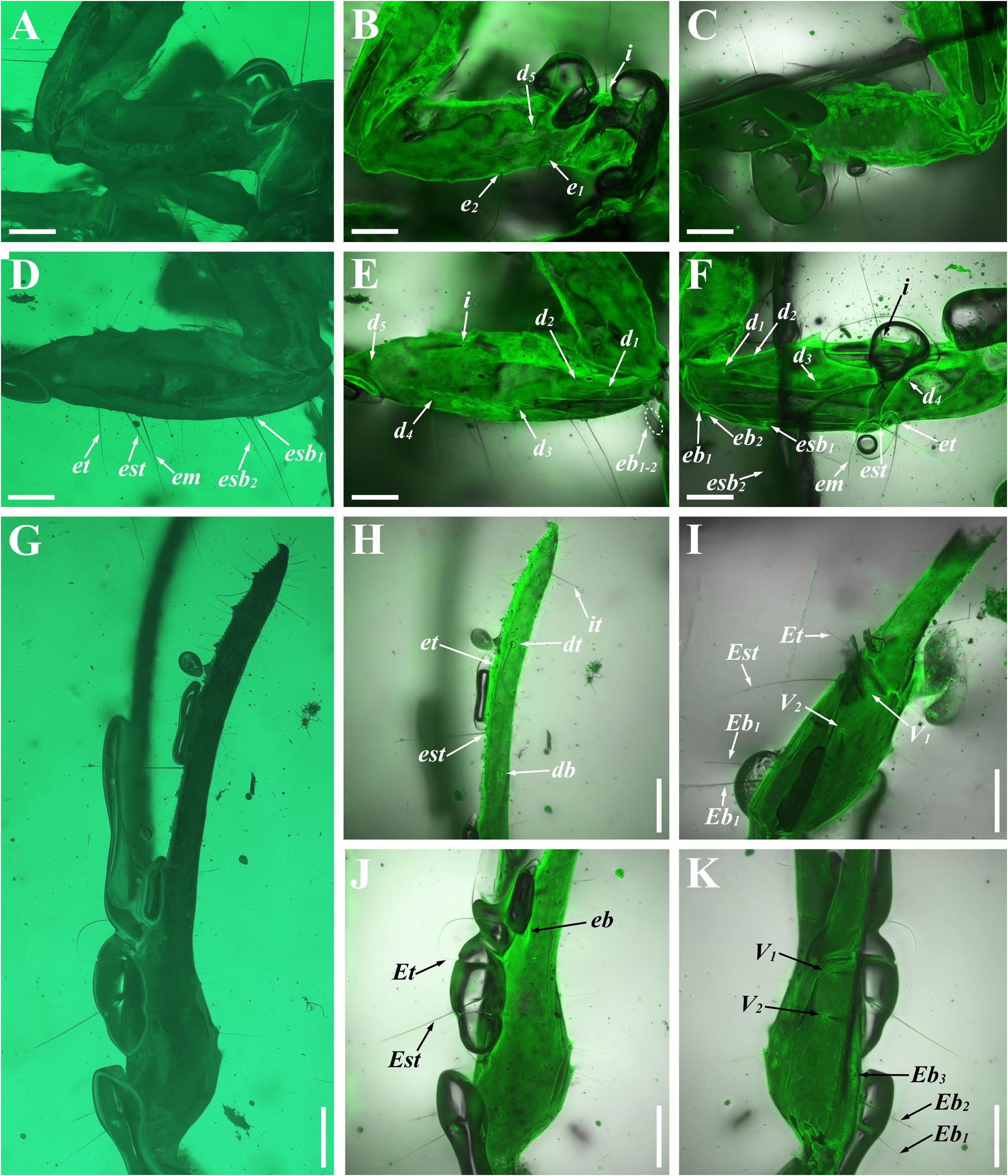
Description. Carapace. Sparsely covered by fine granules without distinct carinae and furrows ( Figs 2A View FIGURE 2 , 6A View FIGURE 6 ); median eyes oval and medium sized, and separated by more than one ocular diameter ( Figs 2A View FIGURE 2 , 6A View FIGURE 6 ); PDMi posterodorsal to PLMa, and ADMi dorsal to PLMa ( Figs 2B View FIGURE 2 , 6B View FIGURE 6 ).
Coxosternal region.Surface smooth with several setae ( Figs 2C View FIGURE 2 , 6E View FIGURE 6 ); anterior margin of coxapophysis I rounded densely covered by fine setae ( Figs 2C View FIGURE 2 , 6E View FIGURE 6 ); lateral margin of sternum very long, and posterior margin of sternum slightly incurved and as long as genital operculum ( Figs 2D View FIGURE 2 , 6E View FIGURE 6 ), posterior depression region small and not obvious ( Fig. 6E View FIGURE 6 ); genital operculum longitudinally divided and composed of two nearly rounded valves ( Fig. 6F View FIGURE 6 ).
Chelicerae.With setae on internal and ventral surface; cheliceral dentition partly visible ( Fig. 6C, D View FIGURE 6 ), including a long dorsal distal (dd) denticle, a small subdistal (sd) denticle, a stout median (m) denticle, a long ventral distal (vd) denticle and two small ventral accessory (va) denticle in movable finger, and distal (d) and subdistal (sd) denticles in fixed finger.
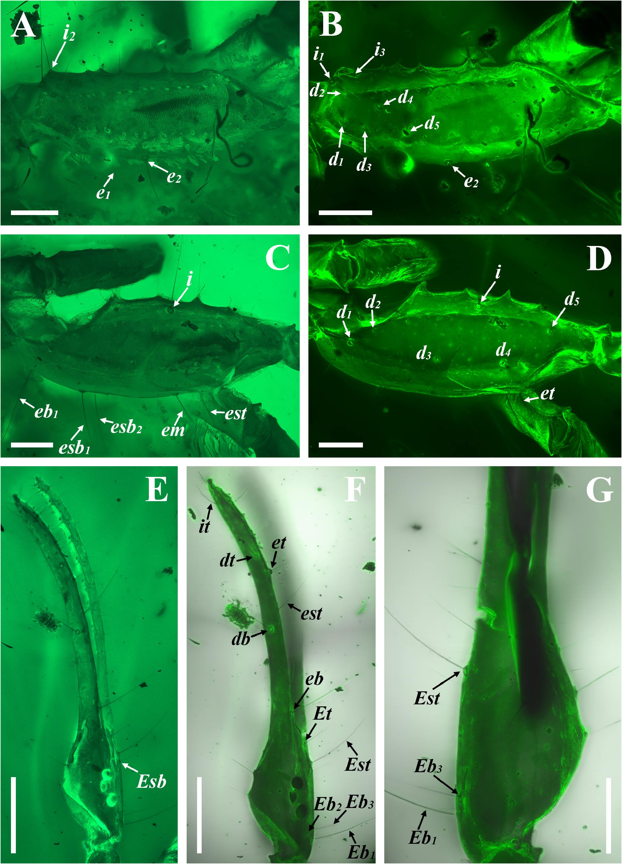
Pedipalps. Femur with five carinae ( Figs 3A–C View FIGURE 3 , 7A, B View FIGURE 7 ): internomedian carina well-developed with about five small tubercles; dorsointernal carina costate with several granules; dorsoexternal carina feebly marked with few granules; ventroexternal carina costate with few small tubercles; ventrointernal carina costate and incomplete. Patella with seven carinae ( Figs 3D–F View FIGURE 3 , 7C, D View FIGURE 7 ): Dorsal Patellar Spur carina(DPSc)andVentral Patellar Spur carina (VPSc) well-developed and each apophysis or tubercle possessing one microseta; dorsointernal, dorsomedian and dorsoexternal carinae smooth and costate; ventroexternal and ventrointernal carinae incomplete and costate. Carinae on chela not evident; Chela relatively slender (Cl/Cw = 6.19 or 6.23, Table 1 View TABLE 1 ); each finger denticle row consisting of about 10–11 (first row with 5) granules and a thick and short seta present beneath each accessory granule.
Trichobothrial pattern ( Figs 3 View FIGURE 3 , 7 View FIGURE 7 ). Pedipalp femur with 10 trichobothria, 5 dorsal, 3 internal and 2 external trichobothria, trichobothrium d 2 straddling dorsointernal carina ( Fig. 7A, B View FIGURE 7 ) and trichobothrium e 1 proximal to trichobothrium d 5 ( Figs 3A, B View FIGURE 3 , 7A, B View FIGURE 7 ). Patella with 13 trichobothria, including 5 dorsal (d 2 petite), 1 internal and 7 external trichobothria; trichobothrium d 3 internal to dorsomedian carina and trichobothrium d 4 external to dorsomedian carina ( Figs 3D, E View FIGURE 3 , 7C, D View FIGURE 7 ). Fixed finger with 1 internal, 3 external, and 2 dorsal trichobothria. Chela manus with 6 external and 2 ventral trichobothria.
Legs. Trochanter bearing a lateral apophysis ( Fig. 5B View FIGURE 5 ). Prolateral and retrolateral pedal spurs present on all legs ( Fig. 2L–O View FIGURE 2 ). Ungues moderately long, dactyl pointed ( Figs 2L–O View FIGURE 2 , 5H, I View FIGURE 5 , 6L, M View FIGURE 6 ).
Pectines ( Figs 2K View FIGURE 2 , 5B View FIGURE 5 ). Basal piece inverted trapezoid with an anterior median furrow ( Fig. 6F View FIGURE 6 ). Pectines with 3 marginal lamellae and 8–9 median lamellae; fulcra present, very small. Short and transparent sensory hairs extending from surface of lamellae. Teeth elongated and terminal one ovoid, peg sensillae very short and sensory area occupied about three quarters of teeth.
Mesosoma. Tergites finely granular, median carina evident on posterior half of tergites III–VI ( Figs 2F View FIGURE 2 , 5A View FIGURE 5 ); dorsolateral carinae observed on tergites IV–VI ( Fig. 2F View FIGURE 2 ); tergite VII with five costate carinae ( Fig. 5A View FIGURE 5 ): paired dorsolateral and lateral carinae, and one axial carina on anterior half. Sternites covered by some fine granules and sparse setae; posterior edge of sternites I–IV incurved ( Fig. 5B View FIGURE 5 ).
Metasoma ( Figs 1A, B View FIGURE 1 , 5C–E View FIGURE 5 ). All segments longer than width and covered by macrosetae ventrally. Segments I–IV with week depression dorsally; segments I and II with ten well marked carinae (paired ventromedian, ventrolateral, lateral, dorsolateral, and dorsal carinae); segments III and IV with eight well marked carinae (paired ventromedian, ventrolateral, dorsolateral, and dorsal carinae) and lateral carinae reduced on segment III; segment V with five carinae (single ventromedian carina, paired ventrolateral and dorsolateral carinae); dorsal carinae on segments II–IV distinctly serrated, other carinae smooth to serrated.
Telson ( Figs 5E View FIGURE 5 , 6I View FIGURE 6 ). Vesicle with ventromedian and ventrosubmedian carinae evident and covered by several macrosetae ventrally, a small subaculear tuberance present on terminal of ventromedian carina and subaculear setal pair significantly evident; vesicle/aculeus juncture moderately wide and shorter than half of vesicle; aculeus with several microsetae proximally and progressively darker towards distal.
Remarks. We add some additional characters to diagnosis, i. e., the DPS of patella and telotarsus armature, which were not mentioned in the original descriptions.

| NIGP |
Naking Institute of Geology and Palaeontology |
No known copyright restrictions apply. See Agosti, D., Egloff, W., 2009. Taxonomic information exchange and copyright: the Plazi approach. BMC Research Notes 2009, 2:53 for further explanation.
|
Kingdom |
|
|
Phylum |
|
|
Class |
|
|
Order |
|
|
Family |
|
|
Genus |