
Microglanis robustus, Ruiz, William Benedito Gotto & Shibatta, Oscar Akio, 2010
|
publication ID |
https://doi.org/10.5281/zenodo.198361 |
|
DOI |
https://doi.org/10.5281/zenodo.6201054 |
|
persistent identifier |
https://treatment.plazi.org/id/F0714421-FFC3-FF84-FF6A-3E31FB3FFD90 |
|
treatment provided by |
Plazi |
|
scientific name |
Microglanis robustus |
| status |
sp. nov. |
Microglanis robustus View in CoL , new species
Figure 1 View FIGURE 1
Holotype. INPA 8053, ( 20.3 mm SL). Rio Tocantins, in small rapids below the municipal district of Jatobal, Tucuruí , Pará, Brazil, 08.vii.1982.
Paratypes. Brazil, Pará: INPA 32885, (2 c&s + 9, 18.4–23.3 mm SL), collected with the holotype; INPA 7943 (2, 20.0– 22.2 mm SL), and INPA 7957 (3, 19.2–21.7 mm SL), Rio Tocantins, rapids in Jatobal, Tucuruí , Pará, Brazil, 08.vii.1982.
Diagnosis. Microglanis robustus can be distinguished from congeners by two putative autapomorphies: (1) the cordiform blotch in the nape (light triangular blotch of stylized heart shape with the tip directed posteriorly in the central portions of the nape, instead of a light band running across the nape) and (2) the neuromasts surrounded by melanophores on trunk, detaching small black points aligned in three series on trunk, four series on head, one on nape, and one posterior to nape. In addition it can be distinguished by the following combination of characters: three wide brown blotches at lateral of body; light brown vermiculations on trunk and caudal peduncle; smaller mouth width 11.4–13.2% of SL ( vs. higher than 13.2% in M. carlae , M. cibelae , M. cottoides , M. iheringi , M. malabarbai , M. nigripinnis , and M. variegatus ,); smaller snout length 9.1–10.7% of SL ( vs. equal or higher than 10.7% in M. cibelae , M. garavelloi , M. malabarbai , M. nigripinnis , M. parahybae , and M. poecilus ); smaller interorbital width 44.4–49.1% of HL ( vs. equal or higher than 49.1% in M. carlae , M. cibelae , M. malabarbai , M. pataxo , and M. poecilus ); shorter head length 25.5–27.8% of SL ( vs. higher than 27.8% in M. cottoides , M. malabarbai , M. nigripinnis , and M. pellopterygius ); smaller predorsal length 35.6–38.5% of SL ( vs. higher than 38.5% in M. eurystoma , M. malabarbai , M. nigripinnis , M. pellopterygius , and M. poecilus ); smaller body width 25.0–27.7% of SL ( vs. equal or higher than 27.7% in M. eurystoma , M. malabarbai , and M. nigripinnis ); longer adipose-fin base length 24.5–28.0% of SL ( vs. less than 24.5% in M. carlae , M. iheringi , M. malabarbai , M. poecilus , M. secundus , and M. variegatus ); longer caudal peduncle length 14.2–17.4% of SL ( vs. equal or less than 14.2% in M. cottoides , M. malabarbai , M. nigripinnis , M. pellopterygius , and M. poecilus ); higher caudal-peduncle depth 13.5–14.7% of SL ( vs. less than 13.5% in M. carlae , M. cibelae , M. cottoides , M. eurystoma , M. leptostriatus , M. malabarbai , M. nigripinnis , M. parahybae , M. pataxo , M. poecilus , M. secundus , and M. variegatus ); 9–11 anal-fin rays; 6–8 serrations on posterior margin of pectoral-fin spine, and 4–8 gill rakers.
Description. Small size, largest examined specimen 23.3 mm of SL. Body proportions presented in Table 1 and counts in Table 2. General view of body in Figures 1 View FIGURE 1 , 2 View FIGURE 2 and 3 View FIGURE 3 . Dorsal profile gently tilted on predorsal region, almost straight from dorsal-fin origin to posterior region of adipose fin, slightly concave on caudal peduncle. Ventral profile gently convex from mouth to anal-fin end, slightly concave on caudal peduncle. Body anteriorly depressed, becoming posteriorly compressed from pectoral-fin insertion. Greatest body depth at dorsal-fin origin usually elliptic in cross section. Greatest body width at pectoral-fin base.
Head short, broad, very blunt, less circular, squarer in dorsal view, gently elliptic in cross section. Small eyes, located anteriorly, more dorsal than lateral, placed in the end of first third of head length, completely covered by skin, without free orbital margin. Very short snout. Anterior nostril tubular, close to superior lip; posterior nostril close to eye, with small flap on anterior portion.
Mouth wide and terminal. Premaxillary tooth patch with rounded lateral margin, without posterior projection. Dentary tooth patch semicircle, longer and wider than premaxillary tooth patch. Teeth small and villiform.
Three pairs of barbels, short and flattened in cross section. One maxillary pair reaching or surpassing pectoral-fin spine base. Two mental pairs, outer pair surpassing base of pectoral-fin spine; shorter inner pair almost reaching half of outer mental barbel length.
Anterior fontanel large and oval, situated between the eyes. Posterior fontanel small and circular, located on posterior portion of parieto-supraoccipital bone and anterior to occipital process. Occipital process short and narrow, contacting anterior nuchal plate. Posttemporo-supracleithrum long, tilted down, addressed for medium region of posterior cleithral process. Posterior cleithral process strong, long and pointed.
Branchiostegal membranes free from isthmus, each composed by nine branchiostegal rays. Gill rakers unbranched, spiniform, and usually small. First branchial arch composed by 1+1+2=4 [1], 1+1+3=5 [8], 1+1+4=6 [5], and 2+1+5=8 [1] rakers.
Dorsal fin situated in front of middle of body, with round margin, formed by one spinelet, one spine, and six branched rays. Dorsal-fin spine straight, smaller than branched rays, smooth at anterior and posterior margins. Base of last dorsal-fin ray vertically aligned with pelvic-fin insertion or a little anterior to it. Dorsal fin when adpressed reaching adipose-fin base. Adipose fin long, with free posterior border. Caudal fin relatively large, emarginated, with upper and lower lobes of same size, 12 [4] to 13 [11] branched rays, 15 [2] dorsal procurrent rays and 10 [2] ventral procurrent rays. Pectoral fin not reaching pelvic-fin base when adpressed. Pectoral fin with one strong spine and five branched rays. Pectoral-fin spine truncate, flat and slightly recurved near tip. Anterior margin of pectoral-fin spine with seven to 11 retrorse serrations proximally, followed by zero to three slightly forked, and by one to three antrorse serrations (total = 12 to 16) (figure 2). Posterior margin with six to eight retrorse serrations, larger than those of anterior margin. Tip of pectoral-fin spine ossified and pointed. Pelvic fin situated at middle of body near vertical line through last dorsal-fin ray; semicircular margin; one unbranched and five branched rays not reaching anal-fin base when adpressed. Anal fin with round margin and 9 [9], 10 [4] to 11 [2] total rays. Anal-fin base almost half of adipose-fin base. Longer rays of anal fin not reaching first ventral procurrent ray of caudal fin. High and elongate caudal peduncle.
Number of vertebrae 27–28. Pleural ribs 6–7. Branchiostegal rays 9. Proximal pterygiophores 9–10 (2 c&s).
Mechano-sensorial system. Lateral line of trunk with incomplete canal with 9 [8], 10 [3], or 11 [4] pores, reaching vertical line through anterior portion of adipose-fin base.
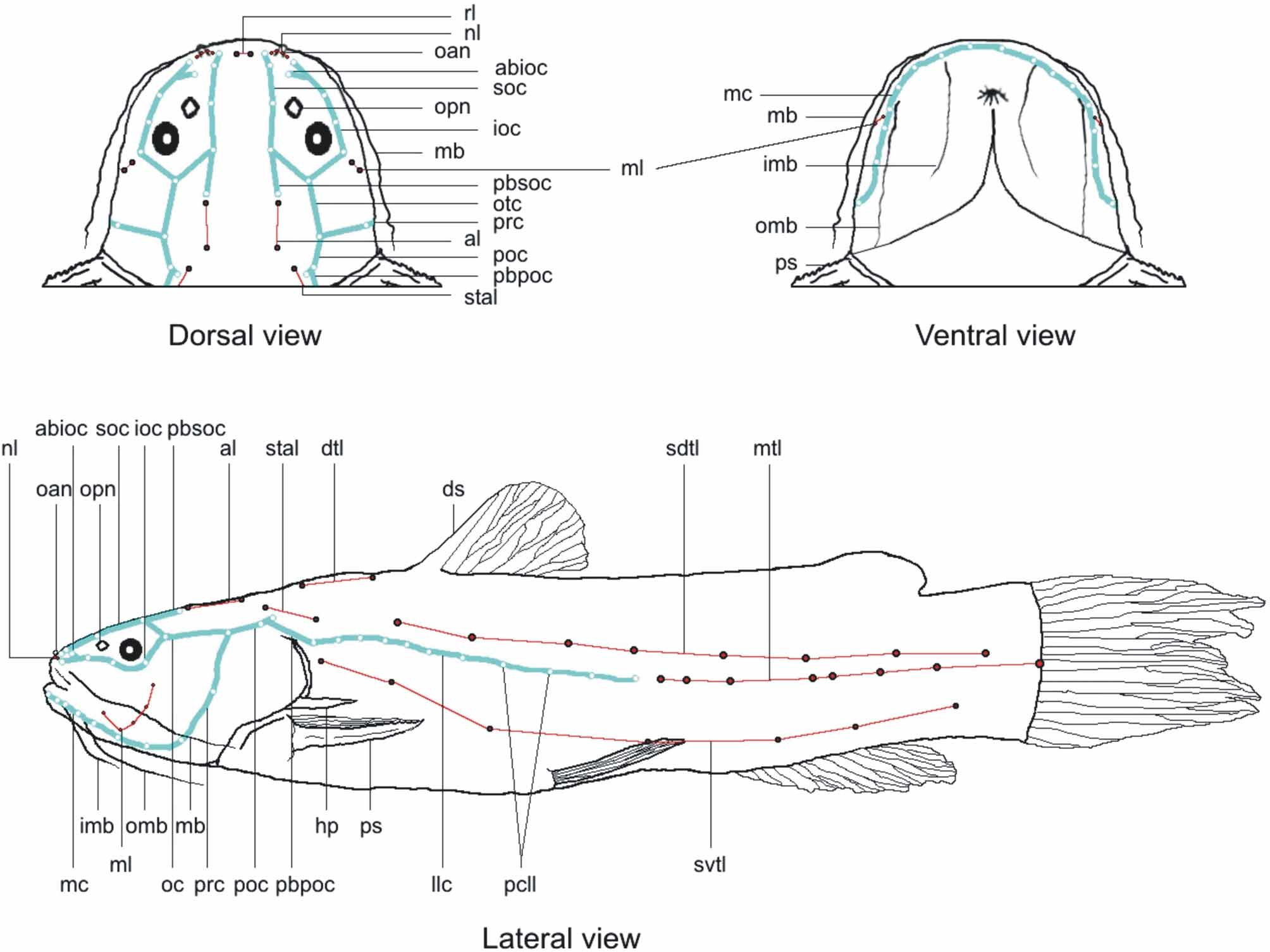
Cephalic sensory canals (figure 3): mandibular canal with eight pores, with last pore situated on lateroventral region of head; preopercle canal connected with mandibular canal, with only one pore, located in the middle of its length, and finishing in the pore of otic canal; infraorbital canal with four pores, beginning on left side of anterior nostril, outlining inferior region of eye, and finishing in last supraorbital pore of postsuperior region of eye. Infraorbital canal with small branch, called antorbital branch, inserted between first and second infraorbital pores, and composed by one terminal pore; supraorbital canal inserted on anterodorsal region of head, beginning on right side of anterior nostril, going over eye and finishing in post-superior portion of eye, at union between end of infraorbital canal and beginning of otic canal. Supraorbital canal with long bifurcation on its fourth pore called parietal branch of supraorbital canal, with a single terminal pore; otic canal with one pore, beginning in junction of infra and supraorbital canals, and proceeding straight until finishing in a single terminal pore; postotic canal beginning after pore of otic canal, continuing obliquely, and presenting two pores close at terminal portion. After first postotic pore there is a bifurcation called pterotic branch, directed ventrally and deprived of pores, connected with main canal of lateral line.
Cephalic neuromasts lines (figure 3): nasal line, located at base of anterior nasal tube, composed by three to four small neuromasts; rostral line with only one neuromast, inserted at medium portion between anterior nasal openings; mandibular line hook-like, beginning at vertical through fourth pore of infraorbital canal, descending anteriorly to ventral region, curving up and ending at horizontal line through mouth angle, with four to six neuromasts; anterior line located horizontally above parieto-supraoccipital bone, anterior to dorsaltrunk line, containing two neuromasts; supratemporal accessory line positioned diagonally in dorso-lateral region above opercle opening, having two neuromasts.
TABLE 2. Meristic counts of Microglanis robustus (* only in c&s specimens).
Trunk neuromasts lines: four superficial neuromasts lines on trunk. Dorsal-trunk line positioned horizontally anterior to dorsal fin, in lateral margin of nuchal plate, composed by two neuromasts; mediumtrunk line, with six to 12 neuromasts, beginning after last pore of main lateral line canal following straight in direction to caudal fin, finishing in a great neuromast inserted little beside base of middle caudal-fin rays; subdorsal-trunk line situated parallel to medium-trunk line, beginning just anterior and below dorsal-fin spine, containing five to eight neuromasts, extending until initial or central portion of caudal peduncle; concave subventral-trunk line, beginning above posterior cleithral process, below lateral line, and finishing in central portion of caudal peduncle, composed by five to seven neuromasts, smaller than ones from other trunk lines.
Color in alcohol. Ground color light brown to slightly reddish. A small cordiform light blotch with vertex addressed posteriorly, on the central portion of nape. Dorsal region of head dark brown, with anterior and posterior nostrils, and pores of cephalic sensory canals light brown. Light blotches of anterior and posterior nostrils united by a light stripe. Upper lip dark and lower lip light. Light-brown barbels with few dark-brown pigments.
Ventral region of body and head light brown with little pigmentation. Color pattern of trunk vermiculated with alternation of three dark-brown blotches with irregular borders. First blotch in form of saddle below dorsal fin, not surpassing the horizontal line through axis of trunk. Second blotch wider, in form of saddle placed after dorsal-fin base to central portion of adipose fin, descending until subdorsal trunk line of neuromasts, and with a great oval light blotch on upper region. Third vertical blotch positioned at end of caudal peduncle, with small tip addressed forward on medium portion. Two circular light blotches enclosed in the upper and lower region of third dark blotch. Dorsal fin with dark brown base and thin dark brown submarginal band across fin rays and membranes. Pectoral and pelvic fins hyaline, with few brown cromatophores. Anal fin hyaline with brown dots, base with large concentration of brown cromatophores on anterior portion. Caudal fin hyaline with concentration of brown cromatophores; two dark brown vertical stripes: a straight basal stripe and another just after middle, following caudal lobes. Melanophores surrounding all neuromasts, forming small black dots distributed in three horizontal series on lateral of body, four on head, one at nape region, and other on post-nape portion.
Distribution. Microglanis robustus is known from two localities of Rio Tocantins: in rapids of Jatobal and in small rapids below Jatobal, in Tucuruí , Pará, Brazil ( Figure 4 View FIGURE 4 ).
Etymology. The name robustus is derived from the Greek, in reference to the truncated body and high caudal peduncle, giving a strong format to this catfish. An adjective.
Ecological notes. This new species was collected by INPA at 1982, in rapids upstream and downstream of the district of Jatobal, Tucuruí , State of Pará, Brazil (figure 4), that were flooded by the construction of Tucuruí dam in 1984. The hydroelectric power plant flooded an area of 2.850 km 2, generating a 40 km wide and 170 km long reservoir along the main river ( Santos et al., 2004). Remarkably, this is the first species of Microglanis captured in rapids. All the other species were reported living among marginal submerged vegetation, depositional substrata of leaves, and/or trunks in calm water stretches of rivers. If this new species of Microglanis used to live restrict in rapids, the disappearance of those environments may be the reason that no other specimen was captured after the flood along 28 years. However, the real impact of the Tucuruí dam reservoir over M. robustus , and the possibility of its extinction, can only become clear with more encompassing samplings in tributaries of Rio Tocantins at the surroundings of Jatobal.


| INPA |
Instituto Nacional de Pesquisas da Amazonia |
No known copyright restrictions apply. See Agosti, D., Egloff, W., 2009. Taxonomic information exchange and copyright: the Plazi approach. BMC Research Notes 2009, 2:53 for further explanation.
|
Kingdom |
|
|
Phylum |
|
|
Class |
|
|
Order |
|
|
Family |
|
|
Genus |