
Koonwarraphis rotundafrons, Martin, Sarah K., Skidmore, Luke I. & Stilwell, Jeffrey D., 2016
|
publication ID |
https://doi.org/ 10.11646/zootaxa.4137.1.7 |
|
publication LSID |
lsid:zoobank.org:pub:EA49068A-884D-4B79-AF5F-82910028EE23 |
|
DOI |
https://doi.org/10.5281/zenodo.6079583 |
|
persistent identifier |
https://treatment.plazi.org/id/EA0487FA-FFDF-FFCB-EA91-2BC92869279E |
|
treatment provided by |
Plazi |
|
scientific name |
Koonwarraphis rotundafrons |
| status |
sp. nov. |
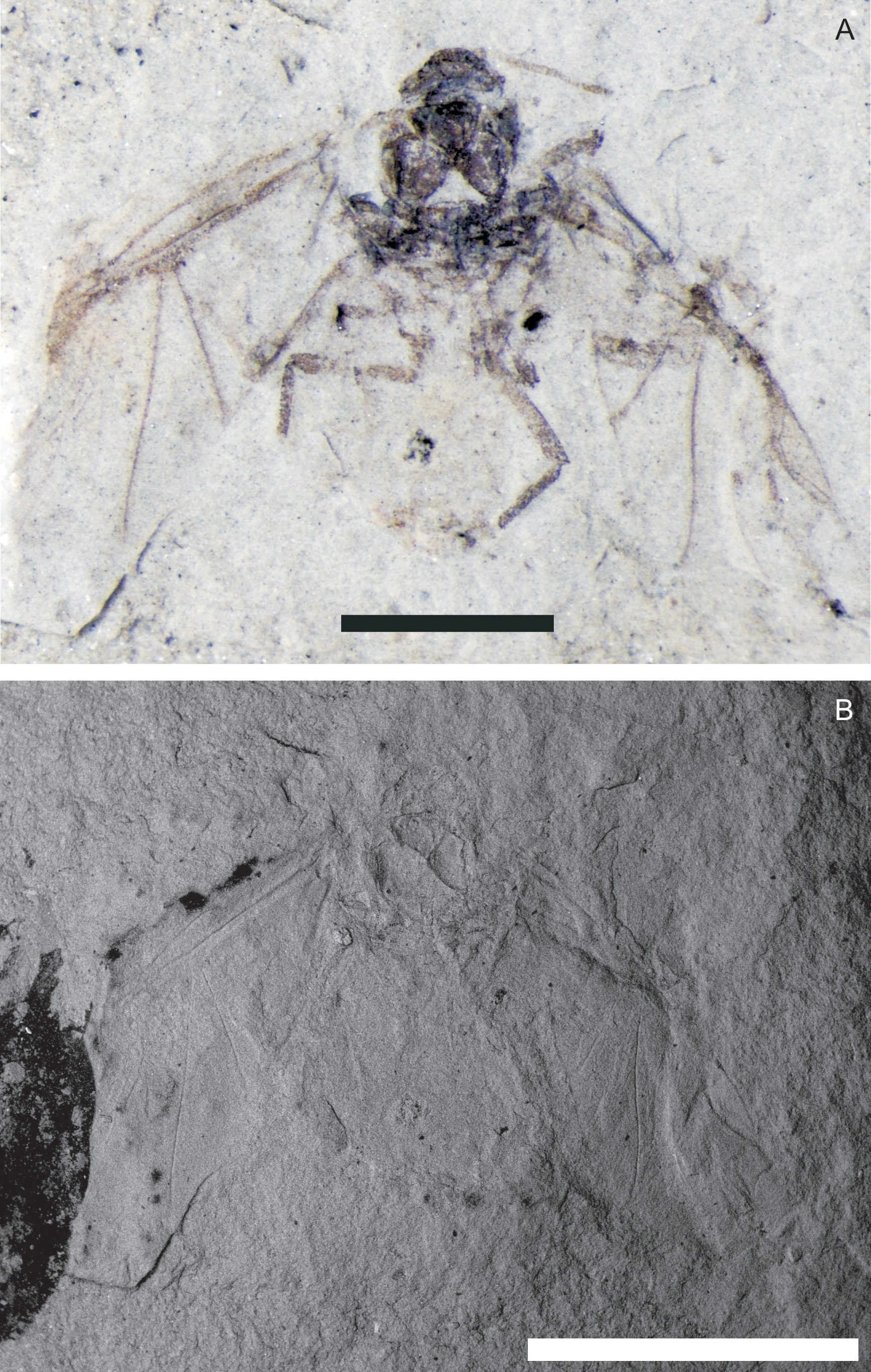
Koonwarraphis rotundafrons sp. nov.
( Figs 2–4 View FIGURE 2 View FIGURE 3 View FIGURE 4 )
Diagnosis. As for genus.
Derivation of name. From the Latin words rotunda ‘rounded’ and frons ‘forehead, face’, in reference to the large, rounded frons characteristic of this fossil.
Material. Holotype: AM F72793 View Materials ; alate adult, preserved in dorsal aspect. No counterpart or other specimens exist.
Description. Body ( Fig. 2 View FIGURE 2 ). 2.5 mm long; forewings displaced and partially crumpled, obscuring some parts of the venation and body features.
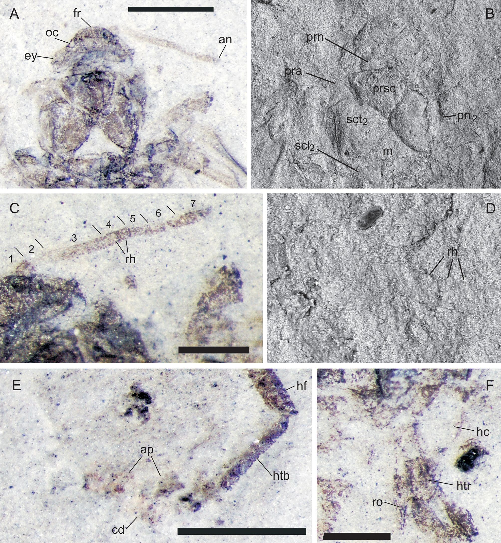
Head ( Fig. 3 View FIGURE 3 A–D,F). Maximum width 0.5 mm, widest at compound eyes; head wider than long, length 0.5x width. Frons enlarged, bulging apically in a gently rounded curve, with prominent ‘v’ shaped ornament positioned in front of ocelli and extending to posterior margin of head; lateral sutures faint, extending from the lateral margins between the compound eyes and ocelli; epicranial suture short and relatively faint, extending forward from lateral sutures but apparently not reaching front of frons. Antenna 0.6 mm long,?7 segments, segment III considerably longer than other segments, processus terminalis apparently absent or similar width to other segments. Although antennal segments are poorly defined due to preservation style, segment III shows lines of large, rounded features, which appear to be secondary rhinaria arranged in transverse rows; faint transverse bands or lines are also observable on antennal segments. Compound eyes placed dorsolaterally adjacent to pronotum, with large obvious lenses; two large round ocelli positioned dorsolaterally between eyes and antennae. Rostrum cylindrical, only faintly preserved; ~ 1.6 mm long, 0.1 mm wide, extending 0.6x length of body, just posterior of hind coxae. Distal end of rostrum apparently narrowed, divided.
Thorax ( Fig. 3 View FIGURE 3 A–B). Pronotum 0.5 mm long; roughly trapezoidal, anterior width subequal to base of head, widening gently to become subequal to mesoscutum at posterior; central portion of pronotum more strongly sclerotized than laterally. Mesothorax anterior edge broader than head, praealare extended out into ‘shoulders’ slightly wider than the remaining mesothorax; mesoscutum, mesopraescutum, and mesoscutellum strongly domed and clearly defined; mesopraescutum and paired mesoscutum plates large and triangular, mesoscutellum narrow, rectangular, apparently divided centrally; membranous area between paired mesoscutum plates and mesoscutellum large, triangular. Metanotum roughly rectangular in shape, narrow; similar in width to mesoscutellum, divided ventrally; other metathoracic segments difficult to discern individually. Combined thoracic segments ~ 0.8 mm long. Fore leg coxae and trochanters not visible; fore femora indistinct. Fore tibiae 0.5 mm long, 0.1 mm wide; fore tarsi short or curled up, tarsal segments indistinct, tarsal claws present. Mid legs indistinct. Both hind legs visible; coxae triangular, 0.1 mm long, 0.2 mm wide; trochanters 0.1 mm long, less than 0.1 mm wide; femora cylindrical, 0.6 mm long, 0.1 mm wide; tibiae cylindrical, 0.4 mm long, less than 0.1 mm wide; tarsi apparently short, tarsal segments and tarsal claws indistinct. Legs apparently short overall, hardly longer than the abdomen.
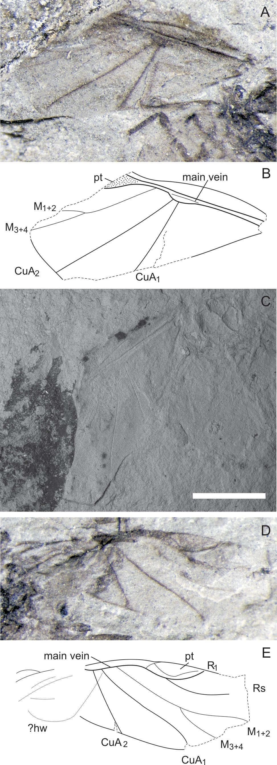
Wings ( Fig. 4 View FIGURE 4 ). Forewing preserved length 2.6 mm; wing triangular, costal margin gently curved, posterior margin straightened, apical margin incomplete. Both forewings missing apex; right forewing with costal region apparently twisted or crumpled, overprinting itself. Costal area of moderate width, R, M, and Cu stems fused basally (main vein), terminating in large, lozenge-shaped pterostigma 1.5 mm from wing base; pterostigma 0.6 mm long, width 0.2 mm. R1 delimits pterostigma distally; Rs emerging from centre of pterostigma margin, gently curving to intersect wing apex. Subcostal cell elongate, maximum width ~ 0.15 mm centrally. M diverging from main vein roughly halfway between pterostigma and CuA1 base, curving parallel to Rs; two visible M veins, M1+2 separating from M3+4 about level with distal end of pterostigma. Main vein distinctly thickened or bulging posteriorly coincident with the cubital vein bases; CuA1 and CuA2 diverge independently from this bulge with bases closely placed, ~1.0 mm from base of wing. Cubital veins diverging, CuA1 curving towards posteroapical margin at an angle of 46° from main vein, CuA2 directed towards posterior margin at an angle of 75° from main vein. Hindwings difficult to discern; left wing likely folded beneath body; right wing apparently confused with the base of the forewing, possibly with one or two straight veins.
Abdomen ( Fig. 3 View FIGURE 3 E). Rounded, as wide as or slightly wider than thoracic segments; maximum width 0.9 mm, length 1.5 mm. Abdomen apparently soft, with no visible abdominal sclerites. Abdomen appears to end with a slightly more sclerotized, bilobed structure or matched plates (?anal plate), with another roughly triangular and distally elongated feature overprinting/overlapping at the centre (?cauda). No obvious ovipositor or siphunculi.
No known copyright restrictions apply. See Agosti, D., Egloff, W., 2009. Taxonomic information exchange and copyright: the Plazi approach. BMC Research Notes 2009, 2:53 for further explanation.