
Mylognathus priscus
|
publication ID |
https://doi.org/ 10.5281/zenodo.1064078 |
|
DOI |
https://doi.org/10.5281/zenodo.6295679 |
|
persistent identifier |
https://treatment.plazi.org/id/E8728790-2E7E-CC24-17E3-FD7AFB97FB88 |
|
treatment provided by |
Jeremy |
|
scientific name |
Mylognathus priscus |
| status |
|
The very singular-looking fish, Chimaera , of the European seas, was represented during the Miocene period in Nebraska, by a genus for which the above name has been proposed. Its former existence is indicated by specimens of dental plates, like those of Chimaera , adapted to the crushing of mollusca and Crustacea, used as food. The specimens, consisting of an upper maxillary and a premaxillary plate , were obtained by Dr. Hayden from the Great Lignite Basin near Long Lake, Nebraska.
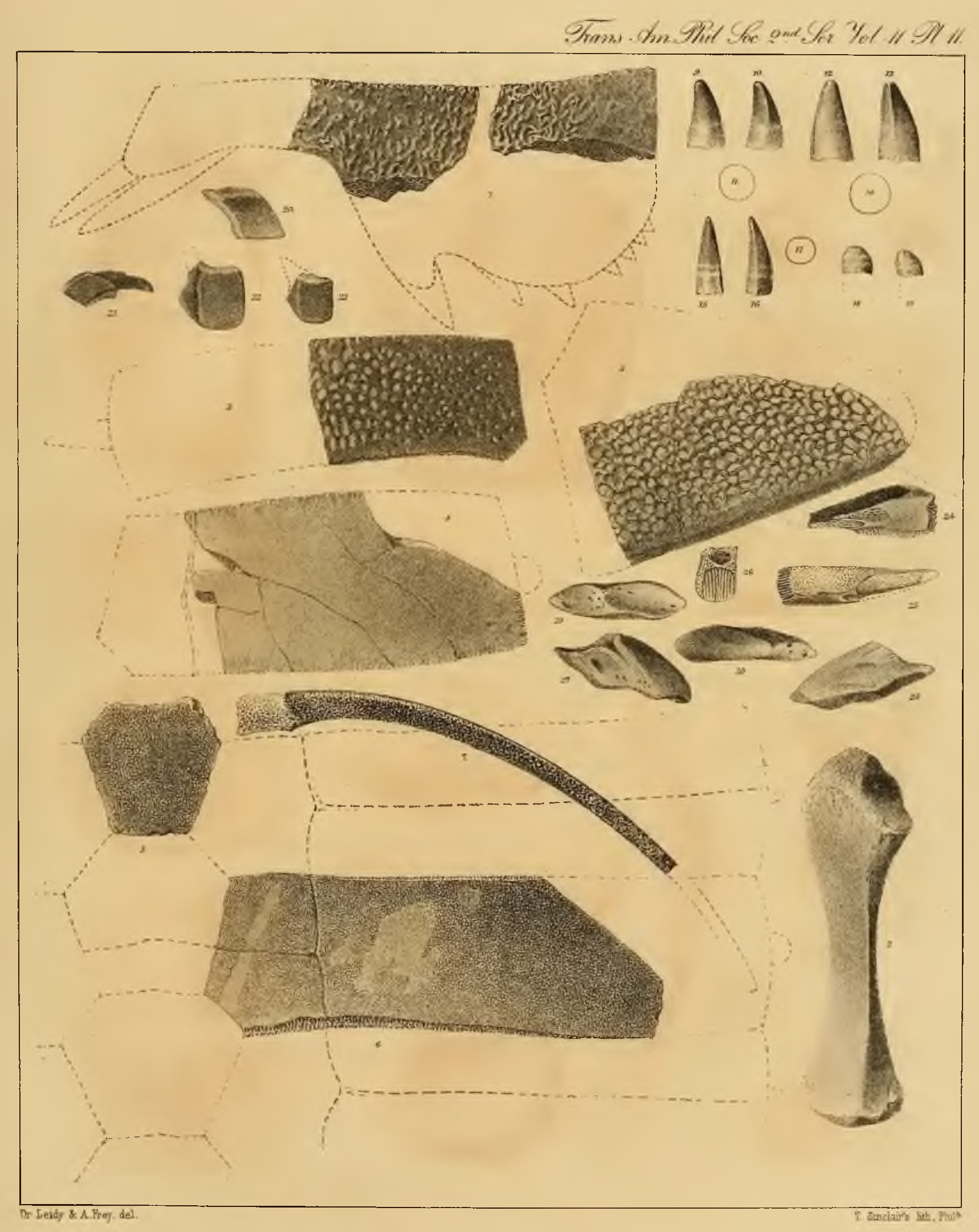
The upper maxillary plate, ( figs. 24, 25, 26, plate 11 View Figure ,) consists of a narrow triangular bone, containing two teeth. The specimen is broken at its two extremities, and when perfect appears to have been a little over an inch in length. Its posterior part is 3 4 3/ lines wide, and about 4 1/2 lines thick. The free convex surfaces of the peculiar porous teeth, occupy nearly the entire length and breadth of the bone, ( fig. 25, plate 11 View Figure ,) and are separated from each other by an oblique, linear tract. The anterior tooth is lozenge-shaped in outline, and when perfect appears to have been about 1/2 an inch in length, and 1 4 3/ lines in breadth. The posterior tooth, somewhat ellipsoidal in outline, appears, when perfect, to have been about 8 lines long, and is three lines wide.
The premaxillary dental plate, ( figs. 27—30, plate 11 View Figure ,) is irregularly lozenge-shaped in its vertical outline antero-posteriorly, is a little over an inch in its long diameter, 5 lines in its depth, and 3 lines in its greatest thickness. Its anterior border is convex, the inner and outer surfaces are vertical, slightly depressed planes, and the crushing surface is concave.
Explanation of Figures, Plate 11.
Figures 24—30. Upper maxillary plates of Mylognathus priscus , of the natural size.
Figure 24. Inner view of the maxillary plate, exhibiting the surfaces of the two teeth projecting below.
Figure 25. Oral or inferior surface of the same.
Figure 26. Posterior extremity of the same, exhibiting the columnar structure of the teeth.
Figures 27, 28. Outer and inner view of a pre-maxillary plate.
Figures 29, 30. Triturating surface and upper view of the same.
No known copyright restrictions apply. See Agosti, D., Egloff, W., 2009. Taxonomic information exchange and copyright: the Plazi approach. BMC Research Notes 2009, 2:53 for further explanation.
|
Kingdom |
|
|
Phylum |
|
|
Class |
|
|
Order |
|
|
Family |
|
|
Genus |