
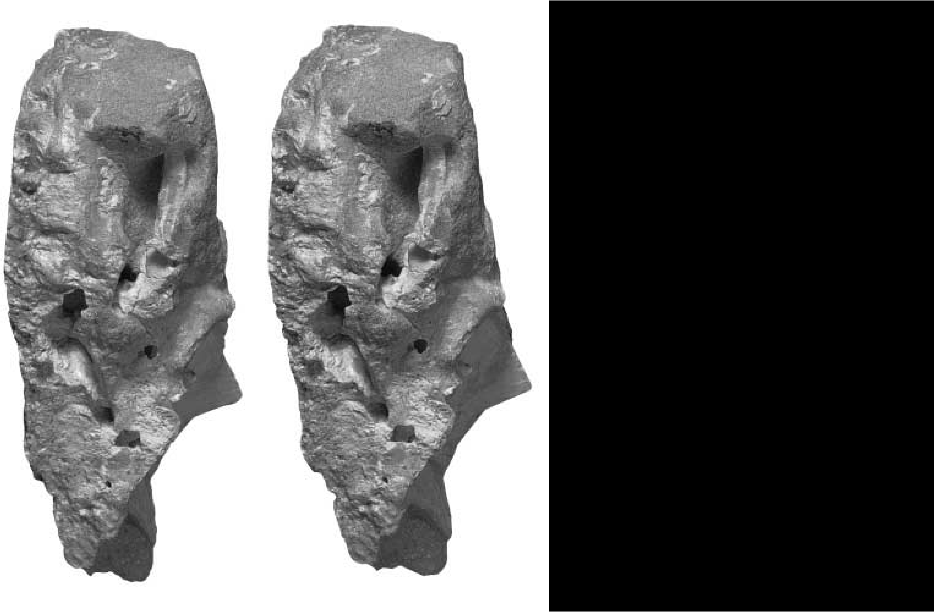
Stagonolepis robertsoni
|
publication ID |
https://doi.org/10.1046/j.1096-3642.2002.00023.x |
|
persistent identifier |
https://treatment.plazi.org/id/E860E61E-FFBC-9C10-FE9B-89E8FFD0FE8E |
|
treatment provided by |
Carolina |
|
scientific name |
Stagonolepis robertsoni |
| status |
|
Walker (1985, 1990) interpreted the lateral ridge on the exocciptal pillar as an indication of ‘some modest development of a subcapsular process’ ( Walker, 1985: 132). That the lateral exoccipital ridge might represent part of the subcapsular process in aetosaurians was followed by Parrish (1994: 200) for Longosuchus meadei and Desmatosuchus haplocerus . However, the details of the development of the embryonic subcapsular process of the crocodilian chondrocranium ( Shiino, 1914) and its relation to ossified structures in the adult skull remain unknown ( Walker, 1990: 33, 107). Gower & Weber (1998: 397) suggested that care must be taken not to associate fossilized structures with hypothetical chondrocranial structures purely through a common terminology. For that reason, the exoccipital ridge is here given a more neutral term and it is stressed that there is no intrinsic evidence that a subcapsular process sensu stricto had developed in embryonic S. robertsoni .
Walker (1985: 132) suggested that in S. robertsoni ‘a secondary tympanic membrane was almost certainly present’. This interpretation was based on the posterolaterally opening, completely framed (with bone) foramen perilymphaticum and the development of a strong lateral exoccipital ridge. In extant crocodilians, these features are associated with a more laterally extensive part of the perilymphatic sac enclosed in bone, and a secondary tympanic membrane. Other suchian and noncrown-group archosaurs bear a closer resemblance in these hard-part features to Sphenodon ( Walker, 1990: 111, Gower & Weber, 1998), which has an incompletely framed (with bone) and posteromedially directed perilymphatic foramen, and no specialized pressure relief window or membrane (e.g. Baird, 1960, 1970 and references therein). Gower & Weber (1998) argued that application of the term ‘fenestra pseudorotunda’, originally coined for the bony secondary tympanic window frame in crocodilians and birds ( De Beer, 1937), requires evidence of a bony subdivision of the metotic fissure. This is lacking in S. robertsoni and in sphenosuchian crocodylomorphs such as Sphenosuchus acutus ( Walker, 1990) . The otic capsule of S. robertsoni clearly approaches that of crocodilians in terms of some derived features, including some of those structures closely associated with the secondary tympanic window and membrane of extant crocodilians. However, Walker’s hypothesis that S. robertsoni possessed a secondary tympanic membrane is based on some possible osteological correlates, such as the lateral ridges surrounding the posterolateral exit for the perilymphatic duct, but not the probably most important correlate, namely the bony subdivision of the metotic fissure. It therefore pushes the limits of a level II inference sensu Witmer (1995a: 28), in which ‘the soft tissue expected to occur in a fossil taxon is found in its extant sister group but not in any other [immediate] outgroups’. A secondary tympanic membrane may well have been present in a large range of extinct archosaurs, but inferring one with confidence is impeded by several immediate and interconnected problems. Finding unequivocal osteological correlates for this soft tissue structure in extant taxa is problematic. Although the metotic fissure becomes subdivided by a bony structure in all extant sauropsids in which a secondary tympanic membrane is present (unlike the situation in Triassic archosaurs), only Sphenodon among major groups lacks a secondary tympanic membrane (and subdivided fissure). Furthermore, the use of extant phylogenetic brackets ( Witmer, 1995a) must be carried out in a controlled way because at least lizards, crocodilians, and birds all seem to have independently evolved a secondary tympanic window (e.g. Rieppel, 1985; Gower & Weber, 1998), and perhaps membrane.
Walker (1990: 111) reported that the lagenar region of S. robertsoni was imperfectly preserved in the available material, but considered that it ‘does not appear to have been significantly elongated’. ‘Significantly’ here means with respect to the relatively short lagenar region of, for example, noncrown-group archosaurs ( Walker, 1990; Gower & Sennikov, 1996; Gower, 1997; Gower & Weber, 1998) and the relatively elongate lagenar recess of Sphenosuchus acutus ( Walker, 1990) and crocodilians (with the associated development of a cochlea). The lagenar region of S. robertsoni remains unknown in detail, but reconsideration and further comparison with other fossil archosaurs suggests that it is premature to remark on the probable length or form of this particular feature. For example, the close association between a recess for the lagena and an incompletely ossified region between the distal end of the ventral ramus of the opisthotic and the basal tubera of basioccipital and parabasisphenoid in as wide a range of taxa as Sphenodon , erythrosuchids, Euparkeria capensis , Batrachotomus kupferzellensis , crocodilians and birds ( Walker, 1990; Gower & Sennikov, 1996; Gower & Weber, 1998; Gower, 2002b), suggests that determining the length of the lagenar/ cochlear recess from external proportions can be problematic. Additionally, the medial view of MCZD 2-4 ( Fig. 3 View Figure 3 ) shows that some matrix remains in place at the anteroventral end of the ventral ramus of the opisthotic. This might be plugging the dorsal end of the lagenar recess, but it has been left in place in order to protect the delicate opisthotic ramus and perilymphatic foramen. Even if it was removed, some distortion in this region (as seen in lateral view) means that it might not be feasible to trace a recess that originally may only have been a gap between elements.
Walker (1961: 125) suggested that the channel transmitting the cerebral branch of the internal carotid into the hypophyseal fossa also transmitted the palatine branch of the facial nerve plus the palatine artery. However, a Vidian canal like that of extant squamates, where a bony channel transmits the palatine artery plus the palatine ramus of the facial nerve, is absent in noncrown-group and crurotarsan archosaurs. Thus, like the situation in Sphenodon ( Säve-Söderbergh, 1947) and that reconstructed for the archosaurs Erythrosuchus africanus (Gower, 1997) and S. acutus ( Walker, 1990: fig. 49), the palatine ramus of the facial nerve (plus palatine artery, if present) of S. robertsoni passed forwards to the base of the cultriform process of the parabasisphenoid along an unenclosed channel between the basipterygoid processes, on the ventral surface of the parabasisphenoid. The palatine artery is absent in extant crocodilians ( Shindo, 1914).
No known copyright restrictions apply. See Agosti, D., Egloff, W., 2009. Taxonomic information exchange and copyright: the Plazi approach. BMC Research Notes 2009, 2:53 for further explanation.