
Parastasia, Westwood, 1841
|
publication ID |
https://doi.org/ 10.11646/zootaxa.5205.6.3 |
|
publication LSID |
lsid:zoobank.org:pub:62DD9430-0B11-46A9-A590-B960F859FE5A |
|
DOI |
https://doi.org/10.5281/zenodo.7327245 |
|
persistent identifier |
https://treatment.plazi.org/id/E853E505-1379-FFEE-FF03-DF9AFA24FBFA |
|
treatment provided by |
Plazi |
|
scientific name |
Parastasia |
| status |
|
Key to species of Parastasia known from Thailand
1 Small to medium in size, total length ˂ 20 mm; elytra with distinct rows of punctures; in lateral view, parameres not bilobed. ................................................................................................... 2
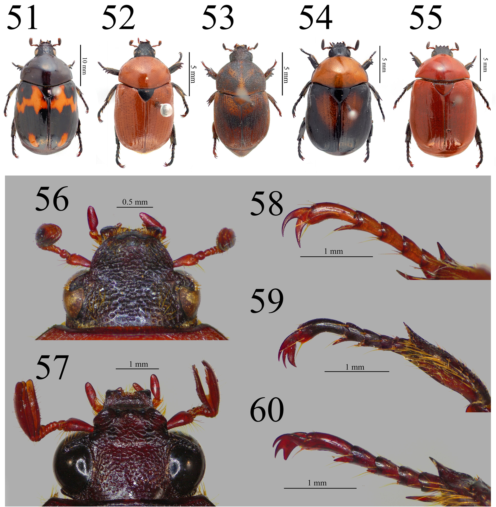
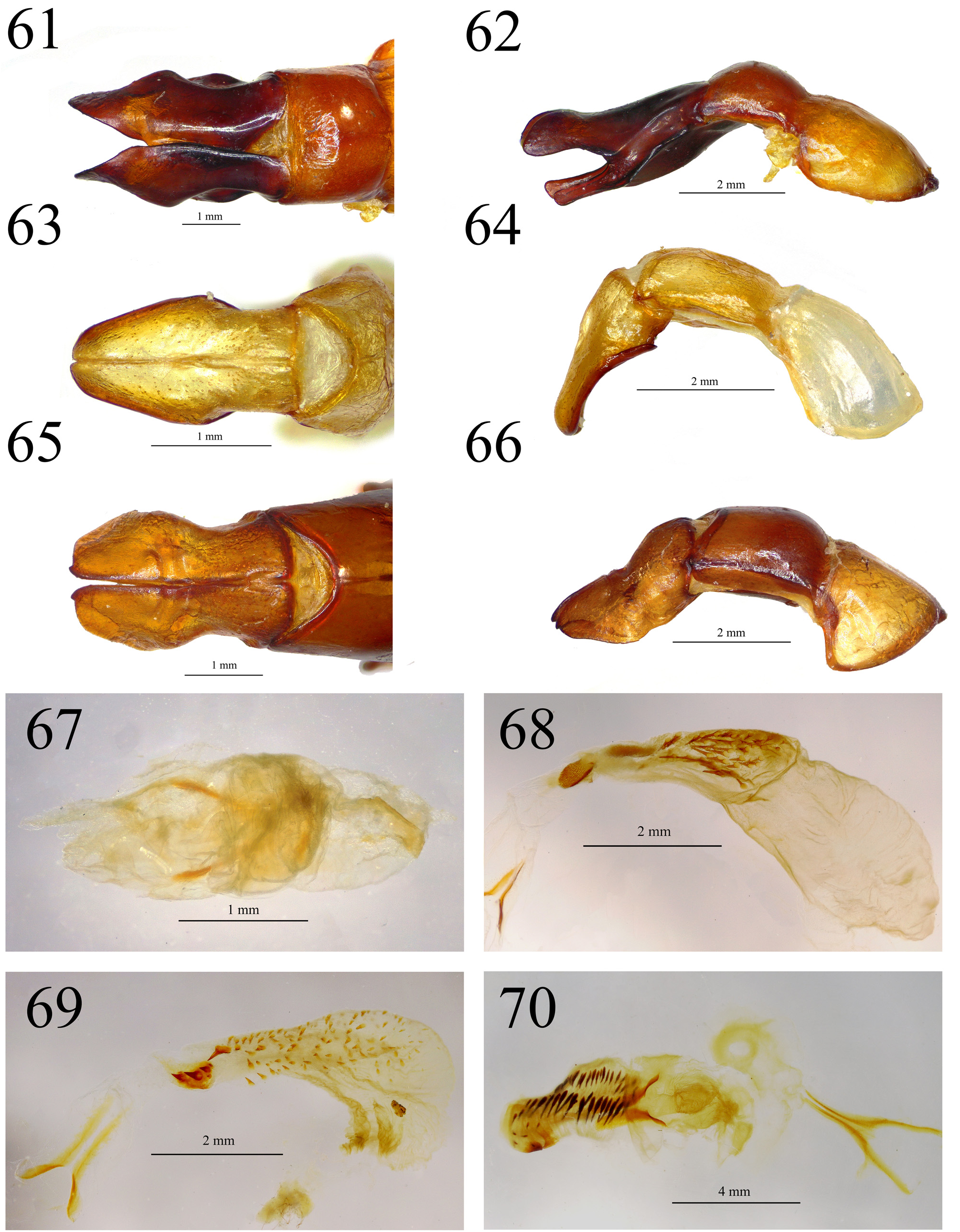
- Large in size, TL ≥ 20 mm; rows of punctures on elytra indistinct ( Fig. 51 View FIGURES 51–60 ); parameres slightly asymmetric, apex bilobed in lateral view ( Figs. 61–62 View FIGURES 61–70 )............................................................ P. birmana Arrow, 1899
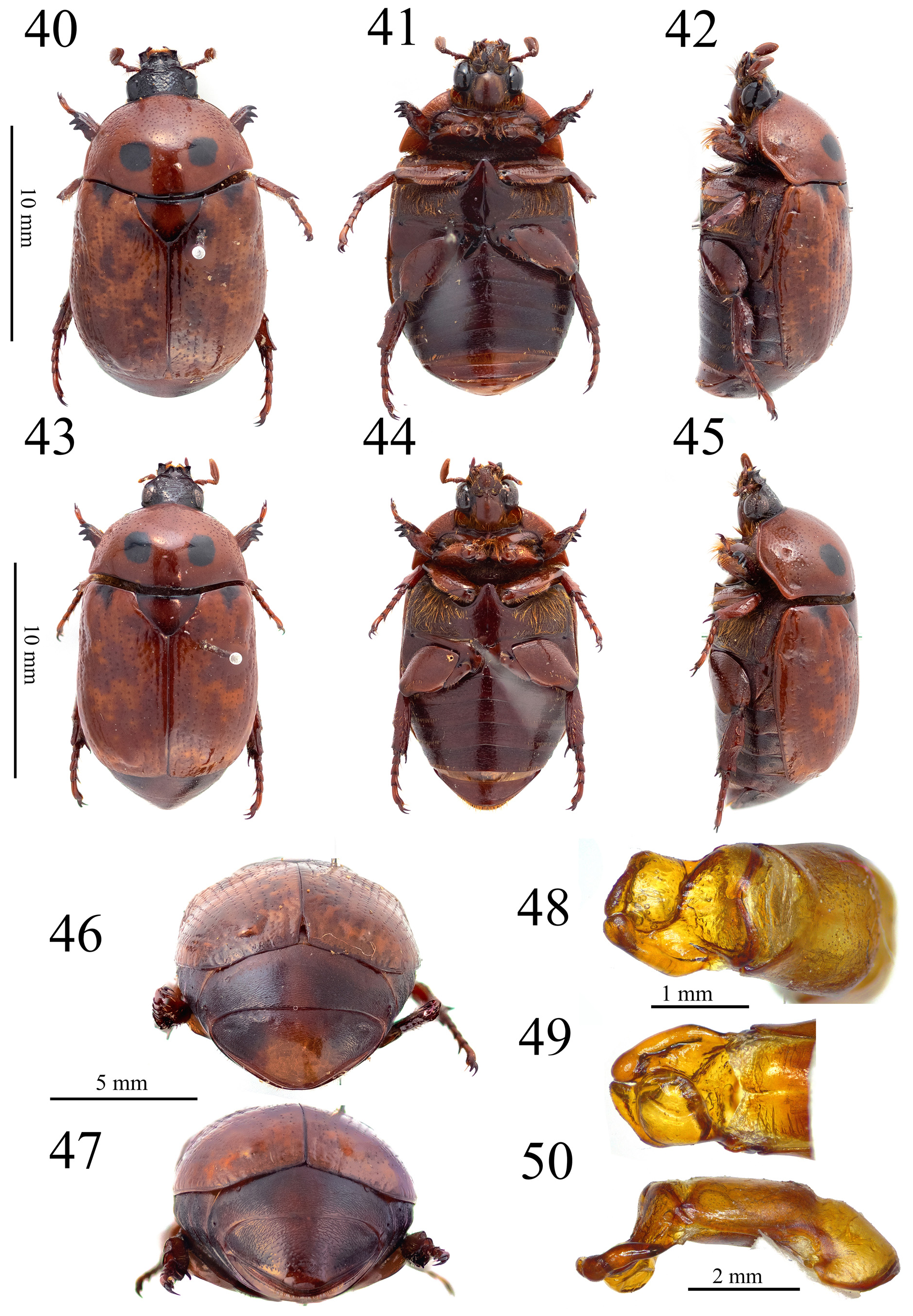
2 All claws simple, sickle-shaped without branching ( Fig. 58 View FIGURES 51–60 ); male aedeagus asymmetric, parameres fused together ( Figs. 48–50 View FIGURES 40–50 )............................................................. P. bimaculata ( Guérin-Méneville, 1843)
- External claw of meso- and metatarsi bifurcate; aedeagus symmetric or asymmetric, but parameres not fused together..... 3
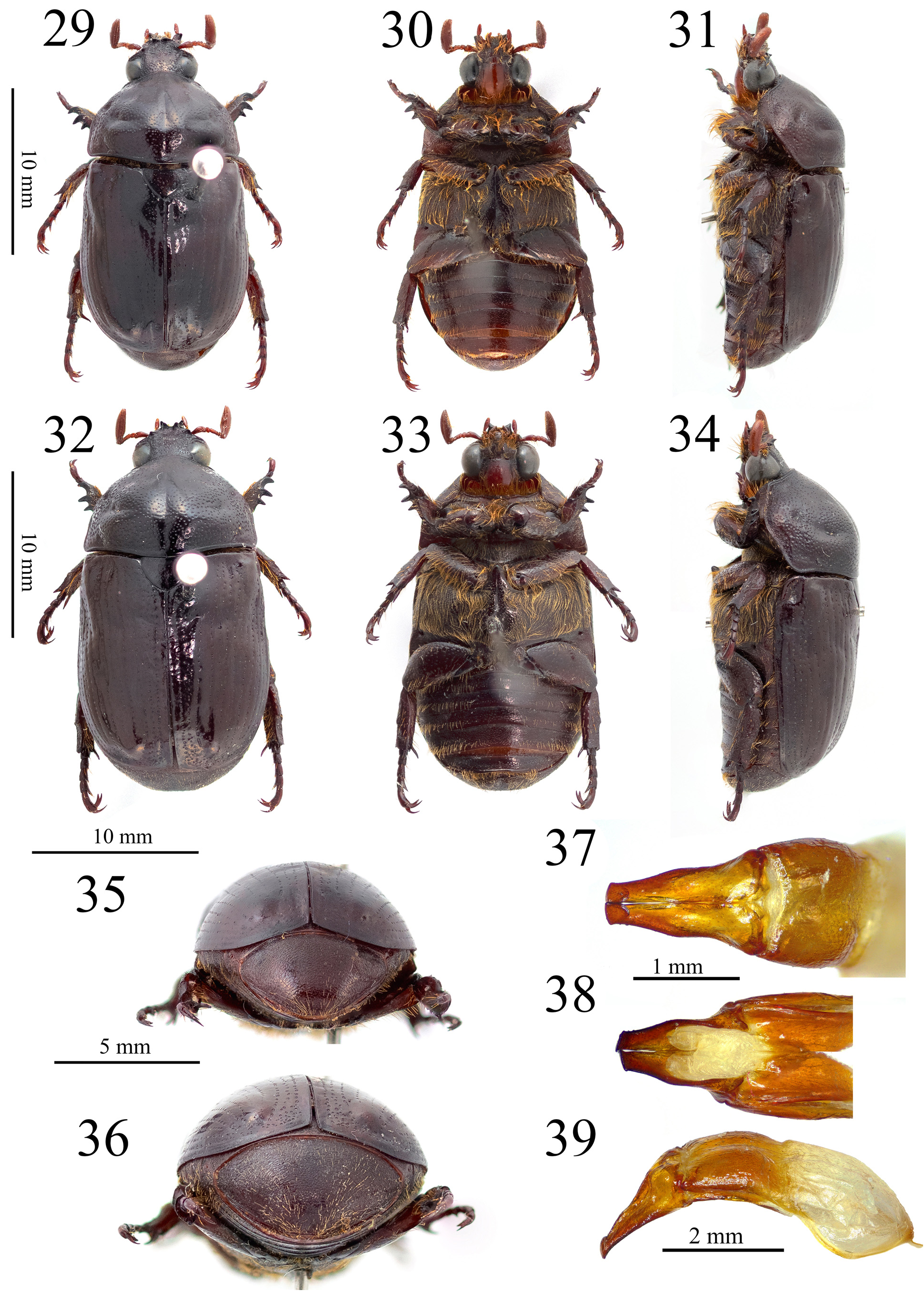
3 Propygidium and pygidium covered with decumbent setae ( Figs. 35–36 View FIGURES 29–39 )......................................... 4
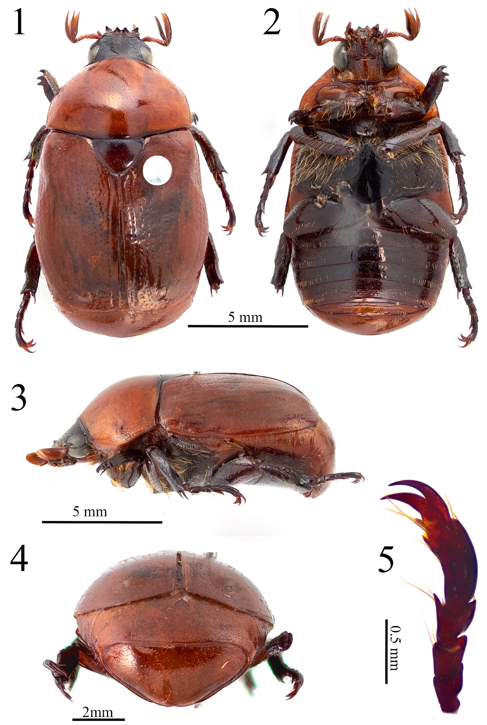
- Propygidium and pygidium without long setae ( Fig. 4 View FIGURES 1–5 )........................................................ 7
4 Eyes narrow, ratio of IOD and HW ˃ 0.67 ( Fig. 56 View FIGURES 51–60 ); antennal clubs distinctly shorter than antennal segment 1–7 combined; mesotarsi of male somewhat enlarged, segment 5 as long as or longer than segment 1–4 combined ( Fig. 56 View FIGURES 51–60 ); median sac of endophallus membranous without any appendages or spines, but sometimes distinctly with sclerotic surface ( Fig. 67 View FIGURES 61–70 )...... 5
- Eyes broad, ratio of IOD and HW ˂ 0.60 ( Fig. 57 View FIGURES 51–60 ); antennal clubs clearly longer than antennal segment 1–7 combined; mesotarsi simple in both sexes, tarsal segment 5 shorter than segment 1–4 combined ( Fig. 60 View FIGURES 51–60 ); median sac of endophallus with numerous spines ( Fig. 68 View FIGURES 61–70 )................................................................. P. bigibbosa Nonfried, 1891
5 Dorsal surface mostly brownish ( Fig. 53 View FIGURES 51–60 ); mesotarsi clearly enlarged in male, segment 5 much longer than segments 1–4 combined............................................................................................ 6
- Dorsal surface mostly orange-reddish ( Fig. 52 View FIGURES 51–60 ); segment 5 of mesotarsi as long as segments 1–4 combined in male......................................................................................... P. anomala Arrow, 1899
6 Ground color of dorsal surface reddish brown to dark brown, posterolateral portions of pronotum with a yellow to testaceous patch; in dorsal view, male aedeagus symmetric. India, Bhutan, China, Vietnam, Laos, and northern Thailand.............................................................................................. P. indica Ohaus, 1898
- Ground color of dorsal surface brownish with dark brown brands on pronotum and elytra ( Fig. 53 View FIGURES 51–60 ); aedeagus curve and asymmetric. Southern Thailand, Malaysia, Indonesia, and Philippines................... P. westwoodii Westwood, 1841
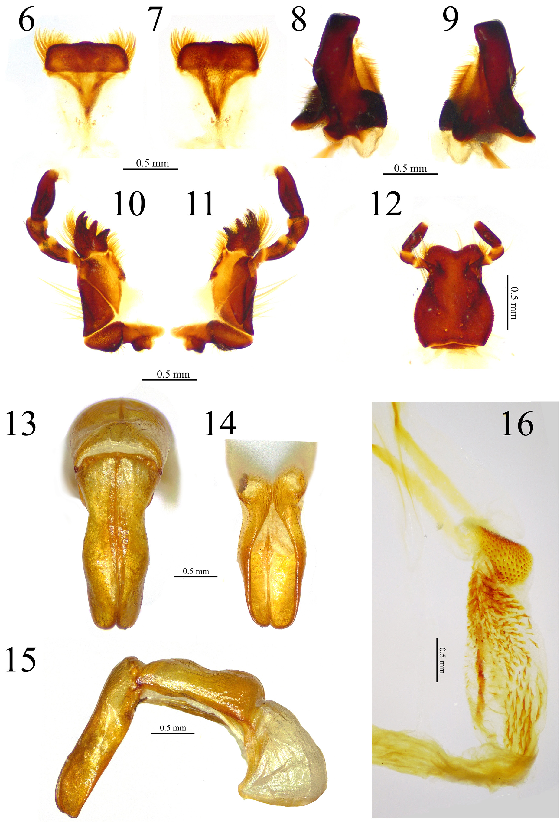
7 Ground color of dorsal surface reddish, elytra unicolor ( Figs. 1 View FIGURES 1–5 , 55 View FIGURES 51–60 ); external margin of parameres sinuate in dorsal view ( Figs. 13 View FIGURES 6–16 , 65 View FIGURES 61–70 ); in lateral view, ventral margin of parameres plain without distinct concavity ( Figs. 15 View FIGURES 6–16 , 66 View FIGURES 61–70 ); anterior portion of median sac of endophallus with small spines, remaining followed by large spines ( Figs. 16 View FIGURES 6–16 , 70 View FIGURES 61–70 ).............................. 8
- Ground color of dorsal surface orange, external and posterior portions of elytra black ( Fig. 54 View FIGURES 51–60 ); in dorsal view, parameres without concavity ( Fig. 63 View FIGURES 61–70 ); in lateral view, ventral margin of parameres thickly marginated and deeply concave one-fifth from base ( Fig. 64 View FIGURES 61–70 ); anterior portion of median sac of endophallus with few large spines, remaining followed by medium spines ( Fig. 69 View FIGURES 61–70 )................................................................. P. masumotoi Wada & Muramoto, 1999
8 Scutellum and ventral surface reddish to dark reddish brown ( Figs. 1–2 View FIGURES 1–5 , 55 View FIGURES 51–60 ); in lateral view, basal of ventral margin of parameres without protrusion.................................................................................... 9
- Scutellum and ventral surface nearly black; in lateral view, basal of ventral margin of parameres clearly protruding backward, process sharp and elongate as spinose formed................................. Parastasia selangorica Kuijten, 1992
9 Rather small in size ( Fig. 1 View FIGURES 1–5 ); first row of punctures on elytra sinuate; metasternal process stout; in dorsal view, apex of parameres roundly convex ( Fig. 13 View FIGURES 6–16 ); in lateral view, dorsal and ventral margins almost straight ( Fig. 15 View FIGURES 6–16 ); median sac of endophallus with a cluster of dense small spines anteriorly, posterior part of median sac with five dark large spines ventrally ( Fig. 16 View FIGURES 6–16 )....................................................... P. spinosa Hongsuwong, Sanguansub & Jaitrong , new species
- Medium in size ( Fig. 55 View FIGURES 51–60 ); first row of punctures on elytra mostly straight; metasternal process sharply produced; in dorsal view, apex of parameres almost truncate with a small carina near internal margin ( Fig. 65 View FIGURES 61–70 ); in lateral view, dorsal margin weakly concave, while ventral margin convex ( Fig.66 View FIGURES 61–70 ); median sac of endophallus with sparse small spines anteriorly, ventral of posterior part without dominantly large spines ( Fig. 70 View FIGURES 61–70 ).................................... sulcipennis Gestro, 1888
No known copyright restrictions apply. See Agosti, D., Egloff, W., 2009. Taxonomic information exchange and copyright: the Plazi approach. BMC Research Notes 2009, 2:53 for further explanation.