
Socoflata, Stroiński & Malenovský & Świerczewski, 2018
|
publication ID |
https://doi.org/ 10.11646/zootaxa.4379.3.3 |
|
publication LSID |
lsid:zoobank.org:pub:485C90BE-2219-423F-A339-51B6744184BC |
|
DOI |
https://doi.org/10.5281/zenodo.5967993 |
|
persistent identifier |
https://treatment.plazi.org/id/E8459174-1314-775F-87D0-FE54C636FCF3 |
|
treatment provided by |
Plazi |
|
scientific name |
Socoflata |
| status |
gen. nov. |
Socoflata gen. nov.
( Figs 1–62 View FIGURES 1–10 View FIGURES 11–16 View FIGURES 17–22 View FIGURES 23–28 View FIGURES29–34 View FIGURES35–44 View FIGURES 45–50 View FIGURES 51–60 View FIGURES61–67 )
Type species. Socoflata histrionica sp. nov., here designated.
Diagnosis. Socoflata gen. nov. can be distinguished from other genera of Flatidae by the combination of following characters: Body small, ovoid, habitus issid-like. Vertex transverse, distinctly shorter than pronotum at midline, with all margins carinate, fore margin truncate. Disc of frons tricarinate, all carinae sharp and well distinct, reaching almost frontoclypeal suture, basally separated. Tegmen short, coriaceous, with sutural angle rounded. Male anal tube, in lateral view, elongate and curved, with ventral margin strongly concave. Male genital style widening apicad; postero-dorsal angle with long and curved capitulum, postero-ventral angle produced into a large tooth. Periandrium bearing 3-armed appendage oriented antero-ventrad; posterior arm simple, oriented dorsoposteriad; median arm stout, diverged into two processes, upper process with 0–3 ventral spine-like ramifications; anterior arm simple, movable. Female gonapophysis VIII stout, laterally flattened, ventral margin sinuate.
Description. Body robust, ovoid ( Figs 1–11 View FIGURES 1–10 View FIGURES 11–16 , 61, 62 View FIGURES61–67 ).
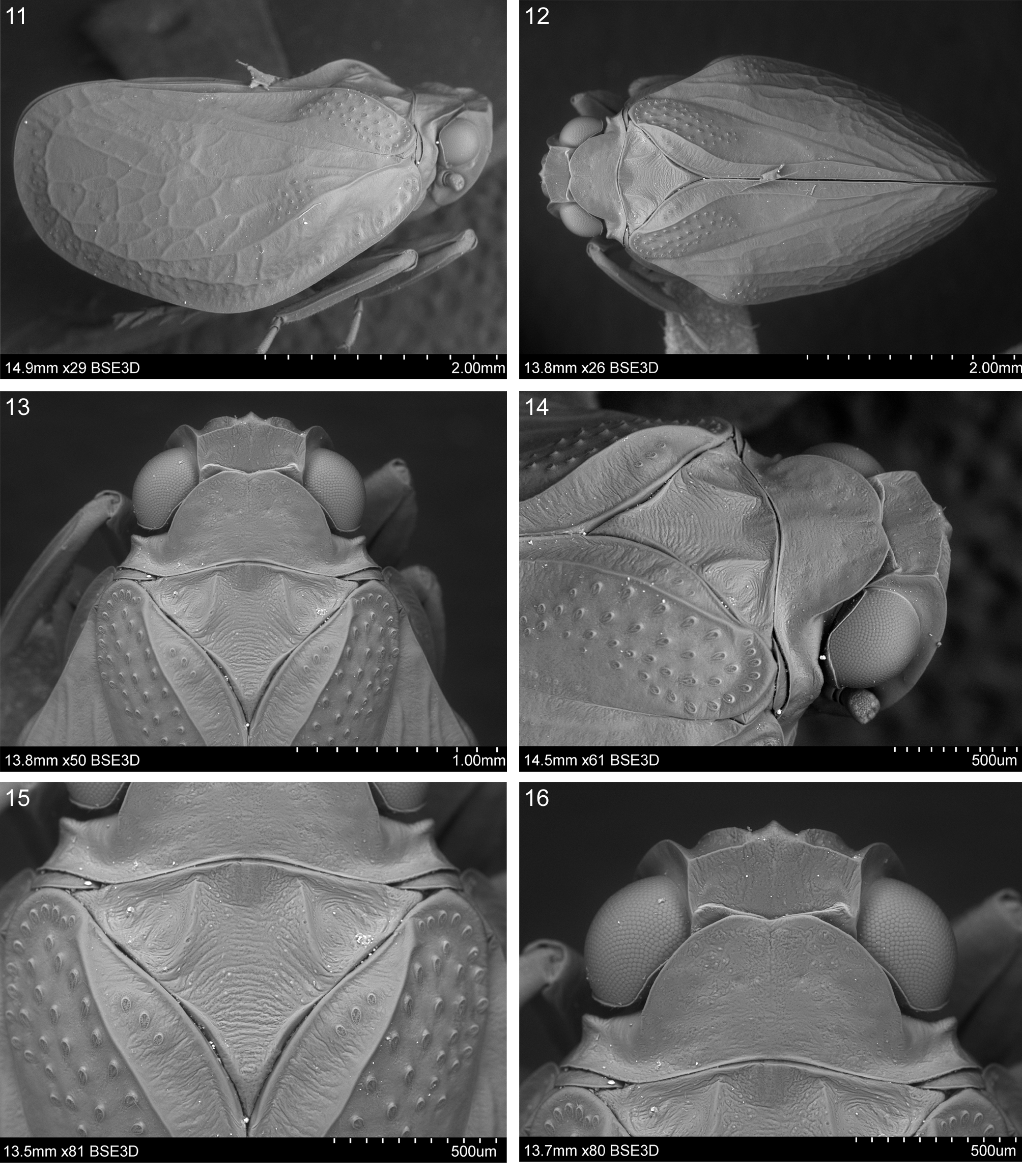
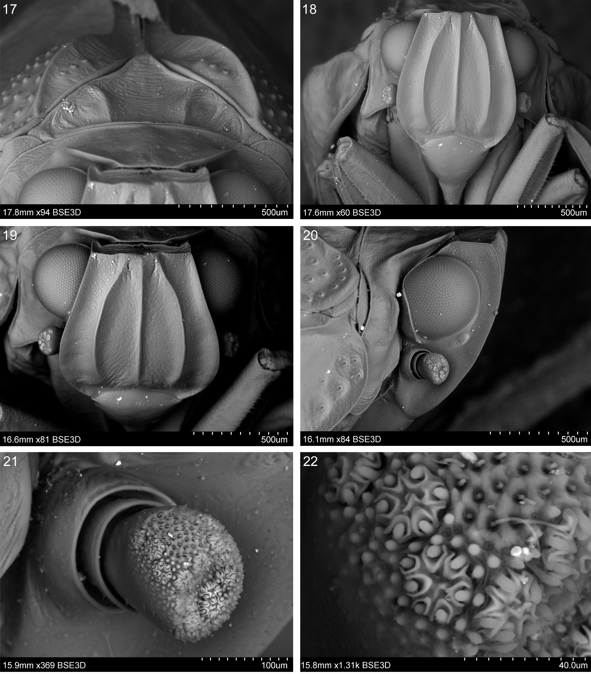
Head truncate, with compound eyes, in dorsal view, slightly narrower than pronotum but almost as wide as mesonotum ( Figs 12–13 View FIGURES 11–16 ). Vertex transverse, distinctly shorter than pronotum at midline, with all margins carinate; posterior margin elevated and medially covered by pronotum; disc of vertex without carinae; anterior margin arcuate, lateral margins almost straight and subparallel, posterior margin medially concave; disc of vertex flattened ( Figs 12–14, 16 View FIGURES 11–16 ). Frons widest at lower third, with upper margin almost straight; lateral margins, in frontal view, strongly carinate and slightly arcuate, broadly curved to frontoclypeal suture in lower third, without incisions; in lateral view, margins of frons and vertex forming a distinct obtuse angle ( Fig. 20 View FIGURES 17–22 ); disc of frons tricarinate, all carinae sharp and well distinct, equal in length, reaching almost frontoclypeal suture, basally separated; lateral carinae almost parallel to lateral margins; frontoclypeal suture arcuate ( Figs 18, 19 View FIGURES 17–22 ). Clypeus without carinae, convex ( Fig. 18 View FIGURES 17–22 ). Rostrum with apical segment shorter than subapical one, apex between hind coxae. Compound eyes hemispherical in lateral and frontal views, with callus at posterior margin. Lateral ocelli present ( Fig. 20 View FIGURES 17–22 ). Antenna located below eye, close to ventral margin of callus, scapus and pedicell together distinctly shorter than diameter of eye; scapus small, ring-like, with sparse setae; pedicell distinctly longer than scapus, club-like, apical part concave, functional area at the top and on dorsal surface with trichoid sensilla type 1, antennal plate organs present on apical concavity and delimiting functional surface ( Figs 20–22 View FIGURES 17–22 ).
Thorax. Vertex, pronotum and mesonotum at the same level ( Fig. 11 View FIGURES 11–16 ). Pronotum slightly shorter than mesonotum at midline, wide, strongly protruded anteriad, with anterior margin exceeding the midlength of compound eyes and forming two small obtuse lobes separated by a shallow median incision; disc with median suture distinct anteriorly (reaching the level of lateral impressions) and lateral impressions; postocular eminences crest-shaped ( Figs 12–16 View FIGURES 11–16 ). Mesonotum with scutellum widely deltoid, wider than long at midline; disc of mesonotum without median carina; lateral carinae separated at base, elevated, weakly curved outwards posteriorly, reaching posterior margin; scutellum elevated, pointed; disc and lateral parts of mesonotum depressed, wrinkled ( Figs 12–15 View FIGURES 11–16 ).
Tegmen relatively short, approximately twice longer than wide, coriaceous, weakly convex, with bulla and distinct venation; costal margin arcuate, costal angle widely rounded, apical margin convex, sutural angle rounded, postclaval sutural margin absent ( Figs 11–12 View FIGURES 11–16 , 24–28 View FIGURES 23–28 ). Costal area with transverse veinlets, terminating anteriorly of the level of clavus apex; postcostal cell basally much wider than costal area, tapering apicad, with several transverse veinlets starting from the level of bulla; apical line present ( Figs 25–28 View FIGURES 23–28 ). Basal cell narrow, all longitudinal veins leaving basal cell separately. ScP+R leaving basal cell with short common stem, with fork before bulla; ScP+RA elevated, passing the top of bulla, terminating at the level of clavus apex. RP obsolete in basal part, with the first fork at nodal line. MP forking posteriorly of CuA fork; first fork of MP1+2 close to MP fork, first fork of MP3+4 situated much more posteriorly of MP1+2 fork, MP ending at apical margin. CuA with the first fork more anteriorly than MP fork, ending at sutural angle. Clavus in anterior part slightly to strongly elevated, convex, in posterior part slightly to strongly concave; Pcu and A1 fusing in apical third of clavus. Tubercles concentrated in the following areas: costal area, bulla—between ScP+RA, RP and MP, basal part of clavus—between Pcu and A1, with single tubercles located also in C1–5 cells ( Figs 25–28 View FIGURES 23–28 ).
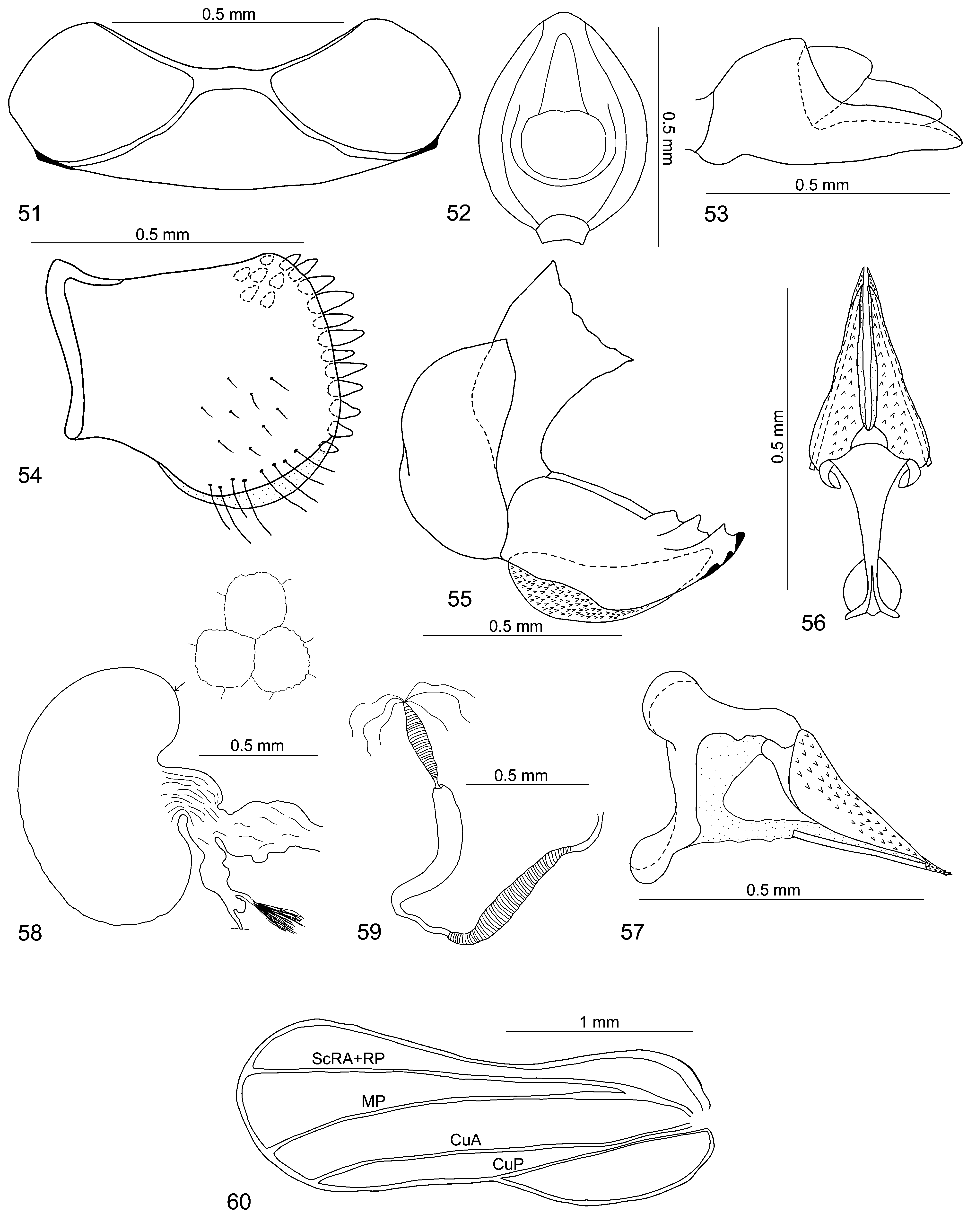
Hindwing well developed, narrow, with anal lobe ( Fig. 60 View FIGURES 51–60 ). ScP+R and MP single, with common stem; CuA single, transverse veinlets absent. Anal lobe without veins.
Pro- and mesotibiae with groove on the external side, about as long as pro- and mesofemora; apical tarsomeres of both fore and middle legs longer than cumulative length of basal and second tarsomeres ( Fig. 23 View FIGURES 23–28 ); metatibia longer than metafemur, with two lateral spines placed close to each other in distal part and apical row of 7–8 welldeveloped spines, external longer than internal; basitarsomere of metatarsus about as long as cumulative length of second and apical tarsomeres, with 8–9 small apical spines U-lined; second tarsomere with two lateral spines and median pad with setae.
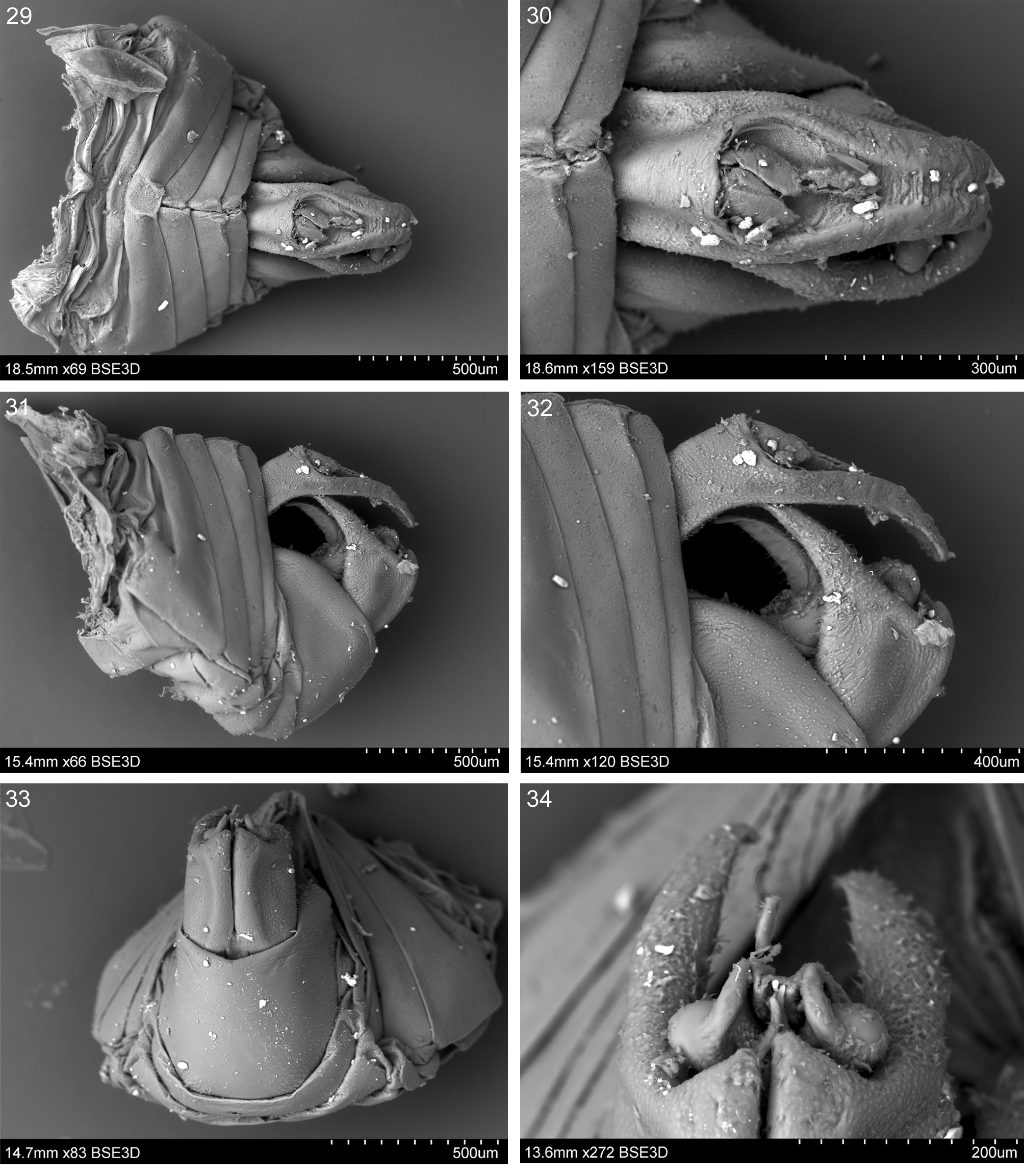
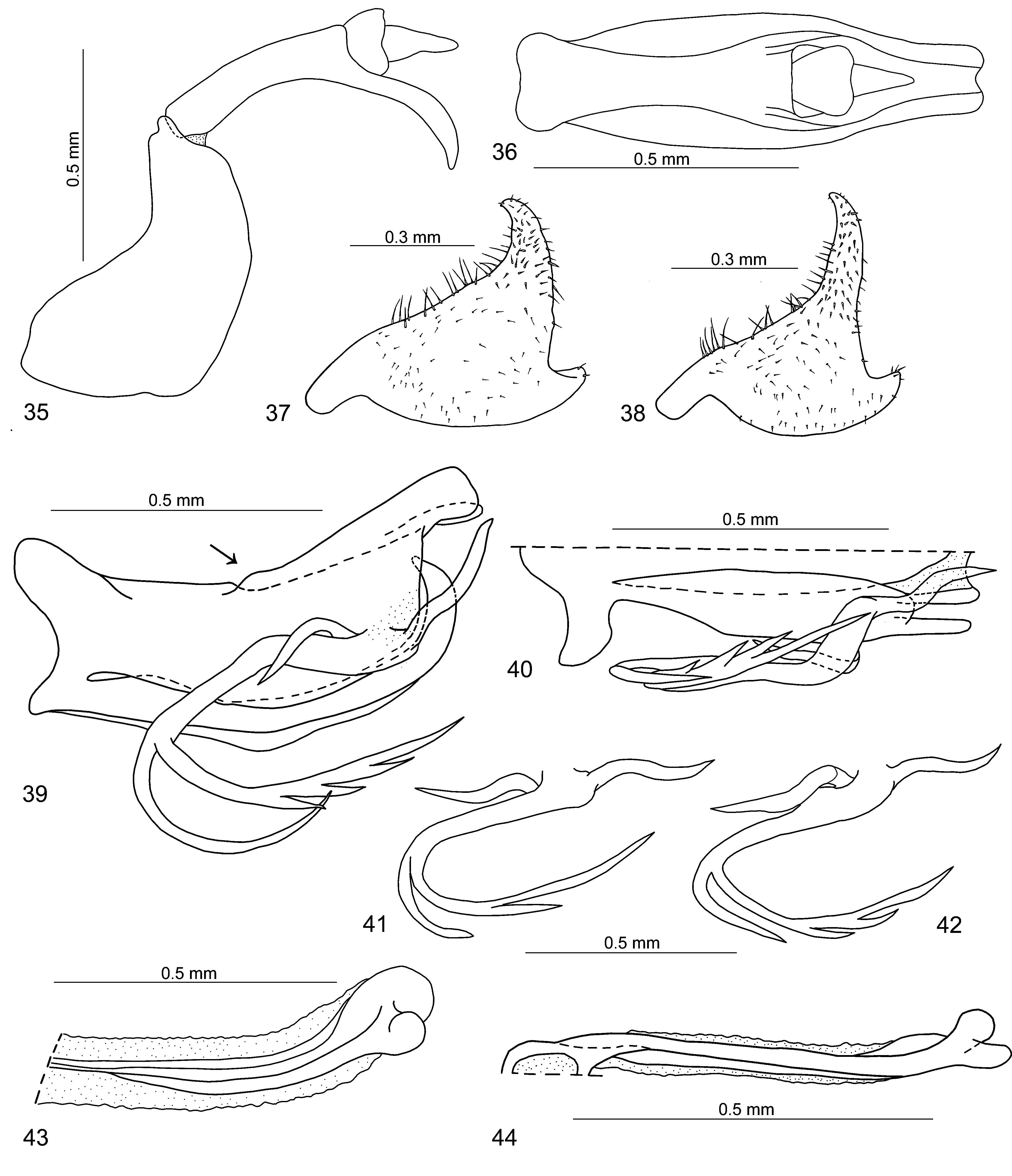
Male terminalia. Anal tube, in lateral view, elongate and curved, with ventral margin strongly concave, dorsal margin weakly convex; basal part wider than apical part; apical part oriented ventrad; anus placed posterior to the middle ( Figs 31, 32 View FIGURES29–34 , 35 View FIGURES35–44 ); in dorsal view, anal tube elongate, bowling pin-shaped; basal part strongly convex medially, apical part medially with groove ( Figs 29, 30 View FIGURES29–34 , 36 View FIGURES35–44 ). Pygofer, in lateral view, with dorsal margin narrower than ventral margin; anterior margin concave, posterior margin convex ( Fig. 35 View FIGURES35–44 ). Genital style widening apicad; postero-dorsal angle with long and curved capitulum, postero-ventral angle produced into a large tooth ( Figs 31– 34 View FIGURES29–34 , 37, 38 View FIGURES35–44 ). Phallic complex: Periandrium elongate, slightly curved, almost as long as aedeagus; lateral split almost reaching base ( Fig. 37 View FIGURES35–44 ). Dorsal part of periandrium, in lateral view, longer than ventral part, with apical prolongation and 3-armed appendage oriented antero-ventrad; posterior arm simple, oriented dorso-posteriad; median arm stout, diverged into two processes, upper process with 0–3 ventral spines; anterior arm simple, movable ( Figs 39–42 View FIGURES35–44 ). Ventral part of periandrium unilobate, apically strongly curved dorsad; ventral side with long median keel, in lateral view, medially broken ( Fig. 39 View FIGURES35–44 ). Aedeagus, in lateral view, long and slightly curved, with apical bulb-like sclerotized appendages; in ventral view, with deep median split, almost reaching base; dorsal and ventral parts of aedeagus membranous ( Figs 43, 44 View FIGURES35–44 ).
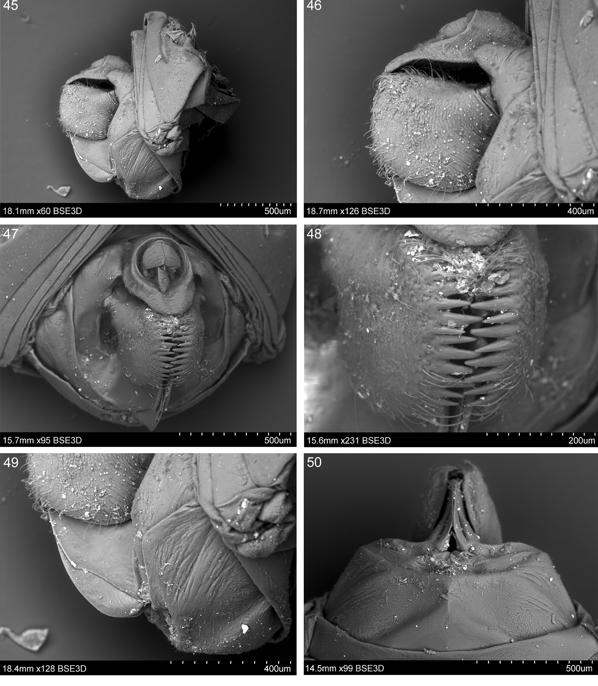
Female terminalia. Pregenital sternite with asymmetrically X-shaped sclerotization ( Fig. 51 View FIGURES 51–60 ); posterior arms of this X shorter than anterior ones, area between anterior arms convex; lateral lobes well developed; posterior margin of pregenital sternite concave, medially with small protuberance; anterior margin regularly convex ( Figs. 50 View FIGURES 45–50 , 51 View FIGURES 51–60 ). Anal tube, in lateral view ( Figs 45, 46 View FIGURES 45–50 , 53 View FIGURES 51–60 ), covering gonoplac and reaching its posterior margin; tapering apicad: basal part wider than apical part, anus placed slightly anterior to the middle, ventral margin almost straight; in dorsal view, anal tube ovoid, widest in median portion ( Fig. 52 View FIGURES 51–60 ). Gonoplac subrectangular, not covering the base of gonapophysis VIII ( Figs 45, 46, 49 View FIGURES 45–50 , 54 View FIGURES 51–60 ); posterior margin with single row of well-developed teeth; teeth of both gonoplacs fitting together in a zip-like manner ( Figs 47, 48 View FIGURES 45–50 ); ventral margin of gonoplac with narrow membranous fold ( Fig. 54 View FIGURES 51–60 ). Gonapophysis VIII stout, laterally flattened, ventral margin sinuate ( Figs 49 View FIGURES 45–50 , 55 View FIGURES 51–60 ); dorsal margin with three lamellate, sharp teeth, ventral margin subapically slightly up-folded, with three blunt teeth oriented exteriad ( Fig. 55 View FIGURES 51–60 ); endogonocoxal process slightly shorter than gonapophysis, tapering apicad, with bluntly rounded apex and spiniferous microsculpture. Gonospiculum as in Figs 56, 57 View FIGURES 51–60 . Bursa copulatrix with single pouch, kidneyshaped, with cells ( Fig. 58 View FIGURES 51–60 ). Spermatheca with ductus receptaculi slightly shorter than diverticulum ductus; ductus receptaculi ribbed, widened subapically; diverticulum ductus smooth, narrow in basal third, widened in apical two thirds ( Fig. 59 View FIGURES 51–60 ).
Etymology. The generic name is a combination derived from “ Socotra ” and “ Flata ” which is used here for a representative of the Flatidae family. Gender feminine.
Distribution. Yemen: Socotra island.


No known copyright restrictions apply. See Agosti, D., Egloff, W., 2009. Taxonomic information exchange and copyright: the Plazi approach. BMC Research Notes 2009, 2:53 for further explanation.