
Psilotreta atrocaudata, Kawase, 2022
|
publication ID |
https://doi.org/10.11646/zootaxa.5195.6.1 |
|
publication LSID |
lsid:zoobank.org:pub:1A42B1B9-9D3F-44D8-B9C7-0D96CB2312ED |
|
DOI |
https://doi.org/10.5281/zenodo.7223854 |
|
persistent identifier |
https://treatment.plazi.org/id/E84587ED-FFAB-0874-E7BC-FB9F62D7FEFE |
|
treatment provided by |
Plazi |
|
scientific name |
Psilotreta atrocaudata |
| status |
sp. nov. |
Psilotreta atrocaudata sp. nov.
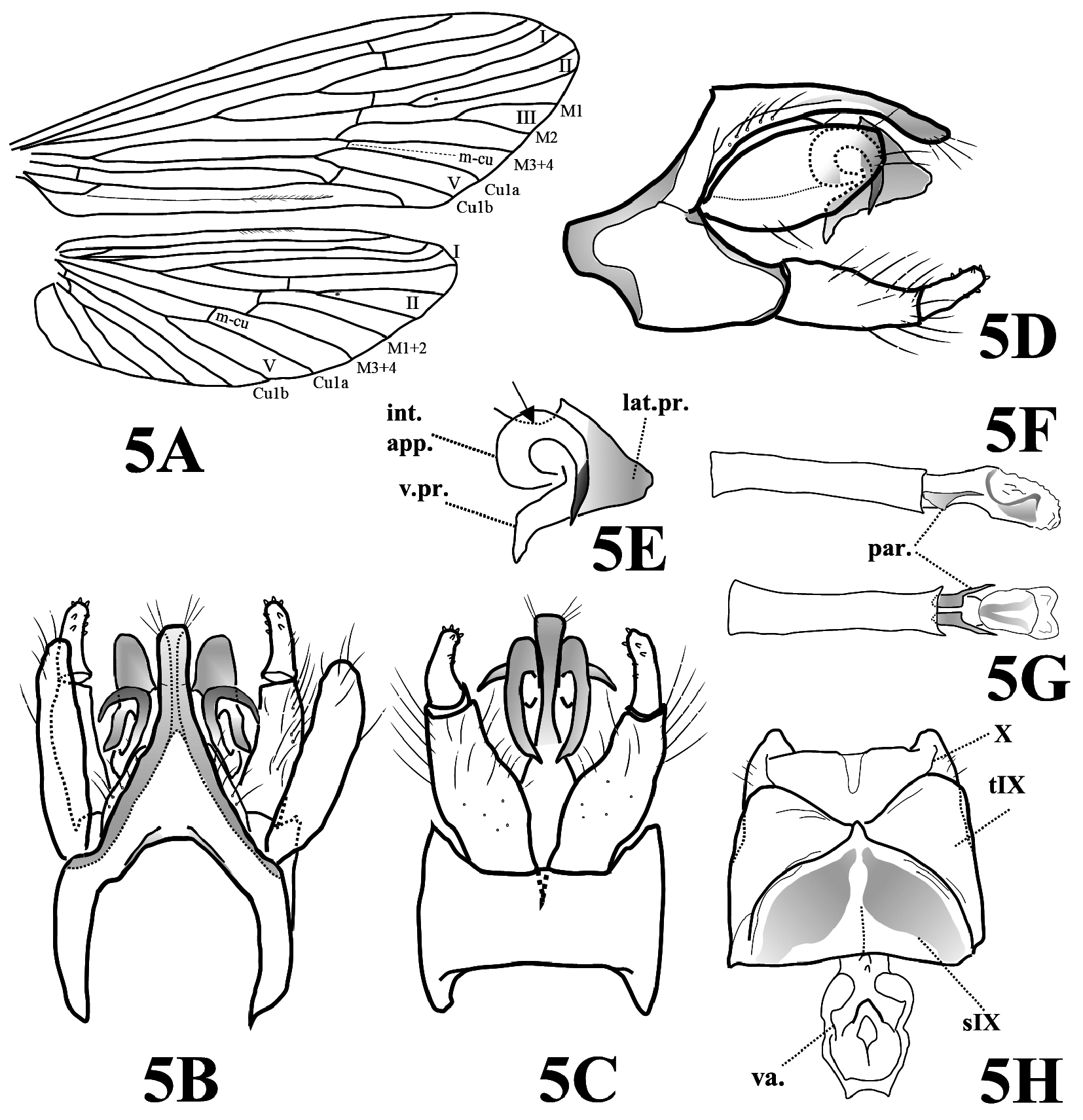
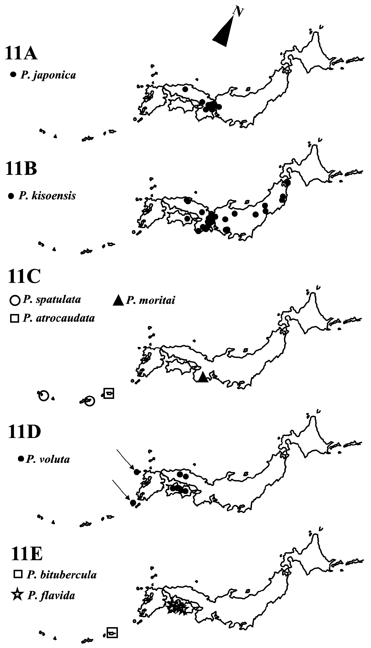
( Figs 5A–5H View FIGURE 5 , 11C View FIGURE 11 )
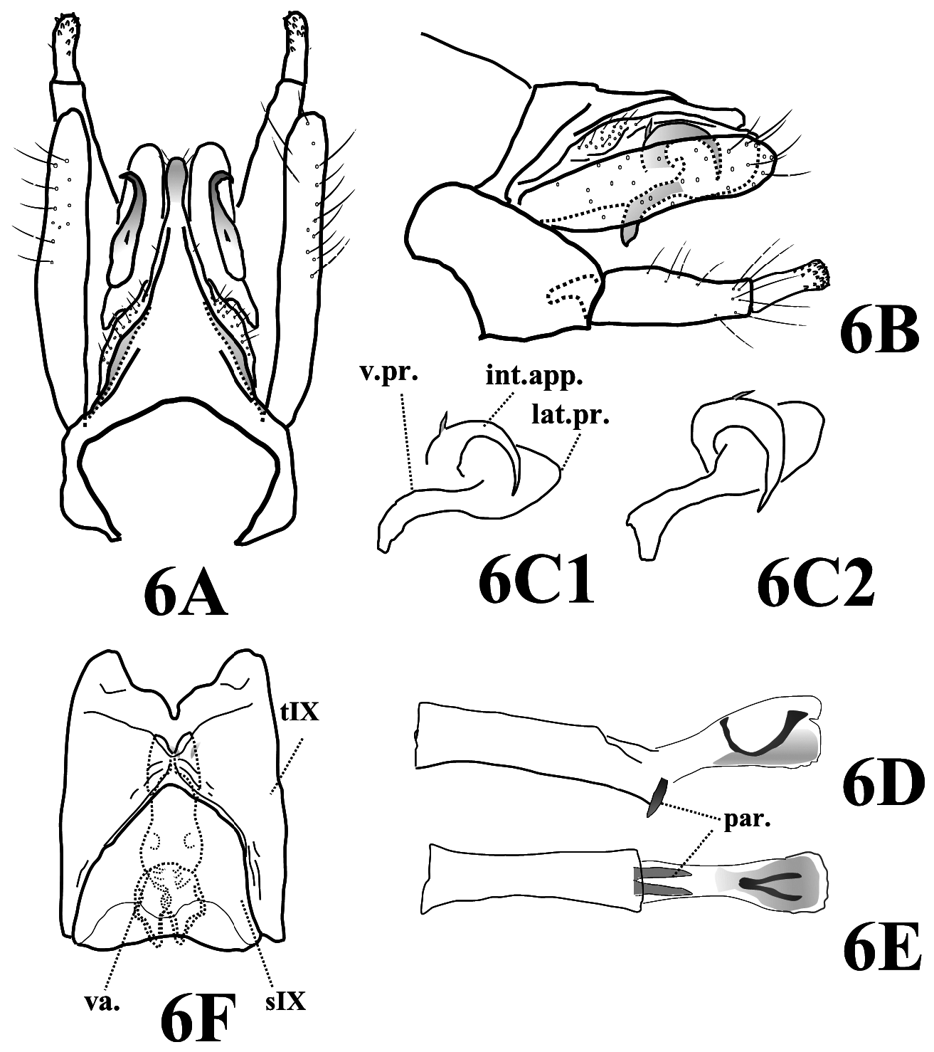
Diagnosis. The male genitalia of P. atrocaudata sp. nov. are most similar to those of P.voluta , but can be distinguished from the latter by the following characters: (1) the median dorsal process of segment X is parallel-sided in dorsal view and broadly dark pigmented caudally in P. atrocaudata ( Figs 5B, 5D View FIGURE 5 ) (the median dorsal process of segment X is constricted at mid-length and pigmented only apically in P. voluta ( Fig. 6A View FIGURE 6 )); (2) lateral processes of tergum X are subtriangular in lateral view in P. atrocaudata ( Fig. 5E View FIGURE 5 ) (lateral processes are oval in lateral view in P. voluta ( Figs 6C View FIGURE 6 1 View FIGURE 1 , 6C View FIGURE 6 2 View FIGURE 2 )); and (3) each paramere bears a distinct mesal protrusion and the acute apex is directed laterad in P. atrocaudata ( Fig. 5G View FIGURE 5 ) (each paramere lacks a mesal protrusion and is straight in P. voluta ( Fig. 6E View FIGURE 6 )). Additionally, the male genitalia of P. atrocaudata also resemble those of P. clyssan Malicky 2014 described from Taiwan but are easily distinguishable from the latter by the broadly dense pigmentation on the median dorsal process and lateral processes, and by the apices of the intermediate appendages that are extended ventrolaterad in P. atrocaudata (that pigmentation is not present and the apices of the intermediate appendages are strongly curved and directed ventrad in P. clyssan ). The female genitalia of P. atrocaudata are most similar to those of P. voluta but can be easily distinguished from them by the horn-like projection on the posterior margin of sternum IX ( Fig. 5H View FIGURE 5 ).
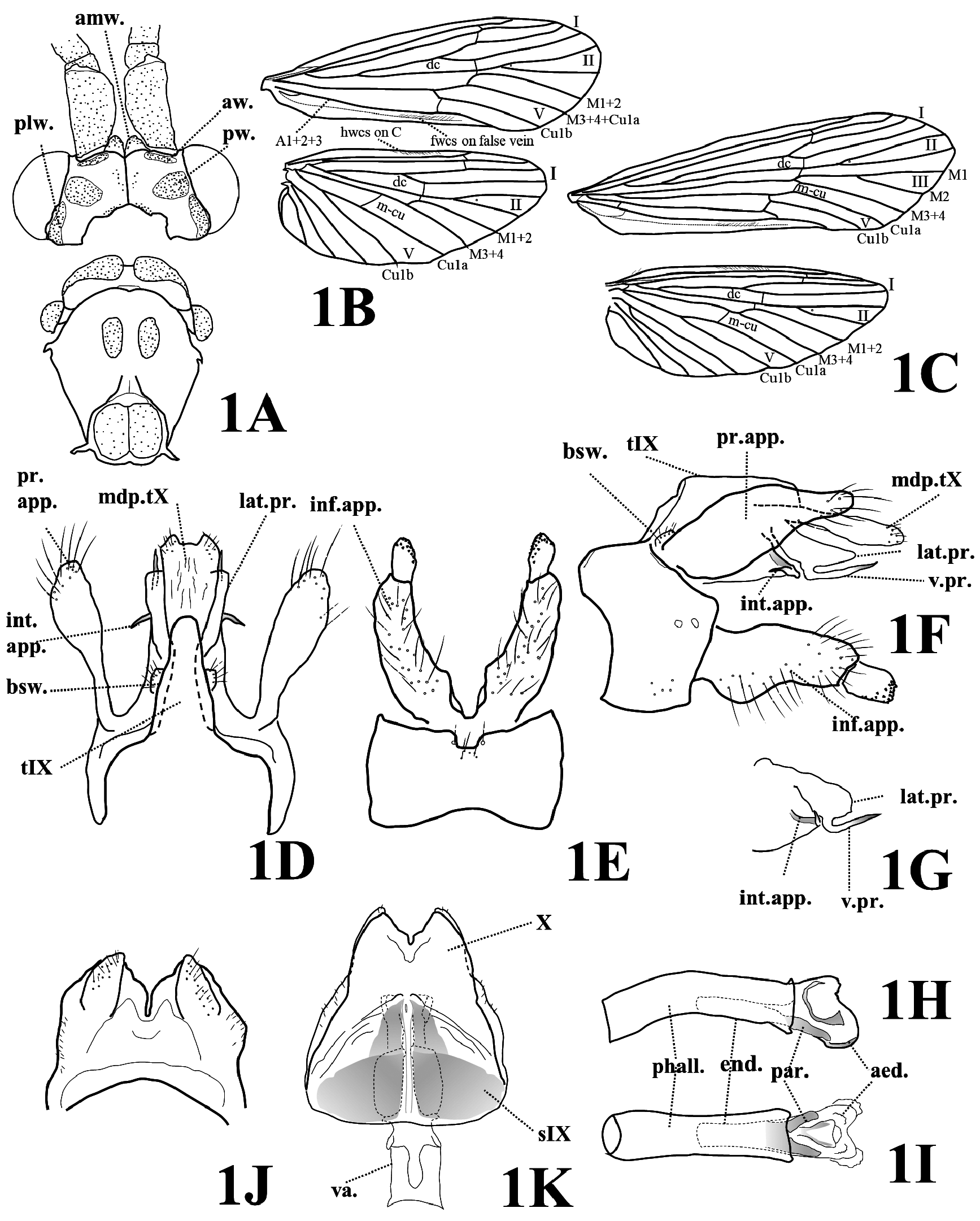
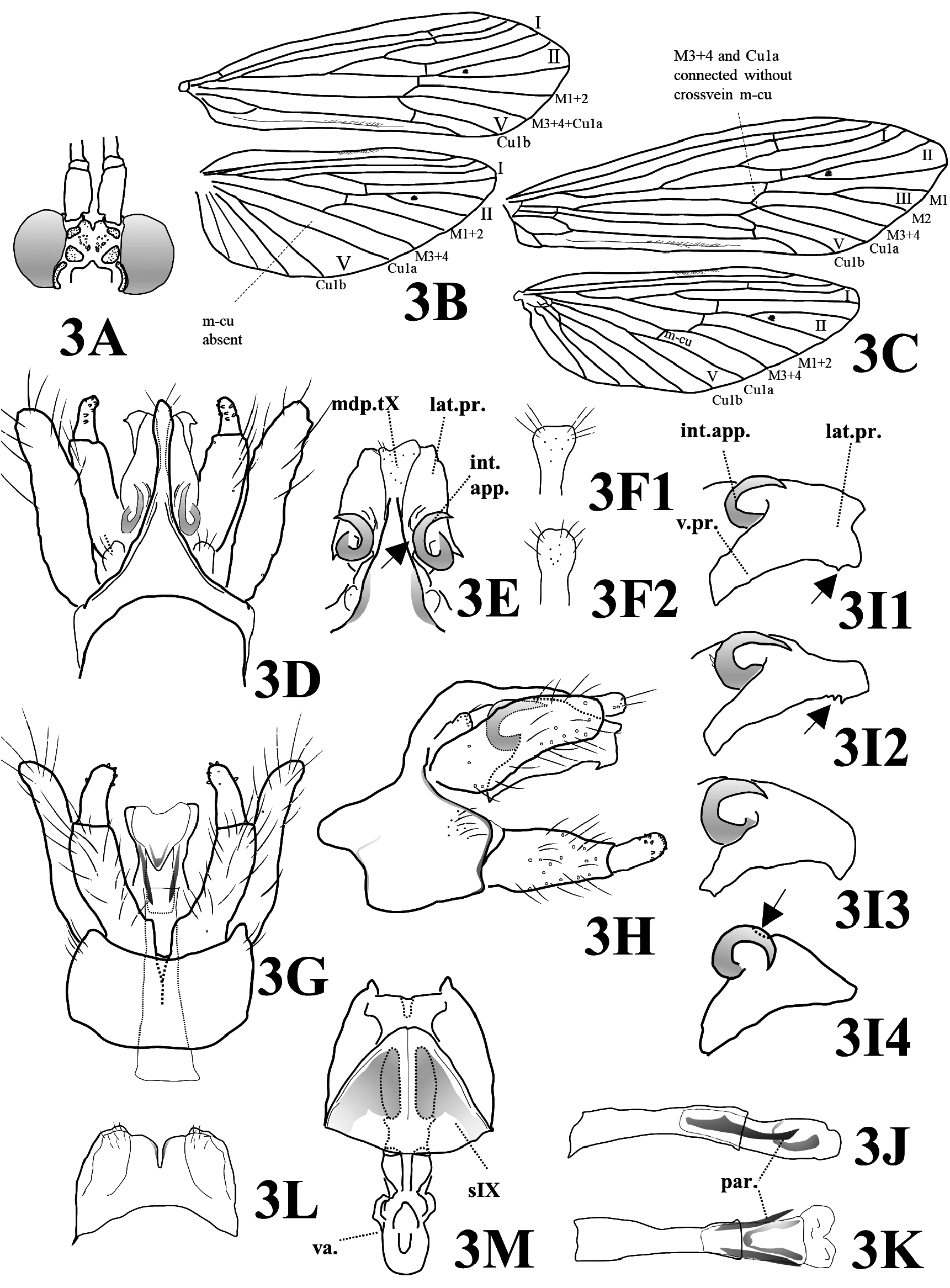
Adult ( Fig. 5A View FIGURE 5 ). Body and antennae brown, wings dark brown in alcohol. Length of each forewing: male 7.9–9.2 mm (mean = 8.4, n = 5), female 9.4–11.2 mm (mean = 10.0, n = 4). General morphology including wing venation similar to that of P. spatulata sp. nov. ( Figs 3A–3C View FIGURE 3 ), but in each female forewing, M3+4 and Cu1a connected by short cross vein m-cu in P. atrocaudata sp. nov. ( Fig. 5A View FIGURE 5 ) (those connected directly without crossvein m-cu in P. spatulata sp. nov. ( Fig. 3A View FIGURE 3 )).
Male genitalia ( Figs 5B–5G View FIGURE 5 ). Tergum IX elongate, subtriangular in dorsal view, with steep sides deeply above basal setal warts ( Figs 5B, 5D View FIGURE 5 ). Basal segment of each inferior appendage approximately same length as preanal appendages, cylindrical, slightly narrower near apex, covered with long setae; apical segment about 0.4 times as long as basal segment, cylindrical with several small brown teeth on apical half ( Figs 5C, 5D View FIGURE 5 ). Preanal appendages elongate, long-oval, not extending beyond posterior margin of lateral processes in lateral view. Tergum X forming median dorsal process thick, parallel-sided in dorsal view (mostly fused with tergum IX); heavily sclerotized, darkly pigmented in caudal half; round apex with several setae in dorsal view ( Fig. 5B View FIGURE 5 ); lateral processes subtriangular, each with dorsal margin partially concave (concavity marked with arrow in Fig. 5E View FIGURE 5 ); ventral projection heavily sclerotized, curved anteroventrad with acute apex; intermediate appendages dark brown, heavily sclerotized, strongly curved, C-shaped ending in acute apex directed ventrolaterad ( Figs 5B, 5C, 5E View FIGURE 5 ).
Phallus with phallotheca long, cylindrical; endotheca with pair of parameres ventrally: each paramere broad at basal half in ventral view, with distinct protrusion mesally in middle, tapering to acute apex directed laterad ( Fig. 5G View FIGURE 5 ); aedeagus membranous with ventral plate weakly sclerotized, phallotremal sclerite V-shaped in ventral view, strongly curved dorsad in lateral view ( Fig. 5F View FIGURE 5 ).
Female genitalia ( Fig. 5H View FIGURE 5 ). Sternum IX wider than long, semicircular with short horn-like median projection posteriorly in ventral view; with pair of leaf-like pigmented areas. Posterior margin of segment X transverse, almost straight with small mesal notch in ventral view, with pair of short fin-like lobes dorsolaterally. Length of vaginal apparatus 2 times as long as sternum IX.
Larva. Unknown.
Holotype. Male (in alcohol), Amami Island, Mt. Yuwan-dake to Materiya falls , Uken-son , Kagoshima Pref., Japan , 28°17'42"N 129°19'56"E, 9.v.2011, T. Ito. ( LBM1410012568 View Materials ). GoogleMaps
Paratypes. Amami Island : 2 males, 3 females, same locality as the holotype, 9.v.2007, T GoogleMaps . Ito ( LBM1410012569 View Materials – LBM1410012573 View Materials ); 2 males, 1 female, Miyama-gawa, Setouchi-cho, 21.iv.2008, T . Ito ( LBM1410012574 View Materials – LBM1410012576 View Materials ) .
Etymology. The Latin species epithet “ atrocaudata ” refers to the dark pigmentation of the median dorsal process of segment X in the male genitalia.
Distribution and habitat. Psilotreta atrocaudata sp. nov. is an Oriental species distributed on Amami Island in the Ryukyu Archipelago, southwestern Japan ( Fig. 11C View FIGURE 11 ). Adults were collected near mountainous streams by light traps or sweeping.
Japanese name. Amami-kiso-tobikera.

| T |
Tavera, Department of Geology and Geophysics |
No known copyright restrictions apply. See Agosti, D., Egloff, W., 2009. Taxonomic information exchange and copyright: the Plazi approach. BMC Research Notes 2009, 2:53 for further explanation.
|
Kingdom |
|
|
Phylum |
|
|
Class |
|
|
Order |
|
|
Family |
|
|
Genus |