
Psilotreta japonica ( Banks 1906 )
|
publication ID |
https://doi.org/10.11646/zootaxa.5195.6.1 |
|
publication LSID |
lsid:zoobank.org:pub:1A42B1B9-9D3F-44D8-B9C7-0D96CB2312ED |
|
DOI |
https://doi.org/10.5281/zenodo.7223834 |
|
persistent identifier |
https://treatment.plazi.org/id/E84587ED-FFA3-087B-E7BC-FD7062DEFB9E |
|
treatment provided by |
Plazi |
|
scientific name |
Psilotreta japonica ( Banks 1906 ) |
| status |
|
Psilotreta japonica ( Banks 1906) View in CoL
( Figs 1A–1K View FIGURE 1 , 9B–9D, 9G View FIGURE 9 , 10A, 10E View FIGURE 10 , 11A View FIGURE 11 )
Odontocerum japonicum Banks 1906 , 110, female, Honshu ( Type locality: Gifu); Ulmer 1907a, 51–52, male, female, Honshu.
Psilotreta japonica: Ulmer 1907b View in CoL , 126, wings of male and female; Tsuda 1959, 143, larva; Parker &Wiggins 1987, 34, male, female, Honshu; Tanida 2005, 556–557, male, female; Kawase 2012, 29–39, wings, male, female, larva, habitat, Honshu; Tanida 2018, 665–667, male, female, larva.
Psilotreta kyotoensis Iwata 1928 , 118, 125, larva, Honshu ( Kyoto). Synonymized by Tsuda (1942).
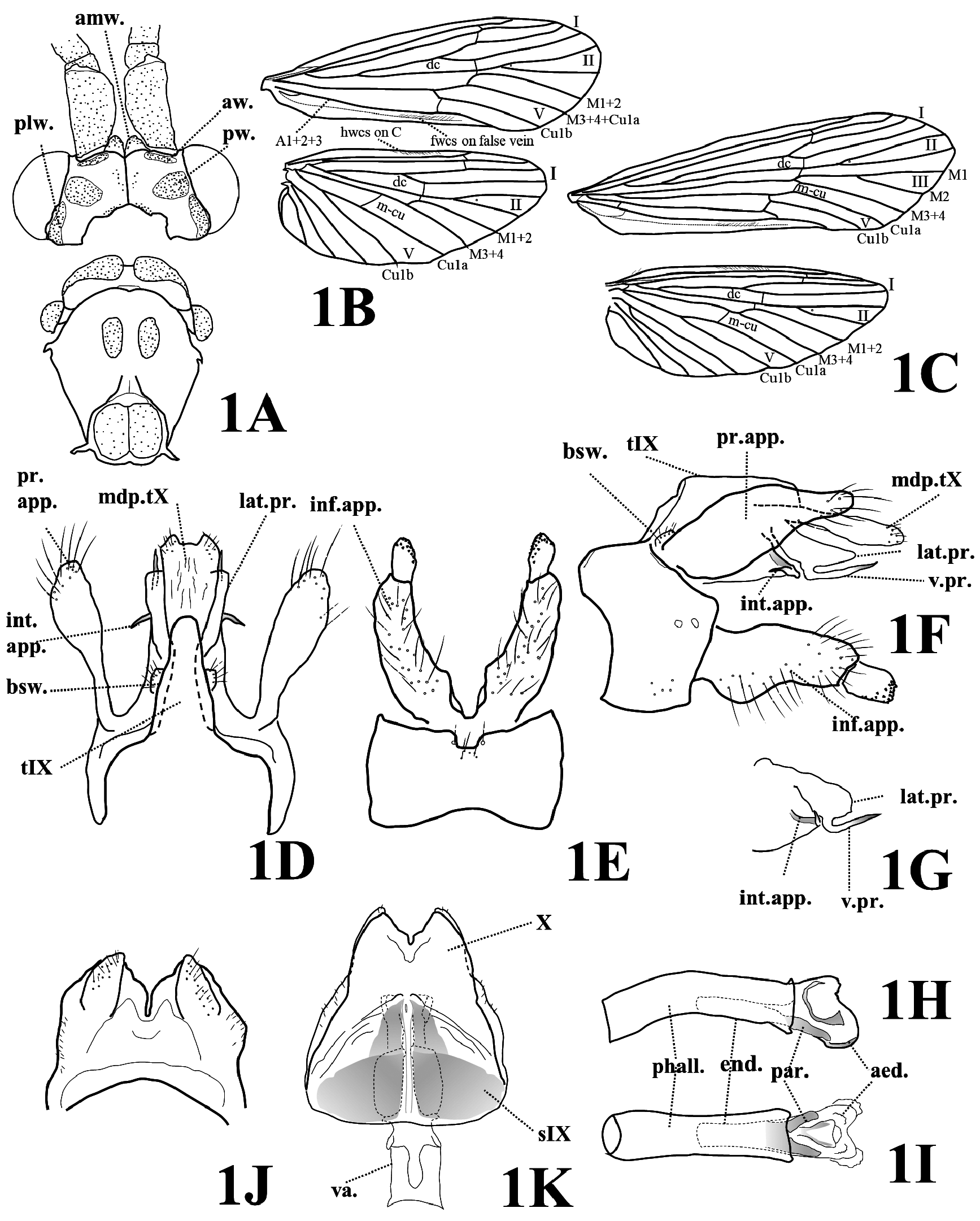
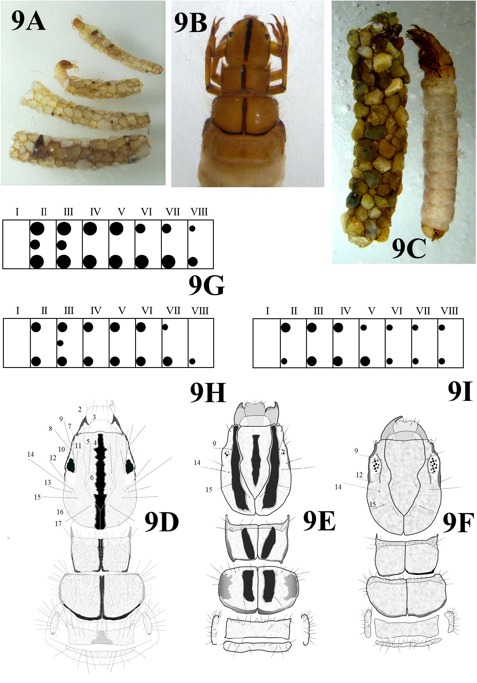
Diagnosis. The male of this species can be easily distinguished from those of other Japanese species by the male genitalia: Each intermediate appendage is very short and needle-like in this species ( Figs 1D, 1F–1G View FIGURE 1 ), but long and curved in other Japanese species. The female genitalia of this species are distinguished from those of other Japanese species by the shapes of paired pigmentations of sternum IX that are long triangular with the apexes directed posteromesad in ventral view ( Fig. 1K View FIGURE 1 ). The larva is unique among known Japanese larvae in having a dark longitudinal median-band from the head to the mesothorax dorsally ( Figs 9B, 9D View FIGURE 9 ), and in having many branches (more than 20 branches on each side of segments II–V) of the dorsal or ventral abdominal gills ( Figs 9C, 9G View FIGURE 9 ).
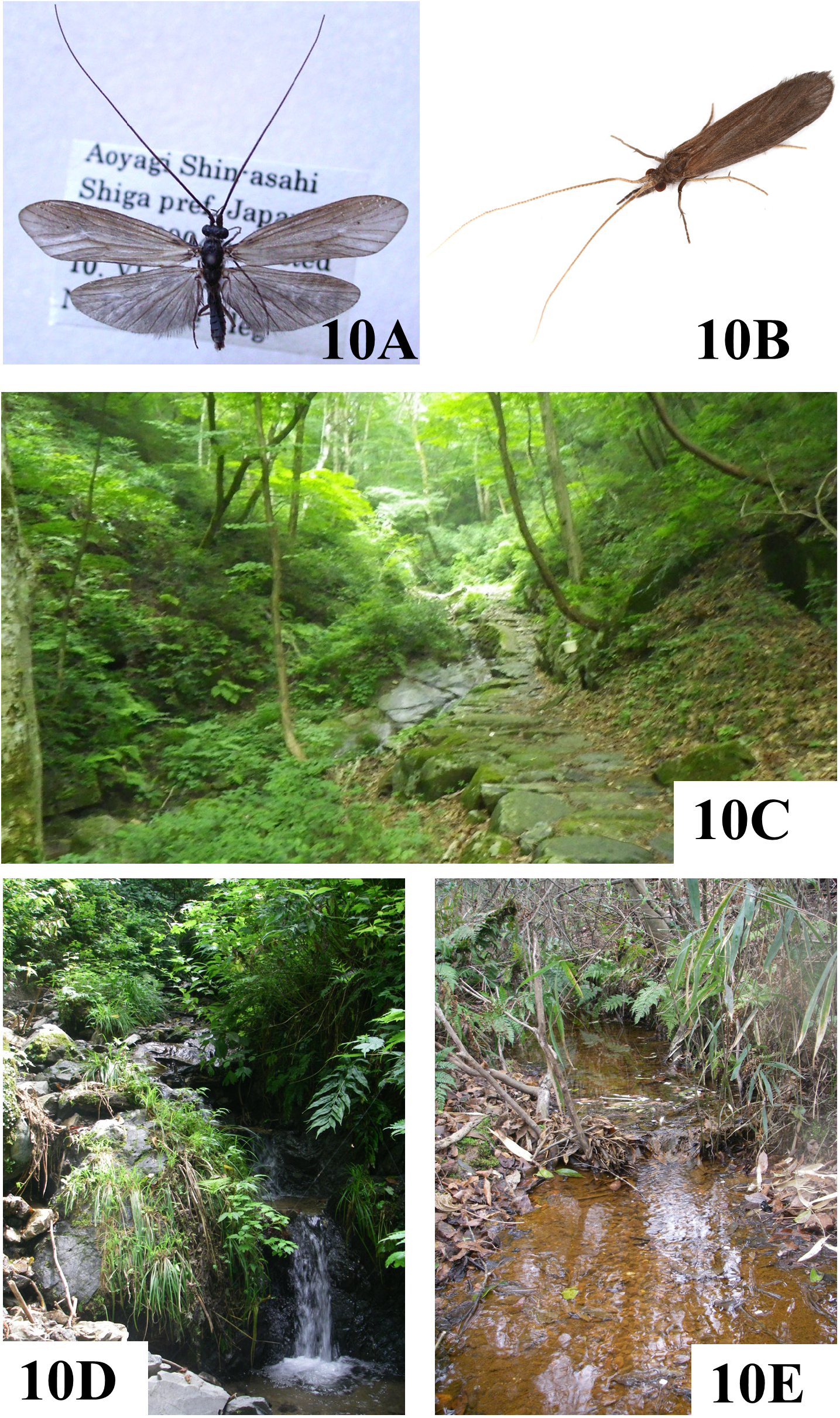
Adult ( Figs 1A–1C View FIGURE 1 , 10A View FIGURE 10 ). General color of body, antennae, and wings black to dark brown ( Fig. 10A View FIGURE 10 ). Length of each forewing: male 10–13 mm (mean = 11.9, n = 15), female 13–16 mm (mean = 15.1, n = 9). Antennae 1.5 times as long as forewings in male, 1.2 times in female. Head with pair of anteromesal, anterior, posterior and posterolateral setal warts; in dorsal view, each anteromesal wart round between antennal sockets, each anterior setal wart narrow, each posterior wart large oval, each posterolateral setal wart narrow and extending ventrad along posterior margin of eye. Venation as in Figures 1B, 1C View FIGURE 1 : in each forewing, fork I rooted on discoidal cell about 1/3 its length in male, 1/ 8 in female; combined anal vein A1+2+3 with forewing coupling setae ventrally on false vein posterior of A1+2+3 (for fore- and hind wings to link on each side) ( Stocks 2010); in each hind wing, fork I rooted on discoidal cell about 1/3 its length in male, about 1/ 4 in female, crossvein m-cu present in male and female; costa with coupling setae dorsally to link with forewing.
Male genitalia ( Figs 1D–1I View FIGURE 1 ). Tergum IX elongate, subtriangular to rectangular in dorsal view, bearing steep sides above basal setal warts ( Figs 1D, 1F View FIGURE 1 ). In inferior appendages, each basal segment as long as preanal appendages, cylindrical with many long setae, slightly enlarged basoventrally in lateral view; each apical segment about 1/3 length of basal segment, apicomesal half with many brown teeth ( Figs 1E, 1F View FIGURE 1 ). Preanal appendages elongate, broadest at basal half and tapered distally in lateral view, extending to posterior margins of lateral processes, each with several long setae near apex ( Figs 1D, 1F View FIGURE 1 ). Median dorsal process of segment X with shallow apical concavity in dorsal view ( Fig. 1D View FIGURE 1 ), and slightly clavate apically in lateral view ( Fig. 1F View FIGURE 1 ). Pair of lateral processes of segment X protruding posteriorly, each with acuminate ventral projection extended posterad; each intermediate appendage very short, needle-like, directed anterolaterad ( Figs 1F, 1G View FIGURE 1 ).
Phallus with phallotheca long, cylindrical; endotheca ventrally with pair of thick parameres ( Figs 1H, 1I View FIGURE 1 ); aedeagus membranous with sclerotized ventral plate, phallotremal sclerite strongly curved dorsad, C-shaped in lateral view ( Fig. 1H View FIGURE 1 ).
Female genitalia ( Figs 1J, 1K View FIGURE 1 ). Sternum IX semicircular, as long as wide, with pair of dark pigmentations along median shallow sulcus in ventral view; each pigmentation subtriangular with acute apex directed posteromesad ( Fig. 1K View FIGURE 1 ). Segment X with pair of fin-like dorsoposterior lobes, setose dorsally in dorsal view ( Fig. 1J View FIGURE 1 ); with deep V-shaped incision in dorsal and ventral views ( Figs 1J, 1K View FIGURE 1 ). Vaginal apparatus longer than sternum IX, anterior sclerite rectangular in ventral view ( Fig. 1K View FIGURE 1 ).
Final instar larva ( Figs 9B–9D, 9G View FIGURE 9 ). Length of final instar larva up to 17 mm. Head and thorax brown to reddish brown dorsally, with dark median longitudinal band from anterior margin of head to posterior margin of mesonotum ( Figs 9B, 9D View FIGURE 9 ). Among primary setae on head, setae 9, 11, 14, 15, and 17 thick, brown; 16 fine, transparent; other setae fine, pale brown; 14 longest, 1.5 times as long as 15. Branched abdominal gills as in Fig. 9G View FIGURE 9 present on each side of following segments (numbers of branches in parenthesis, n = 3): anterior dorsal gills on segments II (20–36), III (22–44), IV (22–34), V (28–29), VI (9–12), VII (9–10), and VIII (6–8); anterior ventral gills on segments II (29–46), III (28–56), IV (28–44), V (21–28), VI (13–24), VII (18–24), and VIII (7–12); anterior lateral gills on segments II (9–16) and III (7–10). Larval case cylindrical, slightly curved and tapered, constructed of coarse fragments of rock ( Fig. 9C View FIGURE 9 ).
Specimens examined. Honshu, Aichi: 1 larva, Yato-cho, Seto-shi , 4.ix.1999, N. Kawase ; 2 larvae, Ichinoi, Maeguma, Nagakute-shi , 8.iv.2010, N. Kawase ; 2 males, same locality, 11.v.2011, H. Nishimoto . Mie: 6 males, 2 females, Washiyama, Kameyama-shi , 9.vi.2006, N. Kawase ; 1 male and 1 female, same locality, 17.vi.2006, H. Morita ; 1 larva, same locality, 29.ix.2006, N. Kawase . Shiga: 3 larvae, Kawashima, Adogawa-cho, Takashima-shi , 27.iv.2003, N. Kawase ; 8 males, 4 females, same locality, pupae collected on 24.v.2004, adults emerged on vi.2004, N. Kawase ; 1 larva, same locality, 30.ix.2007, N. Kawase ; 2 larvae, same locality, 3.xi.2010, N. Kawase ; 1 larva, Shichiri , Ryu-oh-cho, 20.v.2001 (no collector’s name is given) .
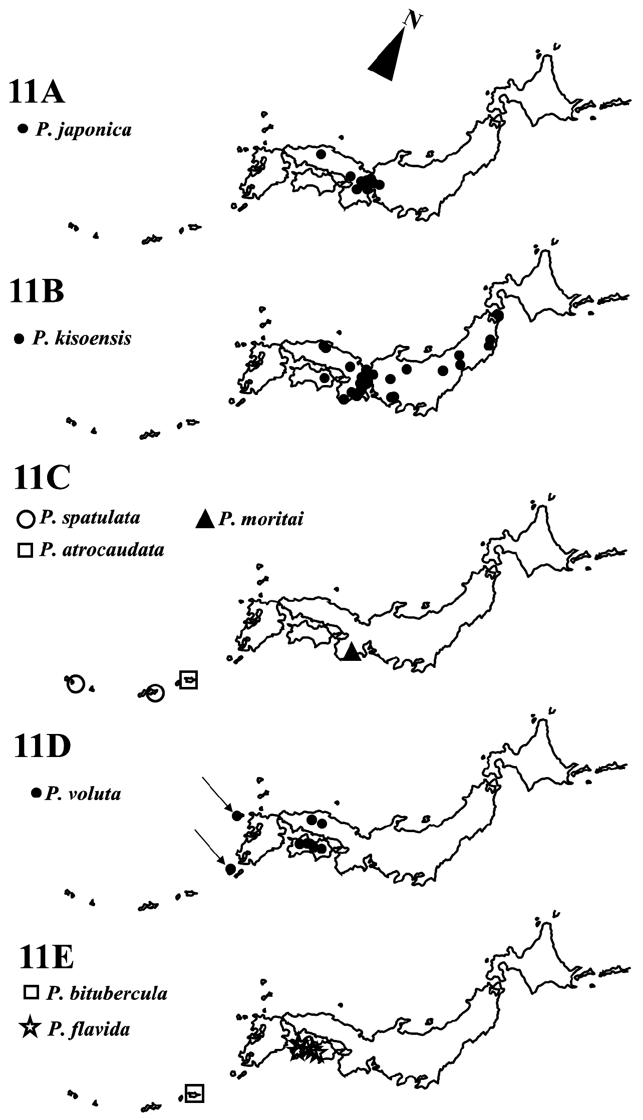
Distribution and habitat. Psilotreta japonica is an East Palearctic species sparsely distributed in central to western Honshu ( Fig. 11A View FIGURE 11 ) and inhabits small water channels in lower hills or delta plains with spring water seeps ( Fig. 10E View FIGURE 10 ).
Japanese name. Hitosuji-kiso-tobikera.
No known copyright restrictions apply. See Agosti, D., Egloff, W., 2009. Taxonomic information exchange and copyright: the Plazi approach. BMC Research Notes 2009, 2:53 for further explanation.
|
Kingdom |
|
|
Phylum |
|
|
Class |
|
|
Order |
|
|
Family |
|
|
Genus |
Psilotreta japonica ( Banks 1906 )
| Kawase, Naoki 2022 |
Psilotreta kyotoensis
| Iwata 1928 |
Psilotreta japonica :
| Ulmer 1907 |
Odontocerum japonicum
| Banks 1906 |