
Heradion intermedium, Chami-Kranon, Thanaphum & Ono, Hirotsugu, 2007
|
publication ID |
https://doi.org/ 10.5281/zenodo.175335 |
|
DOI |
https://doi.org/10.5281/zenodo.6243830 |
|
persistent identifier |
https://treatment.plazi.org/id/E539FB1E-A476-414A-B394-F886FC9AFD72 |
|
treatment provided by |
Plazi |
|
scientific name |
Heradion intermedium |
| status |
sp. nov. |
Heradion intermedium View in CoL spec. nov.
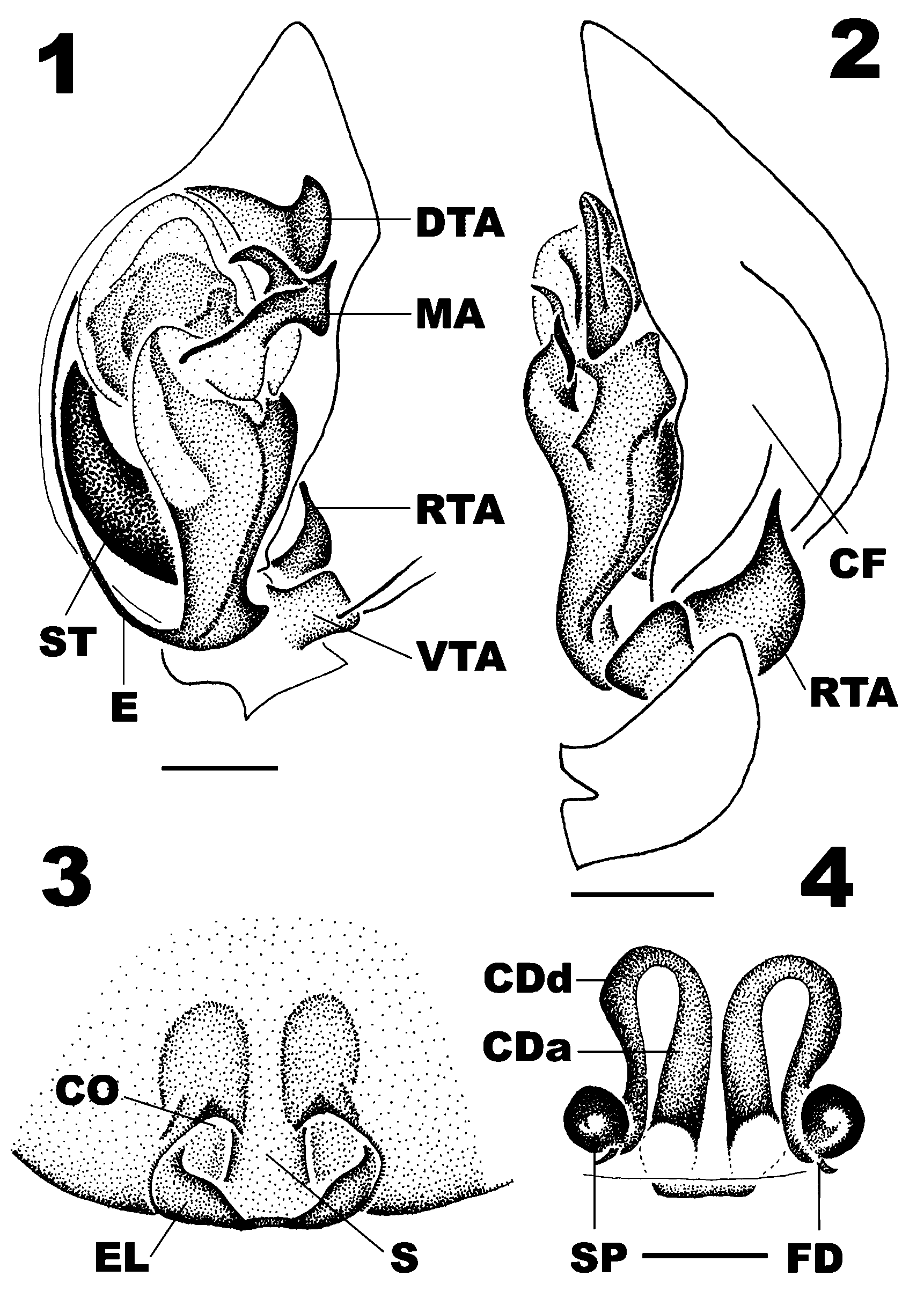
Figures 1–4 View FIGURES 1 – 4 , 7, 10 View FIGURES 5 – 12 , 13–18 View FIGURES 13 – 18
Type material. Holotype: ɗ [ MHNG, SV 03/20], VIETNAM, Lam–Dong Province, Datanla Waterfall, ca. 5 km south of Da–Lat, 11º 54' 02.2'' N, 108º 26' 54.0'' E, 1300 m, evergreen hill forest, 5./11./12.IX.2003, leg. PJ Schwendinger.
Paratypes: 2 ɗ, 3 Ψ, data as holotype [ MHNG, SV 03/20].
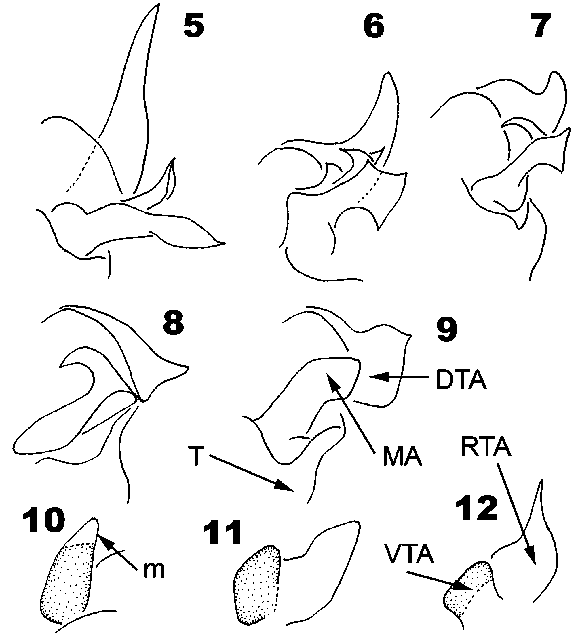
Etymology. Latin, intermedius = meaning in between. The specific epithet refers to the length of the distal tegular apophysis of male palpal organ which is obviously shorter than those of H. naiadis and H. peteri but slightly longer than that of H. flammeum (Ono) comb. nov. ( Figs 5–9 View FIGURES 5 – 12 ).
Diagnosis. Heradion intermedium spec. nov. can be recognized by: the bifurcate median apophysis of males ( Figs 1 View FIGURES 1 – 4 , 7 View FIGURES 5 – 12 ); the distal triangular tegular apophysis ( Fig. 1 View FIGURES 1 – 4 ), with relatively broad base and blunt apex; female with posterior bilateral epigynal lips ( Fig. 3 View FIGURES 1 – 4 ); posteriorly located spermathecae globular ( Fig. 4 View FIGURES 1 – 4 ). Heradion intermedium spec. nov. is most similar to H. naiadis particularly in the general shape of the MA and the sharply pointed retrolateral tibial apophysis ( Figs 2 View FIGURES 1 – 4 , 12 View FIGURES 5 – 12 ). It can be distinguished from H. naiadis by differences in shape of the distal tegular apophysis and elongate internal duct system of female. In H. naiadis , the MA provided with a multibranching projection, clearly visible in ventral view ( Fig. 6 View FIGURES 5 – 12 ). The length of the ascending part of the female internal ducts is shorter in H. naiadis than that in H. intermedium spec. nov. ( Fig. 4 View FIGURES 1 – 4 ). The female spermathecae are globular but reniform in the latter species.
Heradion intermedium spec. nov. belongs to the naiadis group judging from the structures of the male palpal organ. Although it is related to H. luctator (the pernix group) by the strong resemblance of the bifid MA and the comparable orientation of the tibial apophyses, the following differences are obvious: it has a triangular DTA, which is the main character of the species attributed to the naiadis group ( Figs 5–9 View FIGURES 5 – 12 ); the proximal part of tibiae I of H. intermedium spec. nov. is not modified (modification occurs in males of H. luctator ; see also Dankittipakul & Jocqué 2004: 782; Fig. 84).
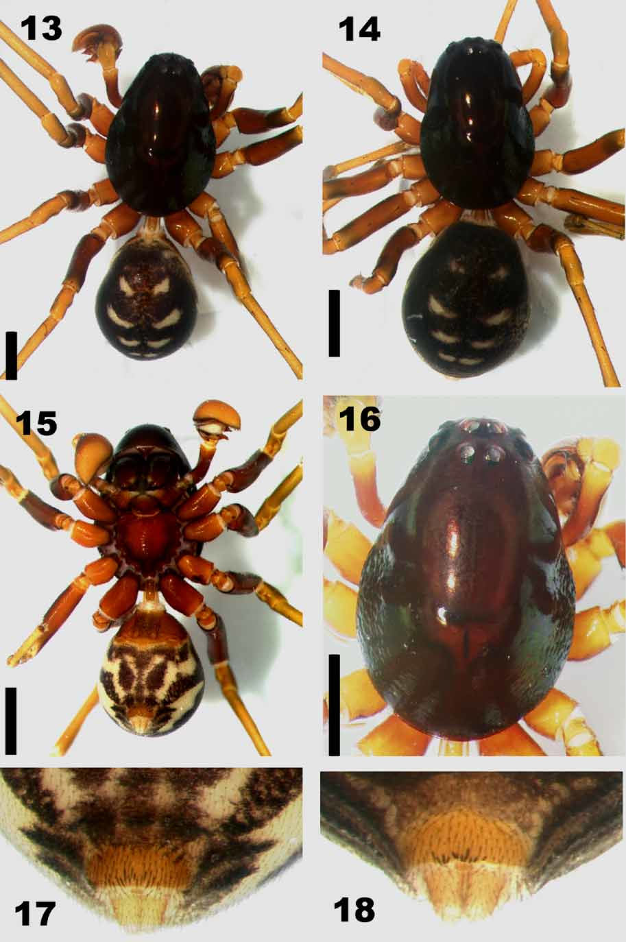
Description. ɗ (holotype). Total length 4.38. Carapace 2.45 long, 1.64 wide. Opisthosoma 1.93 long, 1.46 wide.
Colour. Carapace ( Figs 13, 16 View FIGURES 13 – 18 ) dark reddishbrown; chelicerae dark brown; sternum ( Fig. 15 View FIGURES 13 – 18 ) orangebrown. Legs bicolor: coxae, trochanters, femora and patellae orangebrown; tibiae, metatarsi and tarsi yellow. Dorsum of opisthosoma dark purple with three pairs of pale patches ( Fig. 13 View FIGURES 13 – 18 ), followed by a transverse band; venter pale, provided with markings ( Fig. 15 View FIGURES 13 – 18 ): a pair of teardropshaped patches situated in between a median longitudinal band and a pair of oblique stripes.
Prosoma ( Figs 13, 16 View FIGURES 13 – 18 ). Long oval, widest between coxae II and III, smooth and shiny, except for margin above coxae I and II provided with a small patch of strongly granulate tegument ( Fig. 16 View FIGURES 13 – 18 ).
Sternum ( Fig. 15 View FIGURES 13 – 18 ). Strongly raised, with anterior concavity, accommodating labium and maxillae; lateral margins elevated, provided with pointed intercoxal extensions, fitting coxal concavities.
Opisthosoma ( Fig. 13 View FIGURES 13 – 18 ). Pyriform, longer than wide; slightly sclerotised dorsal scutum narrow and short, extending less than half opisthosomal length. Ventral abdominal sclerotised area ( Figs 15, 17 View FIGURES 13 – 18 ) yellow, slightly sclerotised, clearly delimited, with a group of thin spines located in front of spinnerets ( Fig. 17 View FIGURES 13 – 18 ).
Eyes. AME 0.10, ALE 0.12, PME 0.10, PLE 0.11; AME–AME 0.06, AME–ALE 0.10, PME–PME 0.08, PME–PLE 0.25, ALE–PLE 0.10; MOQ 0.36 long, front width 0.30, back width 0.28. Clypeus 2.03 high. Legs. Relatively long, with elongate tibiae and metatarsi; metatarsal preening bush present on legs IIIV. Measurements: Femora: I: 2.08; II: 1.83; III: 1.18; IV: 2.03. Patellae: I: 0.51; II: 0.42; III 0.34; IV: 0.60. Tibiae: I: 1.76; II: 1.28; III: 0.85; IV: 1.93; Metatarsi: I: 1.35; II: 1.20; III: 1.26; IV: 2.05. Tarsi: I:1.26; II:1.03; III:0.72; IV:1.17. Total: I:6.96; II:5.76; III:4.35; IV:7.78.
Male palp ( Figs 1, 2 View FIGURES 1 – 4 , 7, 12 View FIGURES 5 – 12 ). Palpal tibia with two apophyses; ventrolateral tibial apophysis (VTA) blunt, rectangular when seen from ventral side, narrowed at base in retrolateral view, provided with two macrosetae; retrolateral tibial apophysis (RTA) sharp and pointed, gradually narrowing toward its apex. Cymbial furrow broad and shallow, about half cymbium length, with basolateral fold. Tegulum elongate. Subtegulum strongly sclerotised. Median apophysis (MA) bifid, basal branch partially sclerotised, directed outward; upper branch sickleshaped, slightly curved inward. Distal tegular apophysis (DTA) a simple, sclerotised plate with broad base, gradually raised, forming a triangular structure. Embolic base connected to tegulum, without clear separation; embolus filiform; embolic tip concealed between DTA and MA.
Ψ (one of the paratypes). Total length 4.87. Carapace 2.21 long, 1.48 wide. Abdomen 2.55 long, 1.64 wide.
Colour. As in male but generally larger in size ( Fig. 14 View FIGURES 13 – 18 ); legs orangebrown, femora darker; dorsum of opisthosoma with four pairs of small, pale patches; venter with three dark purple bands running longitudinally between epigastric furrow and spinnerets. Ventral abdominal sclerotised area ( Fig. 18 View FIGURES 13 – 18 ) yellow, strongly sclerotised, clearly delimited, with a short row of spines located on its margin ( Fig. 18 View FIGURES 13 – 18 ).
Eyes. AME 0.10, ALE 0.11, PME 0.11, PLE 0.11; AME–AME 0.05, AME–ALE 0.09, PME–PME 0.08, PME–PLE 0.27, ALE–PLE 0.09; MOQ 0.39 long, front width 0.31, back width 0.30. Clypeus 2.51 high.
Legs. Measurements: Femora: I:1.60; II:1.45; III: 1.34; IV:1.89. Patellae: I:0.57; II:0.58; III:0.54; IV:0.59. Tibiae: I:1.58; II:1.16; III:0.98; IV:1.70; Metatarsi: I:1.15; II:1.18; III:1.30; IV:1.98. Tarsi: I:0.92; II:0.83; III:0.75; IV:1.04. Total: I:5.87; II:5.20; III:
4.91; IV:7.20.
Epigyne ( Figs 3, 4 View FIGURES 1 – 4 ). With large but shallow atrium; median septum (S) broad; posterior margin strongly sclerotised, forming liplike structure (EL); copulatory orifices (CO) situated in the middle of epigyne. Internal duct system elongate, ascending (CDa) then descending (CDd) to the strongly sclerotised posterior globular spermathecae (SP); fertilization ducts simple (FD), originating underneath the spermathecae.
Natural history. Heradion intermedium spec. nov. was collected by sifting leaf and organic litter in evergreen hill forest about 1300 m asl.
Distribution. Known only from the type locality.
| MHNG |
Museum d'Histoire Naturelle |
No known copyright restrictions apply. See Agosti, D., Egloff, W., 2009. Taxonomic information exchange and copyright: the Plazi approach. BMC Research Notes 2009, 2:53 for further explanation.
|
Kingdom |
|
|
Phylum |
|
|
Class |
|
|
Order |
|
|
Family |
|
|
Genus |