
Pirabasoporella chipolae, Zágoršek, Kamil, Ramalho, Laís V., Berning, Björn & Távora, Vladimir De Araújo, 2014
|
publication ID |
https://doi.org/ 10.11646/zootaxa.3838.1.5 |
|
publication LSID |
lsid:zoobank.org:pub:458183F5-78CF-4E00-AEA3-8C4477CF829B |
|
DOI |
https://doi.org/10.5281/zenodo.5617694 |
|
persistent identifier |
https://treatment.plazi.org/id/E46D87BE-FF9E-FFD1-50A9-F9E1FBD8FAA6 |
|
treatment provided by |
Plazi |
|
scientific name |
Pirabasoporella chipolae |
| status |
sp. nov. |
Pirabasoporella chipolae View in CoL n. sp.
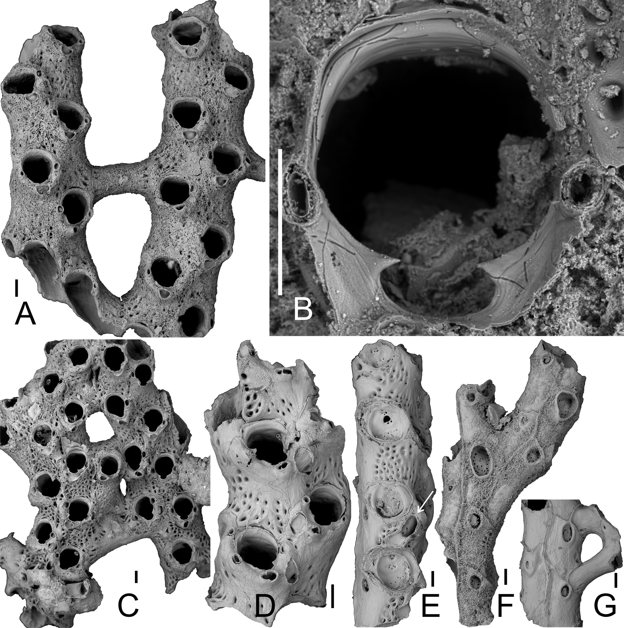
( Figure 6 View FIGURE 6 )
Etymology. Alluding to the type Early Miocene Chipola Formation in Florida.
Material examined. Holotype: USNM 595126, the colony fragment depicted in Fig. 6 View FIGURE 6 A, from the type locality at Farley Creek, Florida; type stratum, Chipola Formation, Early Miocene (sensu Saunders et al. 1986). Paratypes: USNM 595127 to 595131, 5 separate colony fragments. Additional studied material: More than 20 colony fragments in USNM, collection numbers TU LOC. 824, Scolaro collection; plus three fragments on a slide, NHMUK 1968.1.6.7, TU loc.825, leg. R.J. Scolaro.
Diagnosis. Pirabasoporella with numerous frontal-shield pores, a pair of oral spines, large spatulate avicularia occurring on lateral side of some later ontogenetic zooids, and deeply immersed rhizoidal as well as strut pore plates with numerous small communication pores.
Description. Colony rigid, ramose, reticulate with bifurcating branches connected by kenozooidal struts at irregular intervals ( Fig 6 View FIGURE 6 A), branches growing more or less parallel to each other. Struts usually developing chaotically, jointly budded laterally from the strut pore plate, slightly curved or straight, surface smooth or with faint ridges and a row of slit-like areolar pores near each branch contact, struts delimiting rounded-rectangular ( Fig. 6 View FIGURE 6 A) to irregularly oval fenestrules ( Fig. 6 View FIGURE 6 C).
Autozooids subrectangular, arranged in two alternating longitudinal series, not separated by thin raised ridges, but with prominent raised ridges on abfrontal side. Lepralioid part of frontal shield perforated by up to 30 pseudopores, many of which are closed during ontogeny. Primary orifice immersed and ill-defined, condyles indistinct if present; secondary orifice relatively large, oval, longer than wide, with a broadly oval, wide pseudosinus comprising ¼ total orifice length, formed by two short, acute lateral denticles directed medially, the round or oval bases of two large spines situated directly at each lateral orifice margin ( Fig. 6 View FIGURE 6 B).
Avicularia polymorphic: a single, small, suboral avicularium on each autozooid, situated directly proximal to pseudosinus cystid raised distally, oblique to frontal plane, rostrum semielliptical, directed laterally, crossbar complete, without columella; in ontogenetically older zooids a larger spatulate avicularium ( Fig. 6 View FIGURE 6 E) rarely develops proximolateral to orifice towards branch axis, its rostrum curved and directed proximolaterally, distally entirely filled by a flat smooth shelf, crossbar not observed.
Abfrontal side of colony relatively smooth ( Fig. 6 View FIGURE 6 E, F), zooidal borders marked by prominent ridges with deep median suture, each zooid with large, deep, round to oval rhizoid pore of very variable size (36–380 µm diameter; see Fig. 6 View FIGURE 6 F) positioned close to distal margin, usually surrounded by thick prominent rim during ontogeny, sealed at its base by a multiporous pore plate with the pores situated along the margin. An additional pore plate of a similar type present at distal edge of lateral walls of zooids that do not produce a kenozooidal strut.
Ovicells and ancestrula not observed.
BW SD FL FW AL AW AD SPL SDW RPL RPW AAL AAW SAL SAW SL SW mean 543 212 641 339 530 257 178 388 353 117 75 289 81 67 41 49 27 min 423 196 610 248 491 197 151 351 324 54 36 281 78 52 34 34 22 max 594 22 673 438 566 299 210 401 380 206 103 296 85 78 52 60 33 # 17 8 3 3 24 24 44 9 9 10 10 2 2 13 13 10 10 Remarks. Pirabasoporella chipolae n. sp. differs from P. atalaiaensis n. sp. and P. baitoae n. sp. in having oral spines, occasional spatulate frontal avicularia and deeply immersed rhizoidal pore plates with relatively small communication pores. While oral spines are lacking from P. atalaiaensis and P. baitoae and most other jaculinid species, they are present in a few Jaculina species, e.g. J. tessellata , thus their presence in P. chipolae is not extremely unusual.
Preservation of some of the specimens is excellent, and the secondary orifice typical of jaculinids is most apparent in this species (e.g. Fig. 6 View FIGURE 6 B). The proximal pseudosinus is formed by two distinct lateral denticles (sensu Berning et al. 2014) directed medially.
As in P. baitoae , a connecting strut was observed in P. chipolae that originated from two different pore plates on one side of the branch while fusing towards the opposite branch ( Fig. 6 View FIGURE 6 G). Also, ovicells were not found in P. chipolae . Few specimens were studied, however, so their absence is not surprising.
No known copyright restrictions apply. See Agosti, D., Egloff, W., 2009. Taxonomic information exchange and copyright: the Plazi approach. BMC Research Notes 2009, 2:53 for further explanation.
|
Kingdom |
|
|
Phylum |
|
|
Class |
|
|
Order |
|
|
Family |
|
|
Genus |