
Paraluederwaldtia bituberculata ( Mello-Leitão,1922 )
|
publication ID |
https://doi.org/ 10.1080/00222933.2023.2217547 |
|
DOI |
https://doi.org/10.5281/zenodo.8202227 |
|
persistent identifier |
https://treatment.plazi.org/id/E468723D-CB4B-F34F-8671-FF799FAEFEFA |
|
treatment provided by |
Plazi |
|
scientific name |
Paraluederwaldtia bituberculata ( Mello-Leitão,1922 ) |
| status |
|
Paraluederwaldtia bituberculata ( Mello-Leitão,1922) View in CoL
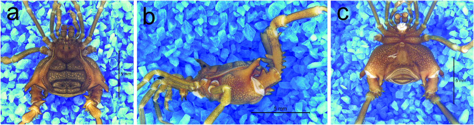
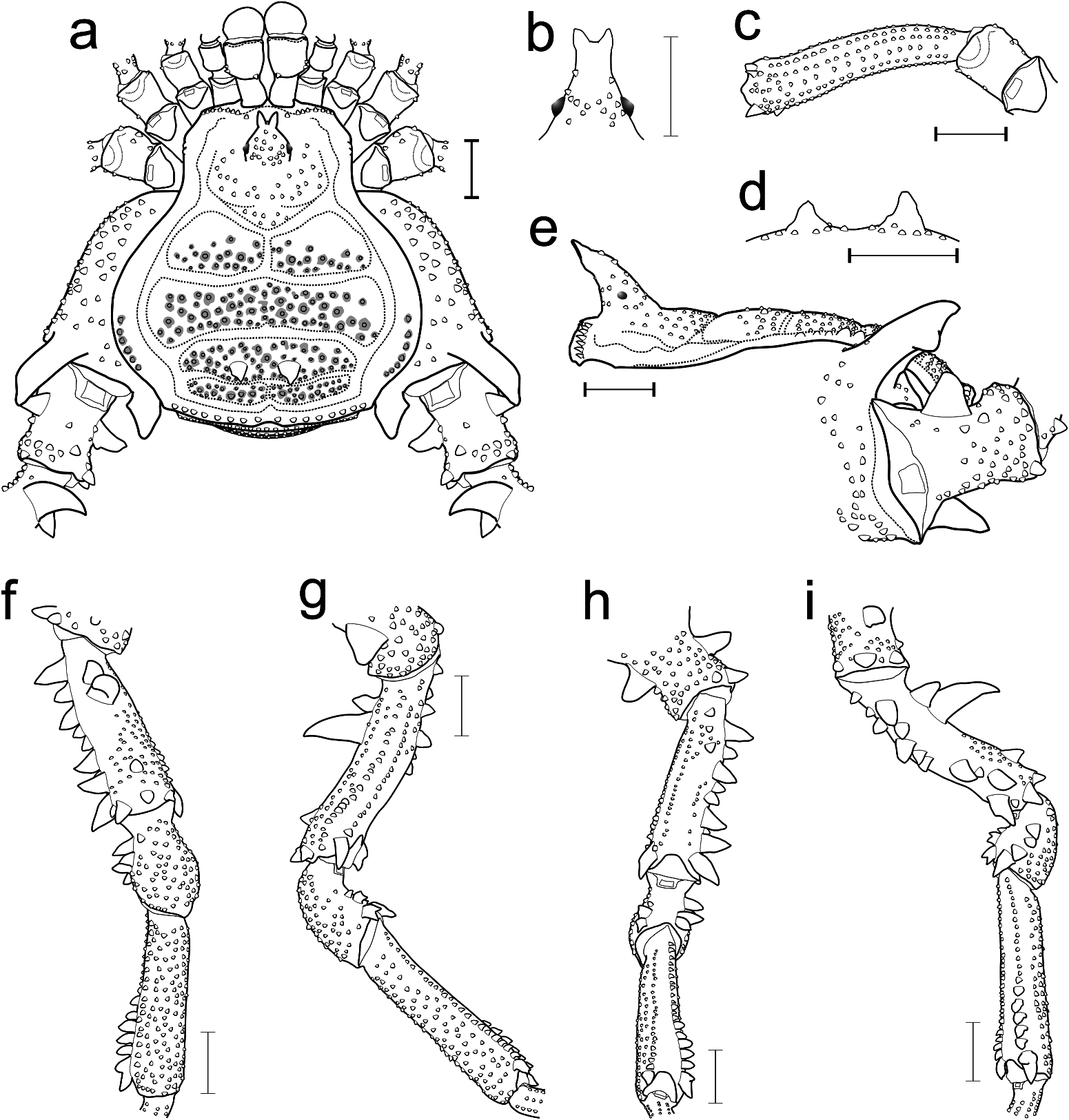
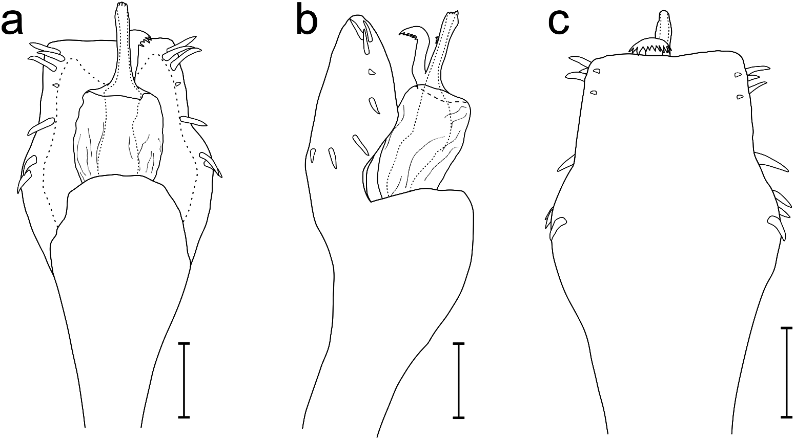
( Figures 10a–e View Figure 10 , 11 a–c View Figure 11 , 12 a–i View Figure 12 , 13a–c View Figure 13 )
Neopucrolia bituberculata Mello-Leitão, 1922: 329 View in CoL .
Neopucrolia bituberculata View in CoL – Mello-Leitão 1923: 116, fig 4.
Paraluederwaldtia bituberculata: Mello-Leitão 1927: 15 View in CoL .
Paraluederwaldtia bituberculata View in CoL – Roewer 1929: 227, fig 19; Mello-Leitão, 1932: 163, fig 86; B. Soares 1944: 286; B. Soares, 1945: 381.
Luederwaldtia bituberculata: Soares and Soares 1954: 269 .
Luederwaldtia bituberculata: Soares and Soares 1970: 340 .
Type data
Neopucrolia bituberculata : ♀ holotype ( MZSP, lost), from BRAZIL, state of São Paulo, Alto da Serra (doubtful data according to Soares 1945; Kury 2003); J lectotype, ♀ paralectotype ( MNRJ 1404, examined, lost in the MNRJ ̾s 2018 fire), from BRAZIL, state of Rio de Janeiro, Itatiaia. J neotype herein designated ( MNRJ 58976), from BRAZIL, state of Rio de Janeiro, Itatiaia, Parque Nacional do Itatiaia, Abrigo Lamego/Piscina da Maromba, 2000 m, 29.xii.2019, Pedroso, D. R. et al. leg.
Records
Without further data.
Geographic distribution
BRAZIL: state of Rio de Janeiro: Itatiaia.
Diagnosis
Paraluederwaldtia bituberculata can be distinguished from Paraluederwaldtia ankeri sp. nov. by the following characters: (1) Ch bulla with proximal margin armed with a spine ( Figures 12a View Figure 12 ); (2) scutal area I with diffused ordinary tubercles on all area extension ( Figures 10a–b View Figure 10 , 11 a View Figure 11 , 12a, e View Figure 12 ); (3) dorsal free tergites with a transversal uniform row of ordinary tubercles ( Figures 10a, e View Figure 10 , 11a View Figure 11 , 12a View Figure 12 ); (4) Ti III tubular and regular ( Figures 10b, e View Figure 10 , 11b View Figure 11 ); (5) Cx IV prodorsal apophysis with its distal portion forming an obtuse angle to the medial body axis ( Figures 10a–e View Figure 10 , 11 a View Figure 11 , 12a View Figure 12 ); (6) Tr IV prodorsal distal portion covered only by ordinary tubercles ( Figures 10a–b View Figure 10 , 11 a View Figure 11 , 12a, f View Figure 12 ); (7) Ti IV dorsally covered by ordinary tubercles ( Figures 10a View Figure 10 , 11 b View Figure 11 , 12f–g, i View Figure 12 ).
Redescription
MNR] 58976 (male) for the external body illustrations and description; DS, measurements: CW 3.0, CL 2.3, AW 5.4, AL 3.4; Leg I–IV measurements in Table 5 View Table 5 ; Right/left tarsal (distitarsal) counts: 6(3)/6(3) – 7(3)/7(3) – 6/x – 6/6. MNR] 58976 (male) for genitalic illustrations.
Dorsum. DS gamma type, as wide as long, with AS lateral margins convex (widest at area II and thickest at area I) and posterior margin convex with a central notch ( Figures 10a–b View Figure 10 , 11 a View Figure 11 , 12a View Figure 12 ). DS anterior portion with two rows of seven acuminated tubercles ( Figures 12a, e View Figure 12 ). Carapace tuberculate on central and lateral regions ( Figures 10a–d View Figure 10 , 11 a–b View Figure 11 , 12a, e View Figure 12 ). Cheliceral sockets shallow, with a small apophysis in the centre ( Figure 12a View Figure 12 ). Ocularium conical (in dorsal view), high (ca. 10× the diameter of the eyes), with an apical curvature for the anterior portion and perpendicularly placed on the anterior portion of the carapace ( Figures 10a–e View Figure 10 , 11 a–b View Figure 11 , 12a–b, e View Figure 12 ). Ocularium armed with a pair of divergent spines (ca. 2× the diameter of the eyes) ( Figures 12a–b, e View Figure 12 ). AS lateral borders with a row of prominent and ordinary tubercles at height of scutal areas II–III ( Figures 10a–c, e View Figure 10 , 11 a View Figure 11 , 12a, e View Figure 12 ). Mesotergum is divided into four clearly defined scutal areas ( Figures 10a–b, d–e View Figure 10 , 11 a View Figure 11 , 12a View Figure 12 ). Scutal areas I and IV divided into left and right halves by a median groove ( Figures 10a–b, d View Figure 10 , 11 a View Figure 11 , 12a View Figure 12 ). Scutal area II posterior-lateral border slightly invades scutal area III ( Figures 10a–b View Figure 10 , 11 a View Figure 11 , 12a View Figure 12 ). All scutal areas are tuberculate, with all tubercles individually covered and surrounded by lighter spots ( Figures 10a–e View Figure 10 , 11 a–b View Figure 11 , 12a, e View Figure 12 ). Scutal areas I and II are only covered by ordinary tubercles ( Figures 10a–b, d View Figure 10 , 11 a View Figure 11 , 12a View Figure 12 ). Scutal area III with a pair of outstanding acuminated tubercles (ca. 9× the ordinary tubercles) ( Figures 10a–e View Figure 10 , 11 a–b View Figure 11 , 12a, d View Figure 12 ). Scutal area IV with a paramedian row of five to six prominent tubercles (ca. 2× the ordinary tubercles) ( Figures 10e View Figure 10 , 12a View Figure 12 ). DS posterior border with a transversal row of prominent tubercles ( Figures 10a, e View Figure 10 , 11a View Figure 11 , 12a View Figure 12 ). Free tergites I–III with a transversal row of ordinary tubercles ( Figures 10a, e View Figure 10 , 12a View Figure 12 ).
Venter. Cx I–III parallel to each other, each with ventral longitudinal rows of 7–12 setiferous tubercles (Cx I rows with comparatively higher and sharper tubercles). Cx II with a retroventral distal row of five acuminate tubercles. Cx III with a retroventral distal row of seven acuminate tubercles. Cx IV much larger than the others (directed obliquely) ( Figure 11c View Figure 11 ). Stigmatic area inverted-Y-shaped, clearly sunken concerning Cx IV̾s distal part ( Figure 11c View Figure 11 ). Cx IV covered by ordinary tubercles ( Figure 11c View Figure 11 ). Intercoxal bridges are well marked ( Figure 11c View Figure 11 ). Stigmata are visible ( Figure 11c View Figure 11 ). Free sternites with a transverse row of ordinary tubercles. Anal operculum covered by ordinary tubercles.
Chelicera. Basichelicerite elongate ( Figures 11a View Figure 11 , 12a View Figure 12 ); bulla well marked, with two ectal and one posterior marginal setiferous tubercles ( Figure 12a View Figure 12 ); hand not swollen.
Pedipalpus. Tr ventral with two geminate setiferous tubercles. Fe with a mesal apical setiferous tubercle and one ventral basal setiferous tubercle. Pa unarmed ( Figure 11a View Figure 11 ). Ti ventro-mesal and ventro-ectal faces with four setiferous tubercles (IiIi). Ta ventro-mesal and ventro-ectal faces with four setiferous tubercles (IIi).
Legs. Tr I–III each with several ventral tubercles. Fe I–II straight ( Figure 11a View Figure 11 ). Fe and Ti I–II with prodorsal, proventral, retroventral and retrodorsal rows of small tubercles. Leg III sub-straight ( Figures 10d View Figure 10 , 11a, c View Figure 11 , 12c View Figure 12 ). Fe III ( Figure 12c View Figure 12 ) and Ti III covered by rows of tubercles. Cx IV significantly expanded transversely (to almost a quarter the size of AW) ( Figures 10a–b View Figure 10 , 11 a View Figure 11 , 12a View Figure 12 ). Cx IV distal portion reaching the scutal areas III–IV longitudinally ( Figures 10a View Figure 10 , 11a View Figure 11 , 12a View Figure 12 ). Cx IV longitudinally tuberculate between prodorsal and ventral faces ( Figures 10a, c View Figure 10 , 11 a–c View Figure 11 , 12a View Figure 12 ). Cx IV with a cylindrical prodorsal apophysis with a subconical apical apex (not covering, in dorsal view, the prolateral basal portion of the Cx IV) and a small accessory blunt branch (not totally visible in dorsal view) ( Figures 10a, c View Figure 10 , 11 a–c View Figure 11 , 12a, e View Figure 12 ). Cx IV with a short retrolateral apophysis, associated with a tiny secondary branch ( Figures 10a, e View Figure 10 , 11a, c View Figure 11 , 12a View Figure 12 ). Tr IV rectangular ( Figures 10a, c–d View Figure 10 , 11 c–d View Figure 11 , 12a, e, h View Figure 12 ). Tr IV dorsal central with a prominent subconical tubercle ( Figures 10a View Figure 10 , 11a View Figure 11 , 12a View Figure 12 ). Tr IV proximal with a conical apophysis on prolateral and retrolateral faces (prolateral wider than retrolateral) ( Figures 10a, d View Figure 10 , 12a, h View Figure 12 ). Tr IV prodorsal distal portion covered by ordinary tubercles ( Figures 12a, f View Figure 12 ). Tr IV with a prominent subconical tubercle on retrodorsal and retrolateral distal portions ( Figures 12a, f, h–i View Figure 12 ). Tr IV ventral face tuberculate ( Figures 12e, g–i View Figure 12 ). Fe IV sub-straight ( Figures 11b–c View Figure 11 , 12e, g–i View Figure 12 ). Fe IV dorsal face with a pair of spines (iI, the second centrally curved to the retrolateral) on the proximal third and a pair of acuminated outstanding tubercles on the distal third ( Figures 10a–b View Figure 10 , 12f, i View Figure 12 ). Fe IV prodorsal face with a row of ordinary tubercles and a reduced spur on the distal portion ( Figures 12f–g View Figure 12 ). Fe IV prolateral face with a row of tubercles (ordinary ones on proximal half, prominent and acuminated ones on distal half) ( Figures 12f–h View Figure 12 ). Fe IV proventral face with a row of ordinary tubercles and a developed spur on the distal portion ( Figures 12g –h View Figure 12 ). Fe IV ventral face with a row of ordinary tubercles on the proximal third ( Figure 12g View Figure 12 ). Fe IV retroventral face with three prominent subconical tubercles on the proximal third, a row of ordinary tubercles on the central and distal thirds and a developed spur on the distal portion ( Figures 12g –i View Figure 12 ). Fe IV retrolateral face with a sinuous row of seven conical spines (IiIiiII) and a reduced spur on the distal portion ( Figures 10d View Figure 10 , 12f, h–i View Figure 12 ). Fe IV retrodorsal face with a subconical spur on the distal portion ( Figures 12f, i View Figure 12 ). Pa IV dorsally tuberculate ( Figures 12f–g, i View Figure 12 ). Pa IV proventral with one prominent conical tubercle and two prominent spines with bifurcated apex on the distal half ( Figures 12g –i View Figure 12 ). Pa IV retroventral with two prominent spines on the distal half ( Figures 12h–i View Figure 12 ). Pa IV retrolateral with two prominent spines on the proximal half ( Figures 12f–g, i View Figure 12 ). Ti IV dorsally tuberculate ( Figures 12f–g, i View Figure 12 ). Ti IV proventral face with a row of acuminated tubercles (prominent on the distal half) and a spur on the distal portion ( Figures 12g –h View Figure 12 ). Ti IV retroventral face with a row of prominent acuminated tubercles on the proximal half, followed by five subconical spines and a spur on the distal portion ( Figures 12f, h–i View Figure 12 ). Mt IV covered by tiny tubercles. Mt IV with proventral and retroventral distal spurs.
Colouration (in vivo). ( Figures 10a–e View Figure 10 ): Ch and Pp glossier background Strong Greenish Yellow (99), with honeycombed reticle in Moderate Olive (107). Carapace background and apex of the spines on ocularium Vivid Orange (48). Ocularium, DS margins and free tergites I–III background Greyish Reddish Brown (46). Mesotergum grooves and paramedian pair of outstanding tubercles on scutal area III Brownish Gray (64). Scutal areas I–IV background Dark Orange Yellow (72), with all tubercles individually covered and surrounded by lighter spots of Brilliant Orange Yellow (67). AS lateral portions Vivid Reddish Orange (34). Cx–Mt I–III and Pa–Mt IV background Brownish Orange (54). Tr I–III with a distal dorsal semicircle of Brilliant Yellow (83). Cx–Fe IV background (and its apophyses and spines) in a combination of Dark Reddish Gray (23) and Dark Reddish Orange (38). Fe IV distal quarter and Pa IV proximal quarter background Deep Orange Yellow (69).
Penis. VP is divided into two regions: distal part trapezoidal (widest basally, with lateral margins curved ventrally), proximal part elliptical ( Figures 13a, c View Figure 13 ). VP ventral surface totally covered with microsetae of type 1. All macrosetae cylindrical, inserted on lateral of VP: MS A1–A3 thick and acuminated in the basal part of the VP ( Figures 13a–c View Figure 13 ); MS B1 short, inserted ventrally, proximal to A3 ( Figures 13b–c View Figure 13 ); MS C1–C3 thick and acuminated, forming a triangle (C2 more dorsal than the others) in the distal part of VP ( Figures 13a–c View Figure 13 ); MS D1 short, inserted on the lateral border of VP, closer to C3 than A1 ( Figures 13a–b View Figure 13 ); MS E1–E2 very reduced, located on the laterodistal flange of VP–E1 between the height of MS C1 and C2, E2 beside MS C3 ( Figure 13c View Figure 13 ). Glans sac arising from the middle bulge on the podium, not extended as a dorsal process ( Figures 13a–b View Figure 13 ). Stylus and its ventral process fused basally (forming a short pedestal) above the glans ( Figure 13b View Figure 13 ). Stylus inserted on pedestal forming a 45° angle, with apical portion swollen and armed with a set of central spines ( Figure 13b View Figure 13 ). Stylus without any expansion or flattening, in situ reaching the distal border of VP ( Figures 13a–c View Figure 13 ). Ventral process twothirds the stylus size, slightly bent to dorsal and with a ventro-apical flabellum ( Figures 13a–c View Figure 13 ). Flabellum curved proximally, scallop-shaped with serrulations and spines, measuring about 35% of the length of the ventral process stem ( Figures 13a–c View Figure 13 ).
Female (MZSP, lost). ( Mello-Leitão 1932: fig. 86): Information about DS and leg I–IV measurements and right/left tarsal counts cannot be accessed.
DS lambda type. Cx IV narrower than male, with the prodorsal apophysis reduced to a conical spine. Tr IV retrolateral face with a conical apophysis on the distal portion. Fe IV sub-straight, arched on the proximal portion towards the dorsal face, and thinner than the male. Fe–Ti IV with armature restricted to acuminated tubercles on the prodorsal, proventral, retroventral and retrodorsal distal portions.
Intraspecific variation: No material was available to analyse intraspecific variation among males or females.

No known copyright restrictions apply. See Agosti, D., Egloff, W., 2009. Taxonomic information exchange and copyright: the Plazi approach. BMC Research Notes 2009, 2:53 for further explanation.
|
Kingdom |
|
|
Phylum |
|
|
Class |
|
|
Order |
|
|
Family |
|
|
Genus |
Paraluederwaldtia bituberculata ( Mello-Leitão,1922 )
| Carvalho, Rafael N. & Kury, Adriano B. 2023 |
Luederwaldtia bituberculata:
| Soares BAM & Soares HEM 1970: 340 |
Luederwaldtia bituberculata: Soares and Soares 1954: 269
| Soares BAM & Soares HEM 1954: 269 |
Paraluederwaldtia bituberculata
| Soares BAM 1945: 381 |
| Soares BAM 1944: 286 |
| de Mello-Leitao CF 1932: 163 |
| Roewer CF 1929: 227 |
Paraluederwaldtia bituberculata: Mello-Leitão 1927: 15
| de Mello-Leitao CF 1927: 15 |
Neopucrolia bituberculata
| de Mello-Leitao CF 1923: 116 |
Neopucrolia bituberculata Mello-Leitão, 1922: 329
| de Mello-Leitao CF 1922: 329 |