
Rhacophorus rufipes
|
publication ID |
https://doi.org/ 10.5281/zenodo.208711 |
|
DOI |
https://doi.org/10.5281/zenodo.5658371 |
|
persistent identifier |
https://treatment.plazi.org/id/E4394C34-D466-F32B-8196-C6B0CE57330F |
|
treatment provided by |
Plazi |
|
scientific name |
Rhacophorus rufipes |
| status |
|
Colour in life (Stage 31; ZMH A10167 View Materials ; Fig. 3 View FIGURE 3 ). The basic colouration of the body dorsum and tail is grey. The pigmentation is diffuse, and there are no sharply defined spots or blotches. The grey background colour is modified with a slight tint of olive on the head and trunk dorsally. There is a superficial layer of small, spindle-shaped epidermal melanocytes and deeper body layers with stellate or irregular polygonal melanocytes. The overall colouration stems from the additive interaction of both layers. For example, around the eyes there are only epidermal melanocytes, whereas the cheeks bear a deeper layer ( Fig. 3 View FIGURE 3 ). In dorsal and lateral views, the colouration is darkest along the abdominal cavity. The red branchial structures and developing forelimbs are partially visible through the skin in lateral view. There are only a few iridocytes that appear in light blue or gold; they are located in small scattered patches between the eye and the spiracle, along the spiracular tube, on the lower cheek, and on the snout close to the oral disc. The spiracular tube is mostly translucent, but clearly visible against the almost black abdominal wall.
The pigmentation of the trunk extends seamlessly onto the muscular part of the tail, but with a more brownish hue. The peripheral areas of the tail fins are without pigmentation and are clear. However, the dorsal tail fin bears pigmentation along its entire base. The ventral fin is almost entirely clear, except for melanocytes in the distal third of the tail, along the neighbouring muscular tail portion. The vena caudalis lateralis is visible as a red line in living specimens due to the erythrocytes in it, but it is not particularly lined with melanocytes.
The ventral body surface is mostly unpigmented and translucent, except for areas below the cheek, where scattered melanocytes in deeper body layers reach the venter. The translucency of the ventral skin is combined with slight iridescence. The gut coil is clearly visible in ventral view. The gut is dark pigmented. The gills and the heart are visible through the clear ventral skin in red. They contrast with the pale sub-buccal region anteriorly and the dark gut coils posteriorly.
The background iris colour is black with dense scattered golden and coppery pigment cells. Around the pupil, the golden pigmentation fuses into a closed ring, which lines the pupil. The skin of the oral sucker is without pigmentation. The colour in preservation shows the same markings as living specimens, however, melanocytes become paler and iridocytes (any silver or golden cells) disappear.
External morphological features (Stages 33–34, n=4, ZMH A 10167 View Materials ). A medium sized tadpole (TTL 24.79 mm at Stage 34; Table 2), with long tail (55–60% of total length). The body shape is depressed and ovoid in lateral view, tapering to the snout. In dorsal view, the body contour is slightly inverse pear shaped. The body is widest at the gill region (posterior to eyes) in dorsal view. Between the gill part of the body and the trunk there is a shallow constriction of the body contour. The body is moderately depressed dorsoventrally.
The tail shape is moderately arched in lateral view, tapering in the posterior two thirds with straight contour lines into narrow, pointed tip; a flagellum is not formed. The muscular part of the tail is moderately high (49–54% of body depth). The dorsal fin starts at the trunk-tail junction, the ventral fin connects broadly to the trunk. The maximum height of the tail (fins included) is 37–43% of the tail length.
The eyes are positioned dorsally, at clear distance from the body contour in dorsal view. The nares are closer to the snout than to the eyes and are spaced moderately wide ( IND = 62–69% IOD). The nares are round. The rims of the nares are elevated posteriorly and flat anteriorly. The spiracle is sinistral. The spiracular tube opens posterolaterally and is below the longitudinal body axis in lateral view ( Fig. 3 View FIGURE 3 ). The medial part of the spiracular orifice is attached to the abdominal wall. Gut coils are concentric. The anal siphon is dextral.
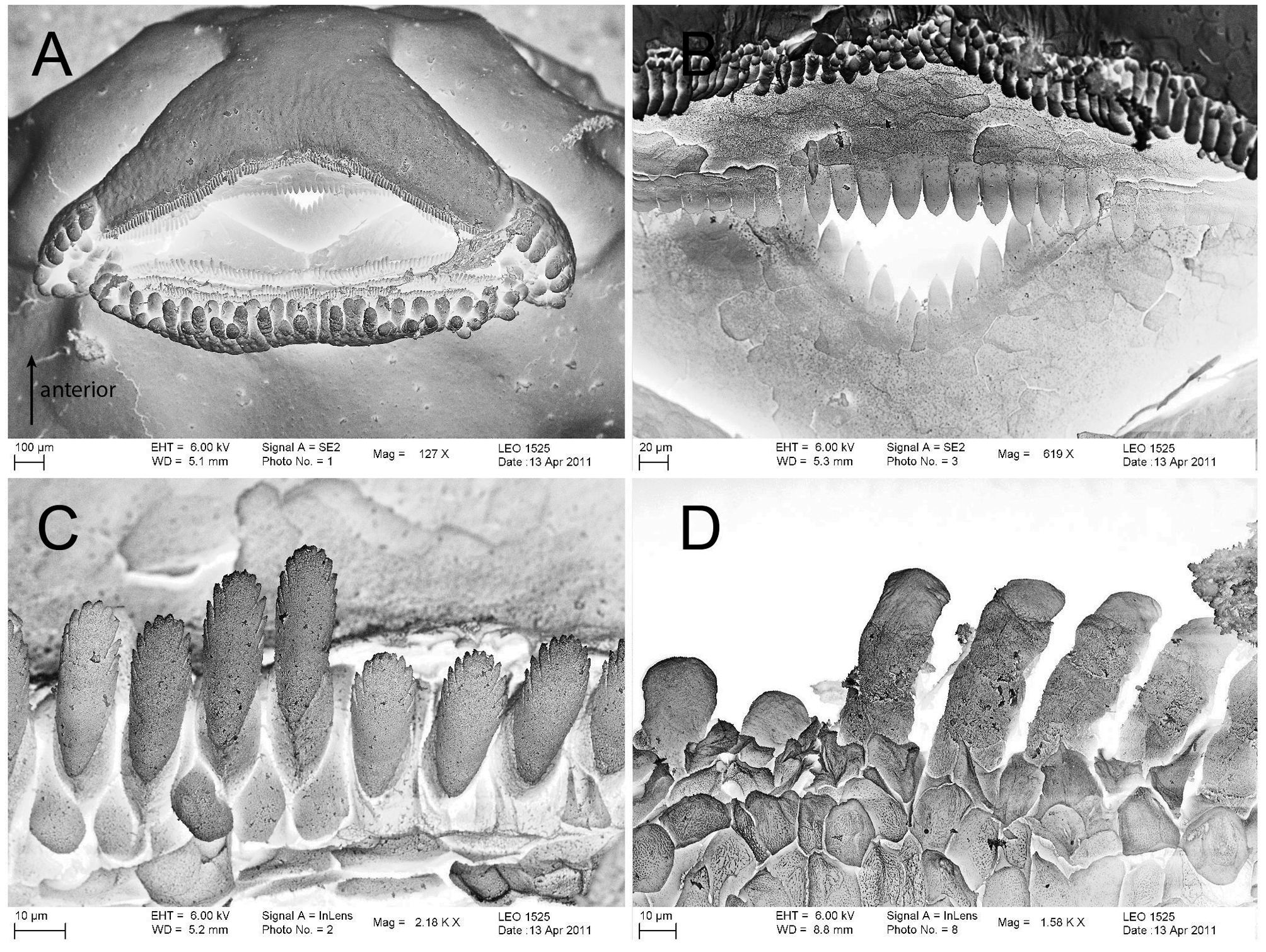
The oral disc is subterminal ( Fig. 7 View FIGURE 7 A). The marginal papillation of the oral disc is present on the lower lip and lateral parts. There is a broad medial gap in the papillae row of the dorsal lip. Marginal papillae are arranged biserially on the lower lip. The lateral part of the upper lip bears a uniserial row of marginal papillae plus a short row of submarginal papillae. The oral disc margins possess lateral indentations between upper and lower lips. Papillae are short (length ≤ 2x diameter), blunt and adjoining ( Fig. 7 View FIGURE 7 A). The labial ridges bear uniserial keratodont rows. The Labial Tooth Row Formula (LTRF) is 4(2–3)/3 or 5(2–4)/3. Generally, distal keratodont rows are long and extend far laterally on both the upper and lower lip, however, the keratodont rows of the divided series become shorter towards the mouth. Row A5, if present, is very short and bears only a few keratodonts (one specimen, Stage 34). The keratodonts are spoon-shaped with fine incisions along their edges ( Fig. 7 View FIGURE 7 C–D). The beaks are well-keratinized and have sharp serrations. The upper beak is almost straight in ventral view, whereas the lower jaw sheath is V-shaped ( Fig. 7 View FIGURE 7 A–B). They are keratinized to about half of the total jaw height.
Variation. The most proximal, fifth keratodont row of the upper lip was present only in the most advanced specimen (Stage 34).
Ecological notes. We encountered Rhacophorus rufipes adults in Kerangas forests (Bornean heath forests) on numerous occasions. Tadpoles were collected from a pool of water created by an uprooted tree, which was partially covered by a fallen tree trunk. Tadpoles of R. rufipes were sighted hovering at the surface of the pool by night. The water was tea-coloured and peaty; its depth was ca. 50 cm, the pool area ca. 1 m 2. The bottom of the pool was covered with leaf litter and a thick layer of humic debris. Adults of R. rufipes , some in amplexus, were spotted in the vicinity of the pool. Other species encountered at the site with R. rufipes included: Calluella flava, Kiew, 1984 , Kalophrynus cf. heterochirus Boulenger, 1900 , Limnonectes malesianus (Kiew, 1984) , Megophrys nasuta (Schlegel, 1858) , Metaphrynella sundana (Peters, 1867) , Microhyla nepenthicola Das & Haas, 2010 , Pelophryne cf. guentheri (Boulenger, 1882) , Philautus kerangae Dring, 1987 , and Polypedates colletti (Boulenger, 1890) .

No known copyright restrictions apply. See Agosti, D., Egloff, W., 2009. Taxonomic information exchange and copyright: the Plazi approach. BMC Research Notes 2009, 2:53 for further explanation.
|
Kingdom |
|
|
Phylum |
|
|
Class |
|
|
Order |
|
|
Family |
|
|
Genus |