
Vanmanenia microcephala, Li & Zhou & Che, 2019
|
publication ID |
https://doi.org/ 10.11646/zootaxa.4603.1.6 |
|
publication LSID |
lsid:zoobank.org:pub:AB9A3D8A-953E-4A14-934C-152AE805F680 |
|
DOI |
https://doi.org/10.5281/zenodo.5610062 |
|
persistent identifier |
https://treatment.plazi.org/id/E438DE7A-FF8F-FFBD-FF7F-FA8CFB0BC025 |
|
treatment provided by |
Plazi |
|
scientific name |
Vanmanenia microcephala |
| status |
sp. nov. |
Vanmanenia microcephala , sp. nov.
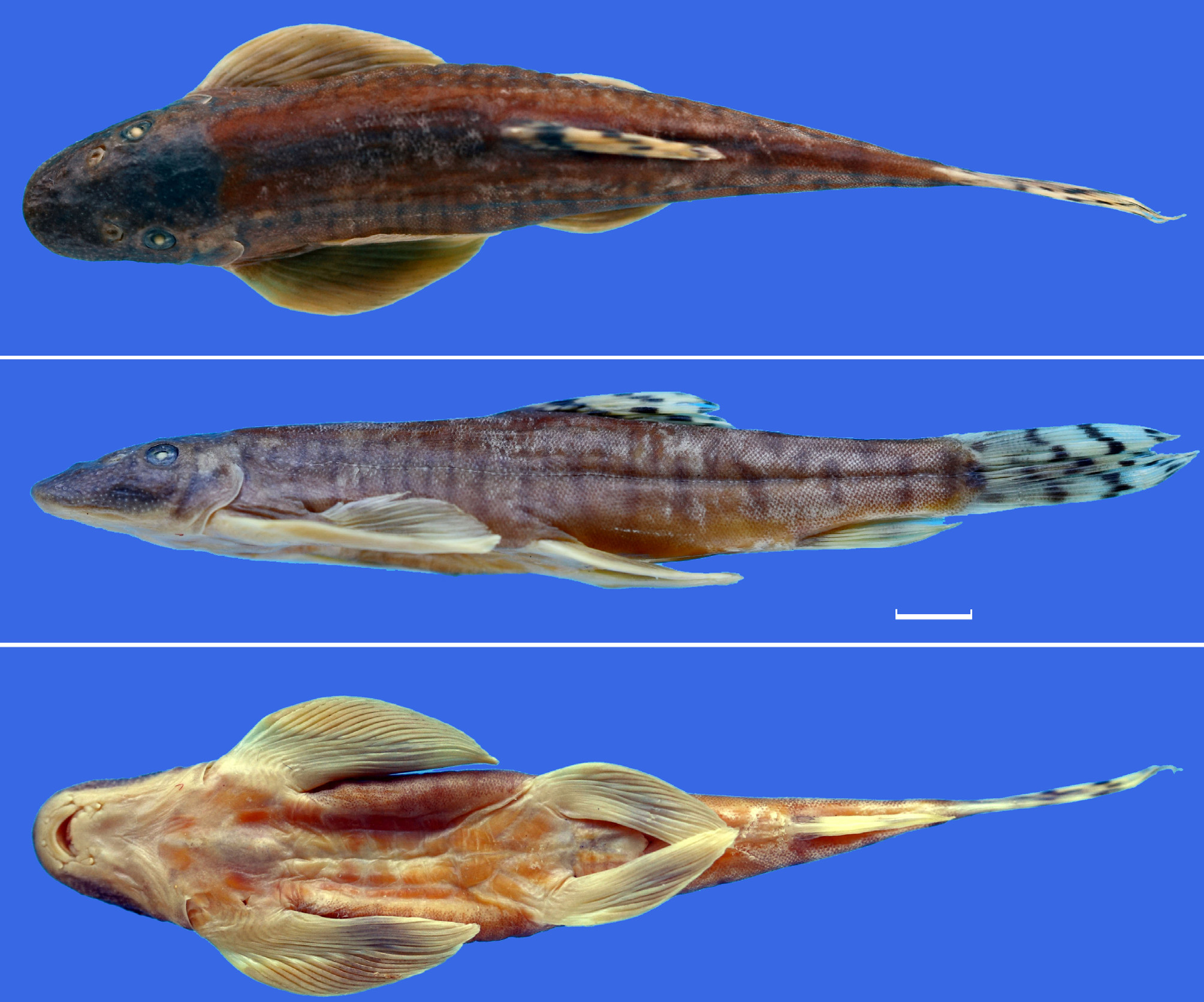
( Fig. 1A View FIGURE 1 , Fig. 3 View FIGURE 3 )
Ƒanmanenia striata (non Chen, 1980): Zheng, Chen & Huang 1982 (in part): 393 (Lancang-jiang drainage, Yunnan); Kottelat 2000: 76 ( China: Yunnan: Yangbi).
Ƒanmanenia tetraloba (non Mai, 1978): Chen 1990 (in part): 85 (Lancang-jiang drainage, Yunnan); Chen & Tang 2000 (in part): 465 (Lancang-jiang drainage, Yunnan).
Holotype. SWFC 0 309282, 66.8 mm SL; China: Yunnan: Yangbi County: Shunbi (25°29'23.12"N 99°59'04.20"E); collected by Y. Yang & J.F. He, 10 Sep. 2003. GoogleMaps
Paratypes. SWFC 0309273–0309281, 0309283–0309290, 17ex., 38.8–73.2 mm SL; other data same as the holotype.
Other material. SWFC 0010005–0010009, 5 ex., 62.2–71.4 mm SL; China: Yunnan: Simao City: Caiyang-he (22°26′50.06′′N, 101°05′08.40′′E); collected by L.X. Han, 15 Nov. 2000. – SWFC 0309291–0309292, 2 ex., 68.5– 72.5 mm SL; China: Yunnan: Yangbi County: Pingpo (25°34′49.86′′N, 100°02′59.37′′E); Y. Yang & J.F. He, 8 Sep. 2003 GoogleMaps . – SWFC 0612003–0612004, 2 ex., 68.2–83.4 mm SL; China: Yunnan: Yunlong County: Jiancao (26°03′45.10′′N, 99°18′34.41′′E); collected by Q. Fu & F.L. Li, 23 Dec. 2006 GoogleMaps . – SWFC 0502117–0502120, 4 ex., 45.9–53.7 mm SL; China: Yunnan: Changning County: Wenquan (24°41′41.78′′N, 99°41′51.04′′E); collected by W. Zhou & X. Li, 12 Feb. 2005 GoogleMaps .
Diagnosis. Vanmanenia microcephala differs from the other species of the barred group in Vanmanenia by the following combination of characters: the lateral side of the body with 14–22 vermiculations with widths smaller than the diameter of the eye (vs. 10–15 regular bars with widths equal to the diameter of the eye in V. tetraloba ); the dorsal side of the head covered with a large black blotch (vs. several short dark brown vermiculations in V. striata and V. tetraloba ); the gill opening smaller and its upper angle level with the lower edge of the eye (vs. the gill opening larger and its upper angle aligned with the middle point of the eye in V. crassicauda , V. serrilineata , V. striata , and V. tetraloba ); dorsal part of the body with 4–6 black saddles (vs. 7–9 in V. striata ); the patch of the caudal-fin base dissociated and not connected to the upper and lower margin of the caudal peduncle (vs. connected to the upper and lower margin of the caudal peduncle in V. tetraloba ); and the head smaller, head depth 45.2–47.1% HL (vs. head larger, head depth 48.1–60.0% HL in V. striata and V. tetraloba ) ( Tables 1 View TABLE 1 & 2 View TABLE 2 ).
Description. Based on holotype and 17 paratypes, maximum standard length 73.2 mm. Morphometric data shown in Table 2 View TABLE 2 . Dorsal-fin rays iii, 7½; pectoral-fin rays i, 15–17; pelvic-fin rays i, 8; anal-fin rays ii, 5½; branched caudal-fin rays 8+7. Lateral line pores 88 (1), 89 (6), 90 (2), 92 (3), 95 (1), 100 (1); scales above lateral line 24–28, scales below lateral line 16–19.
Body elongate, anterior part cylindrical and laterally compressed behind dorsal-fin base. Dorsal profile rising gradually from tip of snout to origin of dorsal fin, then sloping slowly and ventrally to end of caudal peduncle. Ventral margin straight. Head compressed, rostral margin rounded when viewed dorsally. Numerous minute tubercles speckled evenly across cheek. Eyes small, located dorsally and closer to upper angle of gill opening than to tip of snout. Mouth inferior, slightly arch-shaped. Rostral fold thick, separated from upper lip by shallow groove, disconnected from lower lip at corners of mouth and ending at postlabial groove. Rostral fold divided into 3 lobes, with 2 pairs of short barbels flattened at base and located between lobes ( Fig. 1A View FIGURE 1 , rb). One pair of maxillary barbels rooted at corner of mouth, their base thick and slightly longer than outer of rostral barbels. Lips thick; upper lip divided into 3 layers by shallow grooves, inner layer usually slightly saw-toothed; lower lip formed into 4 lobes. Upper and lower lips connected at corners of mouth. Thin horny cutting edge covered on upper and lower jaws ( Fig. 1A View FIGURE 1 ). Gill opening extending to ventral surface of head.
Last unbranched ray of dorsal fin not ossified. Dorsal-fin origin located approximately midway between tip of snout and caudal-fin base. Posterodorsal margin of dorsal fin slightly concave. Anal fin without spine; located posteriorly, its origin closer to caudal-fin base than to pelvic-fin insertion; end of depressed anal-fin rays reaching or exceeding the caudal-fin base. Distal margin of anal fin slightly concave. Pectoral and pelvic fins extending horizontally, without plume fold on their ventral surfaces. Pectoral-fin origin adjacent to gill opening and below eye; distal margin of pectoral fin convex, not reaching origin of pelvic fin. Pelvic-fin origin at vertical behind dorsal-fin origin; distance from pelvic-fin origin to anal-fin origin closer than distance from pelvic-fin origin to pectoral-fin origin; end of pelvic fin extending beyond anus. Anus located at anterior one-third of distance between pelvic-fin insertion and anal-fin origin. Caudal fin forked, lower lobe slightly longer than upper lobe.
Body covered by small scales, except on dorsal side of head and belly in front of anus. Lateral-line complete, located along middle axis of the body extending to caudal-fin base.
Coloration: At time of collection, specimens have brown dorsal surface of body, fading to a lighter brown color ventrally. After fixed in formalin, head and body background color pale yellowish brown; throat, belly, lower part of caudal peduncle whitish. Four to six dark brown saddles across dorsal midline, 14–22 black bars on flank, their width smaller than eye diameter. Bars on front part of flank thin and closely arranged, bars on poster part of flank gradually widen and loosely arranged. Pelvic, pectoral, and anal fins bright orange; dorsal and caudal fins pale yellow. Dorsal-fin rays with 2–3 irregular rows of spots. Mark on caudal-fin base with irregular stripe dissociated and not connected to upper and lower margins of caudal peduncle. Caudal-fin rays with 3–4 irregular rows of spots.
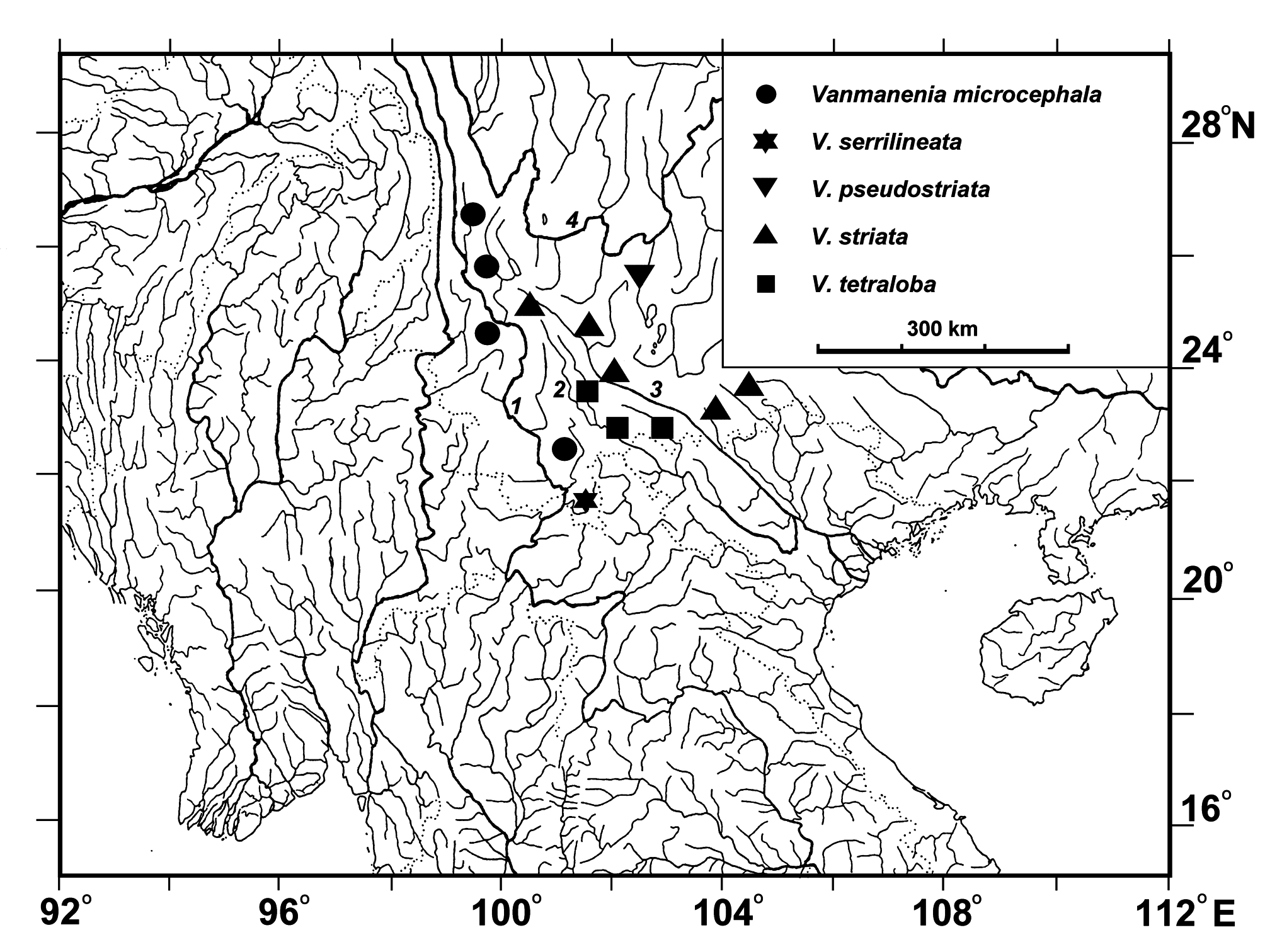
Distribution: Known only from the Lancang-jiang drainage (the upper Mekong River) ( Fig. 4 View FIGURE 4 ).
Habitat and Ecology: Vanmanenia microcephala is omnivorous, mainly feeding on algae attached to rocks and on organic residues and small aquatic insects. Vanmanenia microcephala inhabits rapids, torrents, and waterfalls.
Etymology: From the Latin adjectives micro -, meaning small and – cephala, meaning head, in reference to the small head of this new species. Used as an adjective.
Remark: The type localities of Vanmanenia microcephala and V. striata are close in distance to each other; the former is in China: Yunnan: Yangbi County: Shunbi (the Lancang-jiang drainage), and the latter is in China: Yunnan: Xiaguan City (the Yuan-jiang drainage). However, the type localities belong to two different drainages. Their morphology is remarkedly different (see diagnosis). The photos by Kottelat (2000) show two types of color patterns in the lateral bars of specimens from Yangbi: wide bars and narrow bars types ( Kottelat 2000, Fig. 67). Kottelat (2012) stated that the color pattern of Vanmanenia species strikingly changed with growth. However, we observed specimens with body lengths from 38.8–73.2 mm SL at the same site as those collected by Kottelat (2000), but none were observed with the wide stripe type; thus we cannot confirm whether the color pattern changes based on size.
There are two paired maxillary barbels at the corner of the mouth between the upper and lower lips; the inner ones are very small or papilla-like, which had long been considered diagnostic of the genus Vanmanenia to distinguish its species from allied or closely related genera in Gastromyzontidae ( Chen 1980; Chen & Tang 2000). After comparing specimens of Vanmanenia from the Lancang-jiang, Lixian-jiang, and Yuan-jiang, it was clear that there was only one paired maxillary barbel at the corner of the mouth between the upper and lower lips and no second barbel or papilla at the inner corner. Indeed, there was only a wrinkle formed by the lower lip and the base of the maxillary barbel at the corner of the mouth ( Fig. 1A View FIGURE 1 ). Several new species of the genus Vanmanenia described recently have only one pair of maxillary barbels ( Kottelat 2000, 2017; Yi et al. 2014). Obviously, the two pairs of maxillary barbels at the corner of the mouth is not appropriate to characterize the genus.

No known copyright restrictions apply. See Agosti, D., Egloff, W., 2009. Taxonomic information exchange and copyright: the Plazi approach. BMC Research Notes 2009, 2:53 for further explanation.
|
Kingdom |
|
|
Phylum |
|
|
Class |
|
|
Order |
|
|
Family |
|
|
Genus |
Vanmanenia microcephala
| Li, Xu, Zhou, Wei & Che, Xing-Jin 2019 |
Ƒanmanenia striata
| Kottelat, M. 2000: 76 |