
Dracograllus ngakei, Leduc & Zhao, 2016
|
publication ID |
https://doi.org/ 10.1111/zoj.12324 |
|
publication LSID |
lsid:zoobank.org:pub:175CF665-173D-40DE-AA2F-8417F520AE22 |
|
DOI |
https://doi.org/10.5281/zenodo.10543627 |
|
persistent identifier |
https://treatment.plazi.org/id/A13E7F99-9783-4086-BE97-9E8F134F2F9B |
|
taxon LSID |
lsid:zoobank.org:act:A13E7F99-9783-4086-BE97-9E8F134F2F9B |
|
treatment provided by |
Marcus |
|
scientific name |
Dracograllus ngakei |
| status |
sp. nov. |
DRACOGRALLUS NGAKEI SP. NOV.
( TABLE 2, FIGS 5–8 View Figure 5 View Figure 6 View Figure 7 View Figure 8 )
Type specimens
Holotype male ( NIWA 88385 View Materials ), collected on 12.i.2015, Hataitai Beach , Wellington, New Zealand (41° 18′ 22″ °S, 174° 47′ 58″ °E), intertidal coarse sand and gravel sediments. Three female paratypes, same data as holotype ( NIWA 88386 View Materials , NNCNZ 3196 , 3197 ).
*At level of amphid. a, body length/maximum body diameter; abd, anal body diameter; ant., anterior; b, body length/ pharynx length; c, body length/tail length; CATn, number of cephalic adhesion tubes; cbd, corresponding body diameter; diam., diameter; L, total body length; Max., maximum; Min., minimum; post., posterior; SlATn, number of ventrosublateral adhesion tubes per row; SvATn, number of subventral adhesion tubes per row; V, vulva distance from anterior end of body; % V, V /total body length; 1SlATl, length of anterior-most ventrosublateral adhesion tube; 1SvATl, length of anterior-most subventral adhesion tube.
Etymology
The species name is derived from Ngake, a legendary sea monster (taniwha) in Māori mythology said to have lived in Wellington Harbour and who created the harbour entrance by smashing through the southern cliffs.
Description
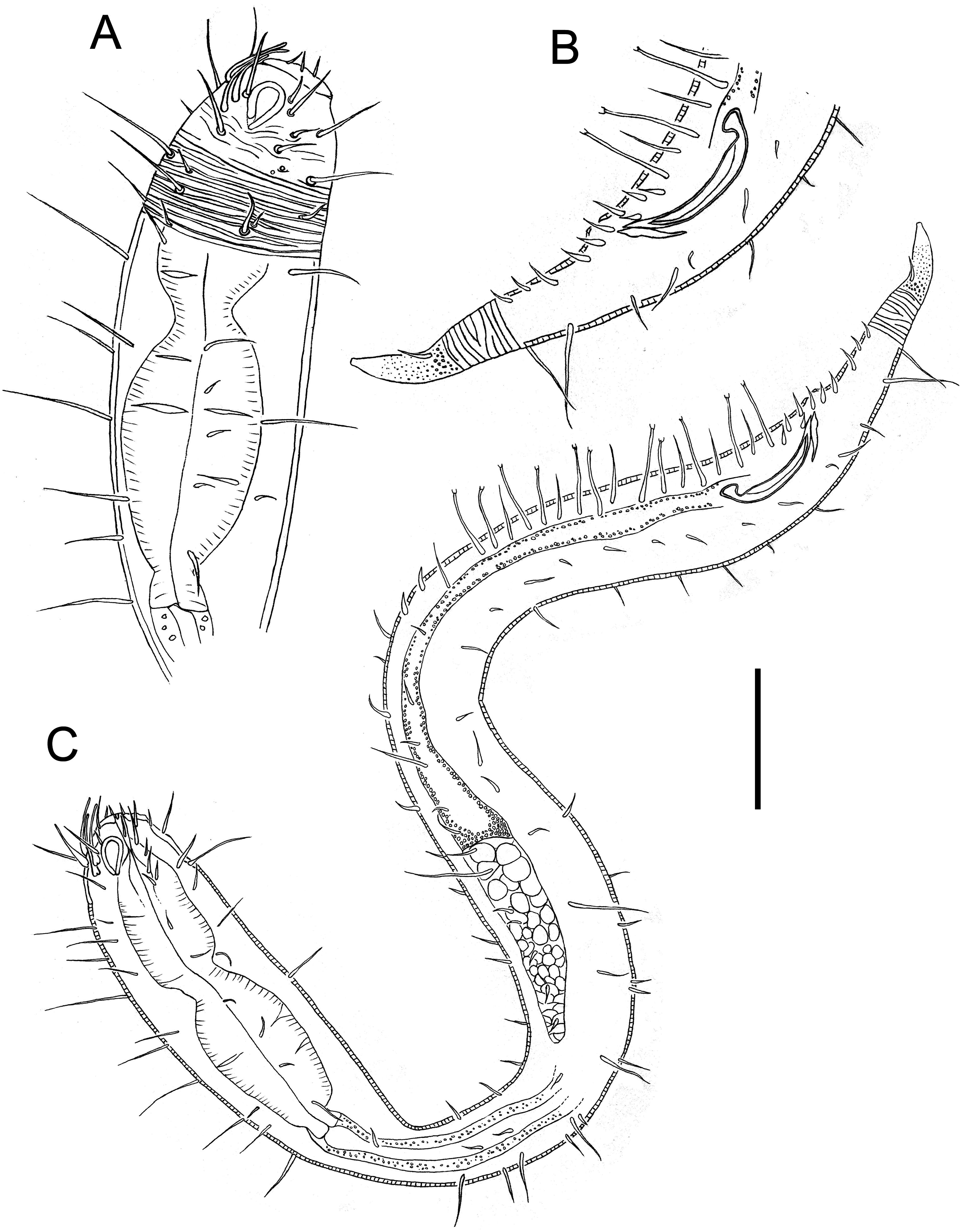
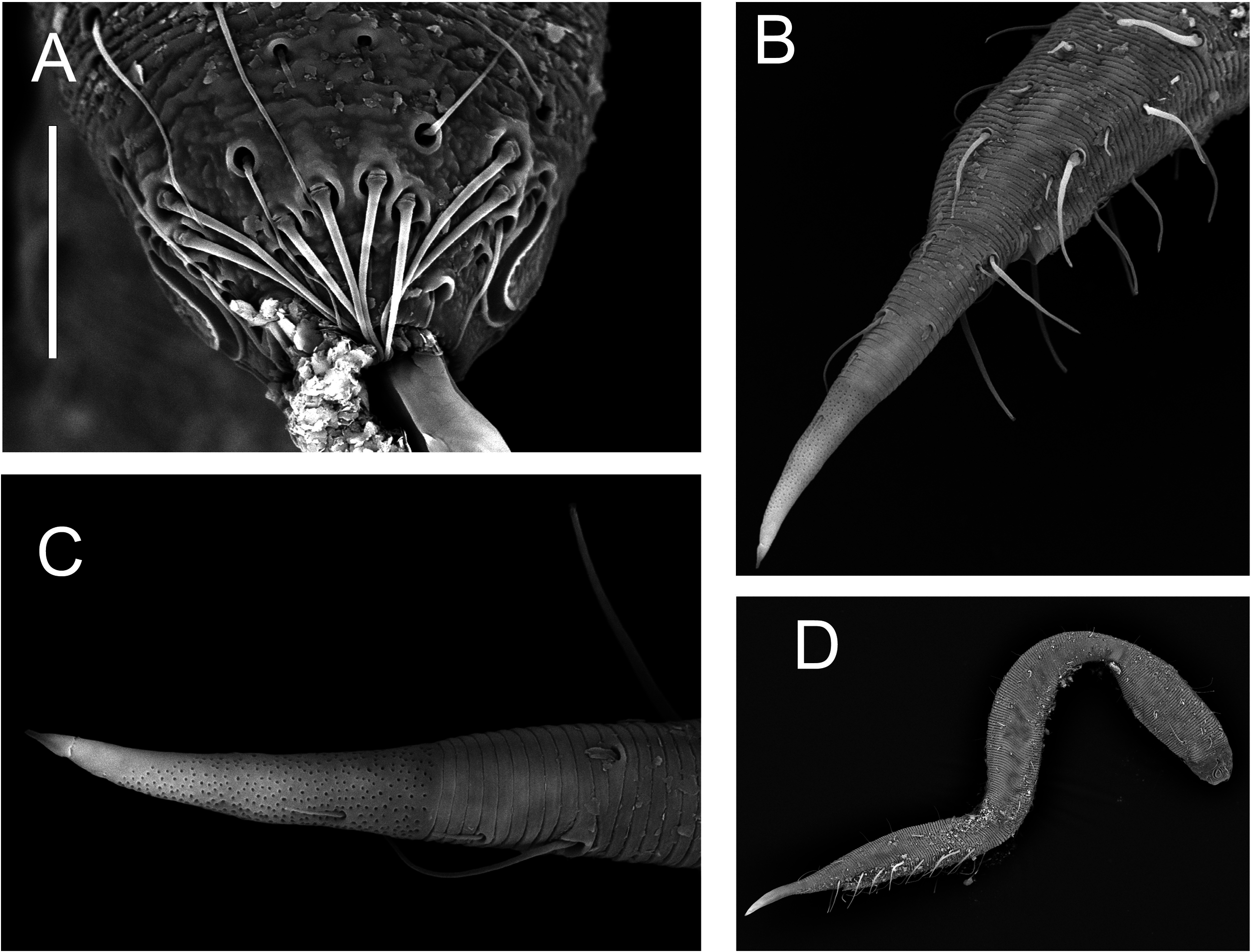
Male: Body partially covered in adhering fine detrital particles; shape typical of genus with pharyngeal region slightly more swollen than mid-body region ( Fig. 7D View Figure 7 ). Swollen pharyngeal region 23% of total body length, set off from rest of body by narrow neck region. Annulated cuticle without vacuoles or spines; no lateral differentiation. Somatic setae arranged in eight longitudinal rows, 5–29 μm in length; longest setae located in pharyngeal and mid-body regions. Most setae with conspicuous cuticularized collar at base.
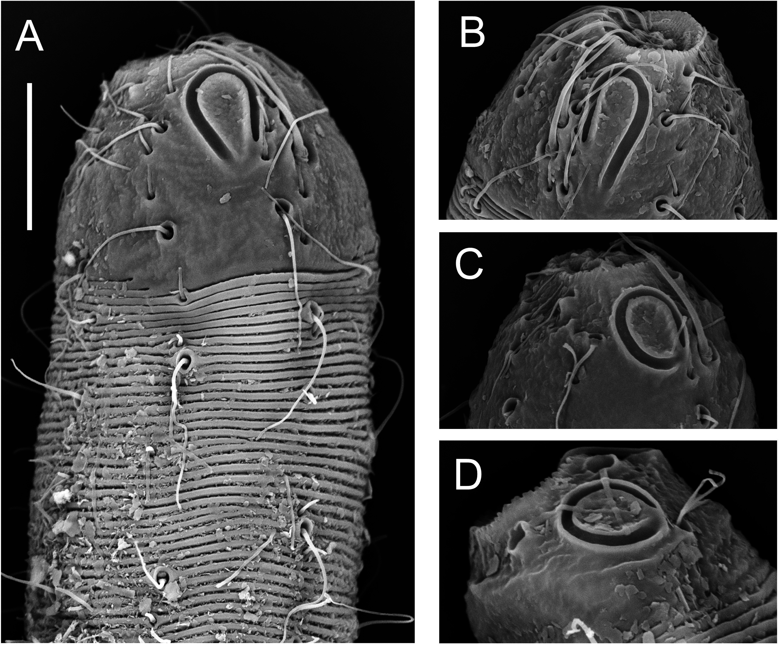
Well-developed rostrum, 25 μm in height and 34 μm at widest point; lip region introverted. Inner labial sensillae not observed; six outer labial setae on lip region, 5 μm long, and four cephalic setae, 13 μm long, situated at anterior edge of amphids. Eight cephalic adhesion tubes dorsally on the cephalic capsule; anteriormost four located in the dorsal sector in a transverse row near mid-level of amphid, and posterior-most four arranged in two dorsosublateral pairs at level of posterior edge of amphid ( Fig. 7A View Figure 7 ). All cephalic adhesion tubes have wide bases and slightly swollen distal ends. Numerous subcephalic setae present on head capsule. Amphideal fovea large, loop-shaped, with two arms of equal length joining at posterior extremity; amphideal aperture similar in shape but with ventral arm slightly longer than dorsal arm and not joining at posteri- or extremity ( Fig. 8A, B View Figure 8 ).
Pharynx dumbbell-shaped with well-developed corpus becoming gradually thinner towards buccal cavity and partially surrounding it. Corpus separated by short isthmus from large posterior bulb; nerve ring at level of isthmus. Cardia short, 7 μm long. Intestine situat- ed dorsally relative to reproductive system.
Reproductive system with single outstretched testis located in anterior portion of swollen mid-body region. Sperm cells globular, 8 × 10–12 μm. Spicules arcuate, 2.1 abd long, with fine central cuticularized projection (lamella); well-developed capitulum and pointed distal end. Gubernaculum small, with pointed proximal end and slightly wider distal end, parallel to spicules. Six pairs of short and stout cloacal setae, three situated anterior to cloaca, one at same level as cloaca, and two posterior to cloaca.
Posterior adhesion tubes slender, with bell-shaped distal ends all located anterior to cloaca and arranged in four rows: two ventrosublateral rows each consisting of 11 adhesion tubes, and two subventral rows each consisting of ten adhesion tubes. Ventrosublateral adhesion tubes alternate with six slightly shorter setae; the anterior-most four pairs of adhesion tubes are followed by one seta each and the following two adhesion tubes are followed by one seta each. No alternating setae observed in subventral rows of adhesion tubes. Two subventral rows of stout setae located anterior to subventral adhesion tubes; two stout setae on right-hand side and four, possibly five stout setae on left-hand side.
Tail conical with non-annulated tail end 29% of tail length. Three pairs of subventral setae and three pairs of subdorsal setae present. Two short subdorsal setae on non-annulated tail end ( Fig. 7C View Figure 7 ).
Females: Similar to males in most respects, but with greater maximum body diameter at or near level of vulva. Swollen pharyngeal region 24–26% of total body length. Amphideal fovea and amphideal aperture as in male; one specimen observed with electron microscopy with unispiral amphideal fovea and aperture ( Fig. 8C View Figure 8 ). Somatic setae arranged in eight longitudinal rows as in males, but many setae appear missing and only cuticular collars are visible. Lip region protruded in two specimens.
Reproductive system didelphic-amphidelphic with reflexed ovaries both located ventrally relative to intestine. Vulva located at mid-body. Cuticular pars distalis vaginae and pars proximalis vaginae surrounded by constrictor muscle; large vaginal glands present. Paravulval setae absent.
Posterior adhesion tubes arranged in four longitudinal rows: two ventrosublateral rows each comprising 13 adhesion tubes with one pair of adhesion tubes located posterior to anus, and two subventral rows each comprising ten to 12 adhesion tubes all located anterior to anus. One or two short setae located between each ventrosublateral adhesion tube. Non-annulated tail end comprises 37–50% of tail length.
Diagnosis
Dracograllus ngakei sp. nov. is characterized by swollen pharyngeal region 23–26% of total body length, pres- ence of eight cephalic adhesion tubes (with anteriormost four located in the dorsal sector in a transverse row near mid-level of amphid, and posterior-most four arranged in two dorsosublateral pairs at level of posterior edge of amphid), loop-shaped amphideal aperture with ventral arm slightly longer than dorsal arm, amphideal fovea of similar shape or with arms of equal length and joining posteriorly (unispiral amphideal fovea and aperture observed in one female), and conical tail with non-annulated tail region comprising 29–50% of total tail length. Male is characterized by 11 ventrosublateral adhesion tubes per row and ten subventral adhesion tubes per row all located anterior to cloaca, two subventral rows of stout setae located anterior to subventral adhesion tubes [two stout setae on right-hand side and four (possibly five) stout setae on left-hand side], spicules 2.1 abd long and six pairs of short and stout cloacal setae; female with 13 ventrosublateral adhesion tubes per row with one pair located posterior to anus, ten to 12 subventral adhesion tubes per row all located anterior to anus, and paravulvar setae absent.
Differential diagnosis
The new species is most similar to Dracograllus timmi , but can be differentiated from it mainly by the cuticle ornamentation (presence of annular ridges and spinelike projections appearing as two rows of fine punctations in Dracograllus timmi vs. smooth annules in Dracograllus ngakei sp. nov.), and also by the number of ventrosublateral adhesion tubes in males (seven to ten in Dracograllus timmi vs. 11 in Dracograllus ngakei sp. nov.). Dracograllus ngakei sp. nov. is also similar to Dracograllus wieseri , but differs in the number of ventrosublateral adhesion tubes (14–17 in Dracograllus wieseri vs. 11–13 in Dracograllus ngakei sp. nov.), number of cloacal setae (four pairs in Dracograllus wieseri vs. six pairs in Dracograllus ngakei sp. nov.), and presence of both long and short setae alternating with ventrosublateral adhesion tubes (vs. long setae only in Dracograllus ngakei sp. nov.). Dracograllus ngakei sp. nov. is also similar to Dracograllus chitwoodi , but can be differentiated from it by the cuticle ornamentation (annules with two transverse of dot-like punctations in Dracograllus chitwoodi ) and number of ventrosublateral adhesion tubes (9–10) and subventral adhesion tubes (8–10) in females.
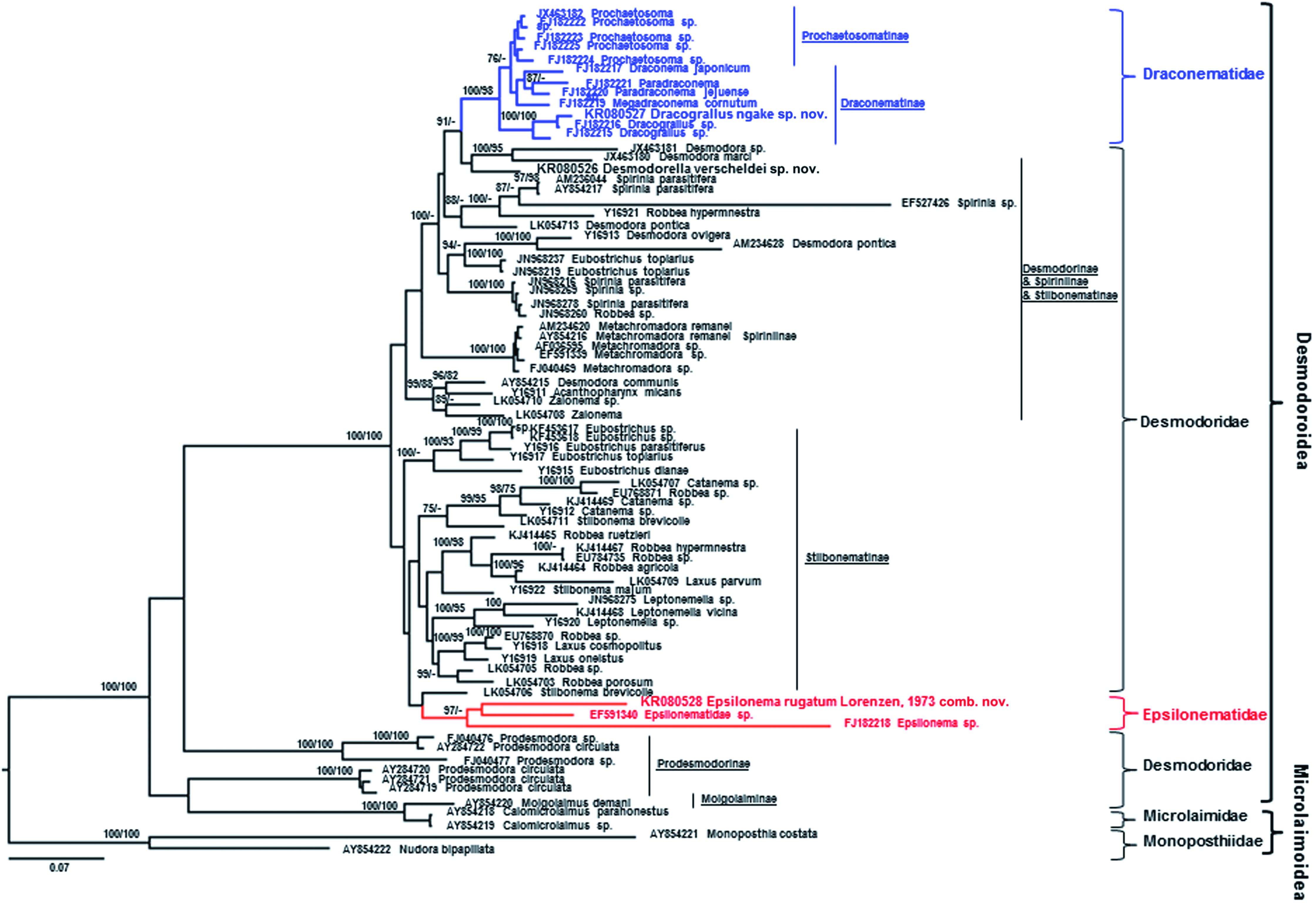
Based on SSU molecular sequences, Dracograllus ngakei sp. nov. differs from Dracograllus sp. ( FJ 182216 View Materials ) by 2% (19 in 785 bp including three gaps) and Dracograllus sp. ( FJ 182215 View Materials ) by 3% (23 in 783 bp including one gap) ( Fig. 14 View Figure 14 ).

| V |
Royal British Columbia Museum - Herbarium |
| SSU |
Saratov State University |
No known copyright restrictions apply. See Agosti, D., Egloff, W., 2009. Taxonomic information exchange and copyright: the Plazi approach. BMC Research Notes 2009, 2:53 for further explanation.
|
Kingdom |
|
|
Phylum |
|
|
Class |
|
|
Order |
|
|
Family |
|
|
Genus |