
Arnapa meja Huber, 2019
|
publication ID |
https://doi.org/10.11646/zootaxa.4546.1.1 |
|
publication LSID |
lsid:zoobank.org:pub:D2C9F49A-9B76-40AE-9A60-CAE9B99BA547 |
|
DOI |
https://doi.org/10.5281/zenodo.5449652 |
|
persistent identifier |
https://treatment.plazi.org/id/E21587DB-FF8C-FFC0-FF11-FE384805F84F |
|
treatment provided by |
Plazi |
|
scientific name |
Arnapa meja Huber |
| status |
sp. nov. |
Arnapa meja Huber View in CoL sp. n.
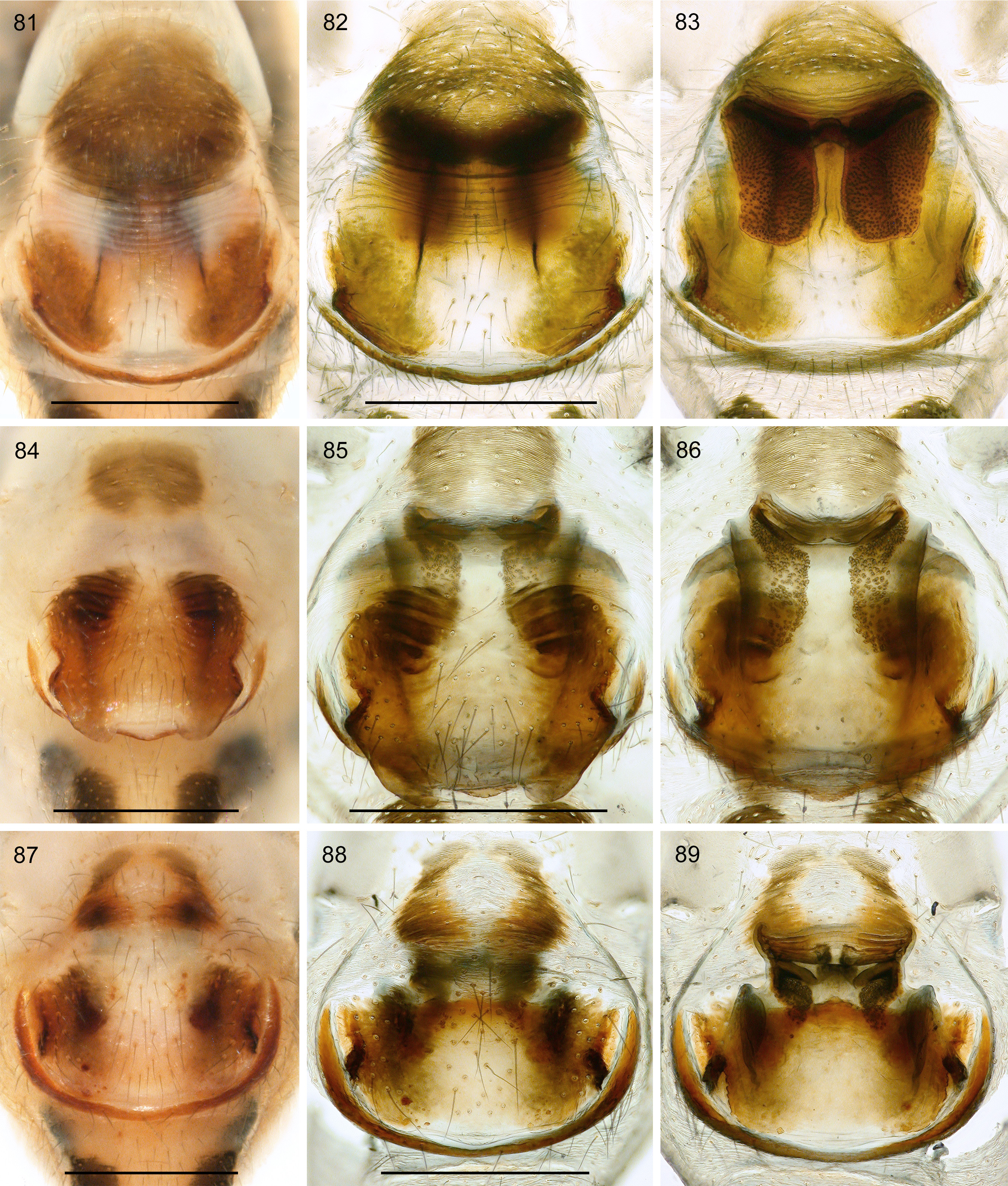
Figs 55–59 View FIGURES 55–59 , 72 View FIGURES 70–74 , 81–83 View FIGURES 81–89
Gen.n. Ind107: Eberle et al. 2018 (molecular data); Huber et al. 2018: fig. 2.
Type material. INDONESIA: ♂ holotype, ZFMK ( Ar 20609), West Papua, Manokwari, Gunung Meja ( 0.860°S, 134.084°E), 190 m a.s.l., 8.xi.2009 ( S. Sutono) GoogleMaps .
Other material examined. INDONESIA: 6♀, ZFMK (Ar 20610), and 1♂ 1♀ in pure ethanol, ZFMK (Ind201), same data as holotype GoogleMaps .
Etymology. The species name is derived from the type locality; noun in apposition.
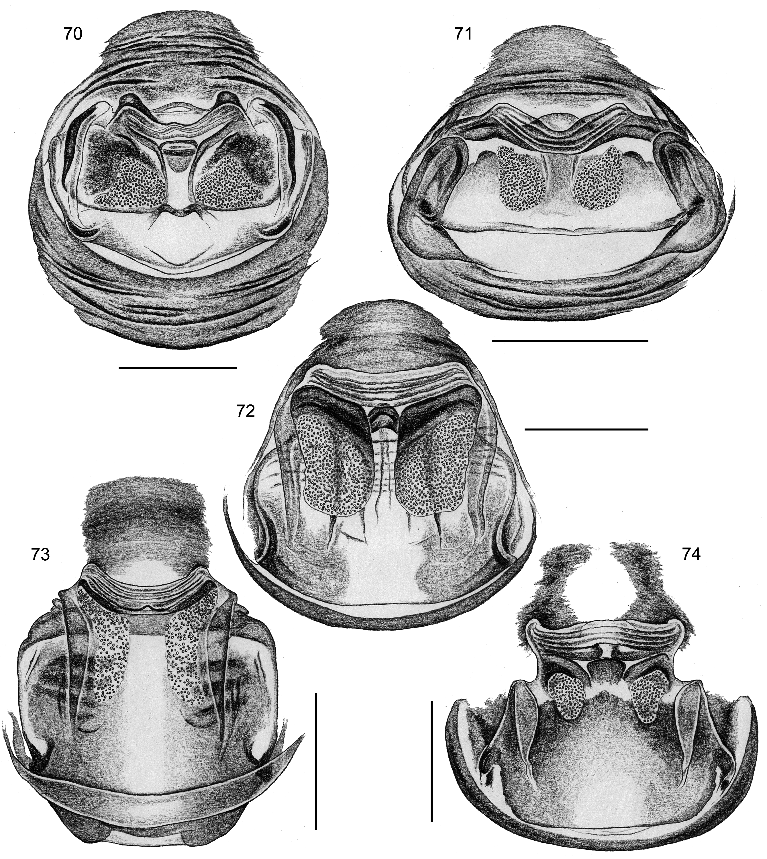
Diagnosis. Distinguished from known congeners by armature of male chelicerae ( Fig. 58 View FIGURES 55–59 ; single lateral row of small apophyses), by presence of whitish projection at basis of bulbal process ( Fig. 59 View FIGURES 55–59 ), by procursus with strong distal sclerite ( Figs 56–57 View FIGURES 55–59 ), by presence of large brown plate in front of epigynum ( Fig. 81 View FIGURES 81–89 ), and by large pore plates bordered anteriorly by large sclerites ( Figs 72 View FIGURES 70–74 , 83 View FIGURES 81–89 ).
Description. Male ( holotype). MEASUREMENTS. Total length 2.2, carapace width 1.0. Distance PME-PME 100 µm; diameter PME 90 µm; distance PME-ALE 70 µm; distance AME-AME 20 µm, diameter AME 40 µm. Leg 1: 32.9 (7.7 + 0.4 + 8.0 + 14.2 + 2.6), tibia 2 missing, tibia 3: 3.2, tibia 4: 4.6; tibia 1 L/d: 100.
COLOR (in ethanol). Carapace ochre-yellow with wide dark brown bands laterally at margin and medially including large part of ocular area; clypeus with pair of dark bands between eye triads and rim; sternum ochreyellow; legs light brown, without dark rings, tips of femora and tibiae whitish. Abdomen gray, dorsally densely covered with dark marks, ventrally with indistinct light brown mark in front of gonopore, dark median band behind gonopore, light brown area and pair of large dark ventro-lateral marks in front of spinnerets.
BODY. Habitus as in close relatives (cf. Figs 39, 41, 43 View FIGURES 39–44 ). Ocular area elevated, thoracic furrow present; clypeus unmodified. Sternum wider than long (0.64/0.46), unmodified.
CHELICERAE. As in Fig. 58 View FIGURES 55–59 , with 10–11 apophyses laterally on each side; without modified hairs; with stridulatory ridges.
PALPS. As in Figs 55–56 View FIGURES 55–59 ; coxa unmodified, trochanter with low rounded process ventrally, femur strongly widened (similar only in A. arfak ), with rounded retrolateral process proximally, with prolateral stridulatory pick (modified hair) proximally, patella ventrally reduced (not closed), tarsus small, procursus with dorsal process proximally, ventral side concave but without distinct pocket, distally with strong retrolateral apophysis and prolateral membranous structures ( Fig. 57 View FIGURES 55–59 ); genital bulb large, with short membranous projection at base of complex process ( Fig. 59 View FIGURES 55–59 ).
LEGS. Without spines; few vertical hairs; with curved hairs on all legs (tibiae and metatarsi, few on femora); retrolateral trichobothrium of tibia 1 at 7%; prolateral trichobothrium present on all tibiae; tarsus 1 with ~35 pseudosegments, distally distinct.
Male (variation). No variation seen. Legs 1 missing in other male examined.
Female. In general similar to male but sternum medially brown. Tibia 1 in 6 females: 5.6–6.7 (mean 6.3). Epigynum anterior plate divided into three sections ( Fig. 81 View FIGURES 81–89 ): anterior bulging brown area, central membranous part with transversal folds, and posterior part brown laterally and whitish medially; posterior plate very short and wide. Internal genitalia with large pore plates bordered anteriorly by massive sclerites ( Figs 72 View FIGURES 70–74 , 83 View FIGURES 81–89 ).
Distribution. Known from type locality only ( Fig. 343 View FIGURE 343 ).
Natural history. This species occupied the same microhabitat as the syntopic Arnapa manokwari (see above). The two species appeared indistinguishable in the field.


No known copyright restrictions apply. See Agosti, D., Egloff, W., 2009. Taxonomic information exchange and copyright: the Plazi approach. BMC Research Notes 2009, 2:53 for further explanation.