
Paeduma Rathbun, 1897
|
publication ID |
https://doi.org/ 10.5281/zenodo.5401414 |
|
persistent identifier |
https://treatment.plazi.org/id/E1673758-3842-FFAF-B6E5-FC75FC7AEE49 |
|
treatment provided by |
Marcus |
|
scientific name |
Paeduma Rathbun, 1897 |
| status |
|
Genus Paeduma Rathbun, 1897 View in CoL
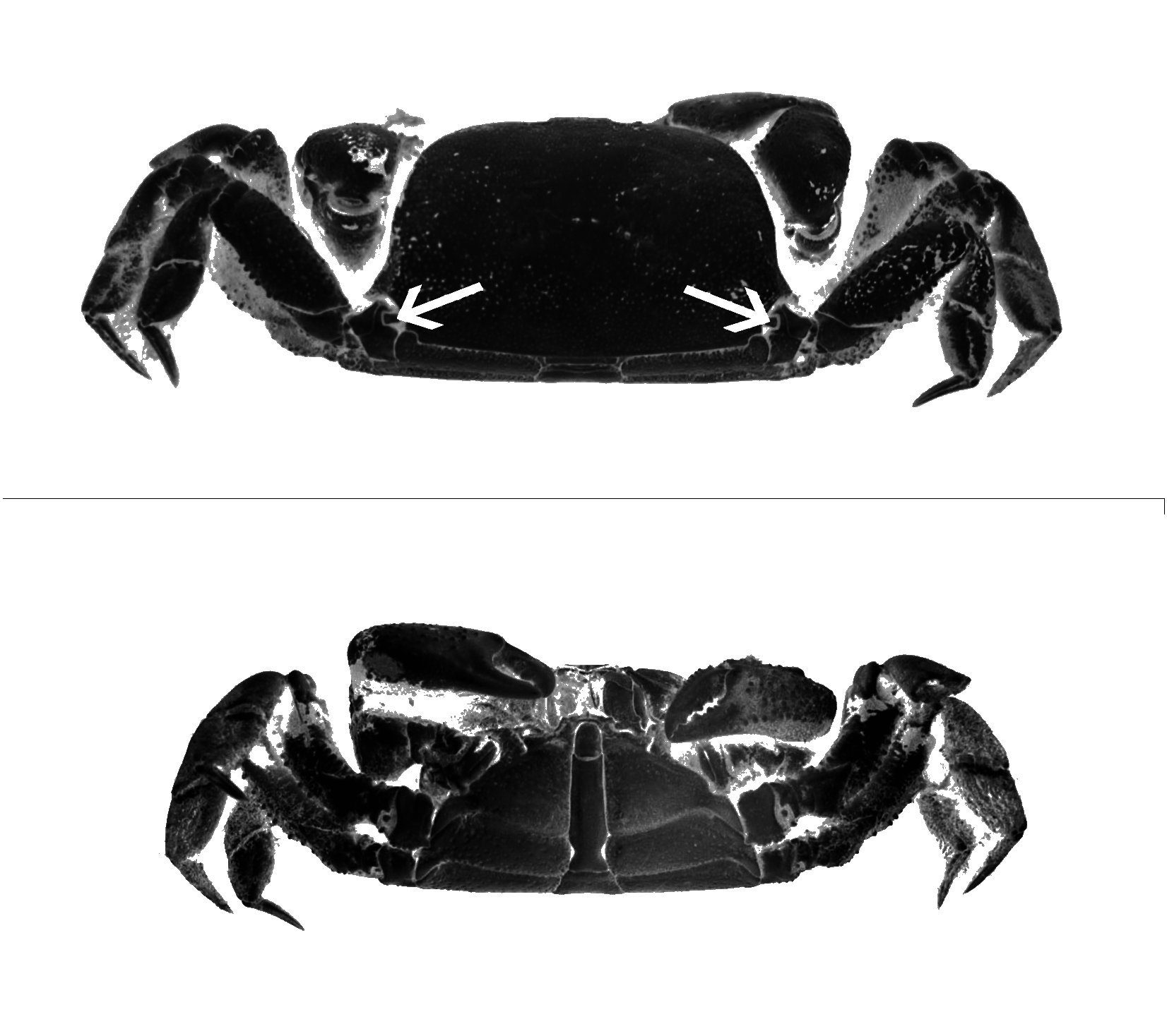
( Figs 1 View FIG ; 2 View FIG )
Amorphopus Bell, 1859: 27-29 View in CoL . — Miers 1886: 275. — Alcock 1900: 293. — Stebbing 1910: 315. — Tesch 1918: 238 (key). — Serène 1968: 93. — Gordon 1971: 108. — Sankarankutty 1975: 4. — Guinot 1979: 114, 215.
Paeduma Rathbun, 1897: 163 View in CoL . — Gordon 1971: 108. — Guinot 1979: 114. — Schweitzer et al. 2000: 55. — Schweitzer & Feldmann 2001 pro parte: 332, 335, 345.
Non Paeduma View in CoL – Manning & Holthuis 1981: 168, 173. — Karasawa 1990: 25. — Huang et al. 2002: 652, table 1. — Schweitzer 2005: 289.
TYPE SPECIES. — Amorphopus cylindraceus ( Bell, 1859) , by monotypy. Amorphopus Bell, 1859 is an invalid junior homonym of Amorphopus Audinet-Serville, 1838 (Insecta) , therefore was replaced by Paeduma Rathbun, 1897 (p. 163), substitute name, from the Greek “rudiment” in allusion to the fifth pair of legs ( Rathbun 1897: 163, footnote). Genus Paeduma : gender neuter according to Manning & Holthuis (1981), thus the type species is Paeduma cylindraceum (but gender masculine according to Huang et al. 2002, who are not followed in the present paper).
EMENDED DIAGNOSIS (male holotype of P. cylindraceum ). — Body thick, nearly cylindrical, narrowing anteriorly, markedly convex, transversely flattened. Dorsal surface with regions indistinct, grooves not marked except for extremely weak traces of cervical and branchiocardiac grooves. Lateral margin arcuate anteriorly, then divergent, posteriorly convergent. Front depressed, relatively narrow (6.4 times in carapace width), with distinct thickened ridge, more advanced laterally than in midline, with very weak, obtuse projection medially. Antennae with articles 2+3 covered by antennules. Epistome relatively developed. Orbits transverse, situated in straight line, not dorsally expanded, rimmed. Eyes movable, small, lying transversally, with cornea small, narrower than stalk. Buccal cavity with sides convergent anteriorly. Mxp3 of “normal” type (i.e. operculiform), broad, nearly filling entire field. Endopod with broad ischion and merus; anteroexternal margin of merus oblique; propodus, carpus and dactylus slender; palp cylindrical; dactylus longer than propodus but extending only two-thirds of ischion, close to its border. Exopod wide, with long but concealed flagellum (not shown in Fig. 2C View FIG ). Thoracic sternum very wide. Sternites 1-2 forming a narrow, triangular piece extending between bases of mxp3, clearly separated from sternite 3; sternite 3 distinct but not delimited by suture; sternite 4 much developed; sternites 5-7 similarly developed, high; sternite 8 not aligned with preceding sternites, strongly reduced, only visible dorsally as small, ornamented plate inserted between sternite 7 and abdominal somite 1. Episternites 4-5 similarly elongated, pointed; episternites 6-7 similarly rounded; episternite 7 forming projection overhanging posterolateral angle of carapace. Sutures 4/5 to 6/7 nearly parallel, equidistant. Sternal grooves or trenches absent. Sterno-abdominal cavity elongated, reaching sternite 3. Male abdomen very long, extending beyond bases of mxp3, extremely narrow, specially at level of somite 6 and telson; strong constriction at level of abdominal somite 6 opposite to middle part of thoracic sternite 5. Somites 1, 2 free, approximately similar in size; somites 3-5 fused, forming distally-truncated triangle; somite 6 as extremely long, linear, undivided piece; its proximal part markedly constricted; telson elongated, rounded at tip. Gonopods concealed under abdomen, shape unknown (dry condition of specimen). G1 supposedly relatively slender (because of narrowness of abdomen), not recurved posteriorly or doubled recurved into a 8-shaped figure; G2 probably small. Chelipeds markedly unequal in male, robust, thick; large cheliped with palm nearly as long as wide, gap between fingers; fingers armed with blunt teeth; small cheliped with closer fingers. Dactyli on each side with striae on inner surface. P2-P4 short, rather similar in size, shape. P5 not visible on the outside, reduced to a vestigial coxa in males, absent in females. Stridulating apparatus of two striated parts. Pterygostomian region with an oblique row of rather thick, short, spaced sticks (about 14), progressively diminishing in size to show externally as rounded granules; dactylus provided with numerous thin, long, closed striae on whole length of inner surface (except for apex).
GEOGRAPHICAL DISTRIBUTION. — Galápagos Islands ( Garth 1946, 1991; Hickman & Zimmerman 2000) or possibly Pacific coast of South America.
REMARKS
Bell’s Amorphopus (= Paeduma ) was established without indicating locality and without figures, which may explain why Paeduma cylindraceum was never reported since its description.
The P5 was considered a “mere rudiment, in the form of a minute tubercle inserted in a little notch at the base of the first joint of the fourth pair, and scarcely discernible by the naked eye” ( Bell 1859: 28). Bell did not believe that a leg could vanish completely in a decapod. De Haan (1835: 35, under Hexapus sexpes Fabricius, 1798 ) correctly remarked: “Nullum indicium quinti paris, neque sub abdomine ulli reconditi” (= “no sign of a fifth pair, or is any hidden under the pleon”). Stebbing (1910: 315) corrected Bell’s statement “six pairs of legs beside the claws” to “three pairs of legs”.
Two of Bell’s statements need comment.
1) The location of the P5 rudiment showing as a tubercule at the base of the P4 does not correspond to the normal place of the coxa of an appendage (always articulated on the sternite). This location, which might be interpreted as the result of the displacement of the P 5 in the reduction processus of sternite 8, does not match with the interpretation of an hexapodid P5 vestigial coxa that is concealed under the abdomen ( Saint Laurent 1989; Guinot, Tavares & Castro unpublished data).
2) A similar “tubercle” was seen by Bell (1859: 29) at the base of the P 4 in dorsal view of Hexapus sexpes (Fabricius, 1798) figured by De Haan (1835: pl. D, pl. 11, fig. 6). The small figure of De Haan (1835: pl. 11, fig. 6) does not allow to be sure of the presence of such a tubercle, but the presence in H. sexpes of a structure similar to that of P. cylindraceum is confirmed here.
Bell’s “mere rudiment” on the P4 coxa of P.cylindraceum ( Figs 1A View FIG ; 2D View FIG ) actually corresponds to the external portion of the apodeme of the P4 coxa, i.e. the apodemal platelet. It is exposed in a notch of the proximal border of the P4 coxa, and shows externally as a calcified strip extending through the arthrodial cavity as usual. The apodemal platelet is clearly visible in the dry holotype of P. cylindraceum , as well as in a number of hexapodids (for instance in Hexapus sexpes ). The supposed P5 rudiment seen by Bell (1859) definitely is not a vestige of P5, but corresponds to a portion of the P4.
Actually, apodemal platelets are present on P2- P4 coxae in all the hexapodid genera that were examined, although this could not be confirmed in the holotype of P. cylindraceum because of its dry condition. The apodemal platelets are also visible ventrally on the basis-ischion of P2-P 4 in the P. cylindraceum ’s holotype as well as in other hexapodids. Such apodemal (coxal and ischio-basal) platelets are present to a variable extent on P2 to P 5 in other eubrachyuran families. It seems, however, that the coxal platelets are particularly well visible on the pereopods of the Hexapodidae , and on the P 4 in particular. Authors such as Huang et al. (2002) who have discussed other species of Paeduma , viz. P. orientalis , unfortunately have not provided enough information.
As previously mentioned, Manning & Holthuis (1981) gave erroneous characters for Paeduma because they combined the characters briefly provided by Bell (1859) for P. cylindraceum , the type species, and those shown by the two other species of Paeduma , P. orientalis and P. chuenensis . The latter two are herein placed in Hexalaughlia n. gen. Manning & Holthuis (1981) indicated for Paeduma “third and fourth and fourth [sic] and fifth male abdominal somites fused” (p. 168, in the key) and “third and fourth and fifth and sixth somites fused” (p. 173, 175). We put another interpretation, hypothetically: abdominal somites 3-5 fused in Paeduma and in Hexalaughlia n. gen.
Manning & Holthuis’ (1981) assertion that the gonopods of Paeduma are slender and recurved posteriorly is applicable to the gonopods of Hexalaughlia n. gen. The gonopods of Paeduma ( P. cylindraceum ) are unknown because of the dry condition of the
A
B
holotype, but they are probably neither recurved posteriorly nor doubly recurved into an 8-shaped figure as in Hexalaughlia n. gen. Bell’s (1859) description of P.cylindraceum does not mention pterygostomian stridulating striae, which explains why this character is missing in the generic diagnosis of Paeduma provided by Manning & Holthuis (1981: 173: “pterygostomian region lacking oblique striae”). Manning & Holthuis attributed to Paeduma two other species, P. orientalis and P.chuenensis , that lack a stridulating apparatus. In contrast, Manning & Holthuis (1981: 177) did characterize Stevea williamsi by the presence of stridulating pterygostomian striae, a condition confirmed herein.
Paeduma cylindraceum ( Bell, 1859) View in CoL ( Figs 1 View FIG ; 2 View FIG )
Amorphopus cylindraceus Bell, 1859: 27 . — Serène 1968: 93.
Paeduma cylindraceus – Rathbun 1897: 163. — Gordon 1971: 108. — Crane 1981: 3. — Manning & Holthuis 1981: 173.
MATERIAL EXAMINED. — Holotype, ♂ 15.3 × 23.5 mm, dry and in good condition, with Bell’s handwritten labels: “ Amorphopus ” (placed above specimen) and “ Am: cylindraceus male sign. Gallapagos [sic] Mr Cuming” (placed below specimen). Registration number: OUMNH 15693 (S. De Grave pers. comm.).
DESCRIPTION OF DRY MALE HOLOTYPE
Granules absent on dorsal surface of carapace, present only on lateral borders, more developed at level of P4; with numerous pits except medially. Large cheliped with palm much inflated; outer surface covered by coarse, rounded granules, more numerous on inferior half near superior border; proximal superior border of dactylus coarsely granulated, prehensile margin armed with two distinct blunt teeth; fixed finger curved, granulated, prehensile margin armed with strong proximal tooth and less distinct teeth; marked gap between fingers. Small cheliped much reduced; no marked gap between pointed fingers; outer surface of palm covered by coarse, blunt granules, closer on inferior half near superior border; dactylus with superior bor- der coarsely granulated except distally, prehensile margin armed with several triangular teeth; fixed finger with two rows of strong granules, prehensile margin armed with several triangular teeth. Thoracic sternum ornamented with marked granules along border of sterno-abdominal cavity and sutures; surface punctate. Sutures 4/5 to 6/7 equidistant; sternites 1-2 advanced between mxp3; sternite 3 distinct but not demarcated by suture; sternite 4 well developed, with latero-anterior projections; sternites 5 to 7 inflated, of about same size. Sternite 7 tilted, its posterolateral corner (episternite 7) forming a marked projection which fits with a notch on border of carapace (interlocking mechanism carapace/sternum). Sternite 8 present but extremely reduced and concealed under carapace, except for small plate exposed dorsally, calcified, ornamented. Pereopods 2-4 with margins of meri ornamented with salient, blunt granules. Setae on surfaces of P2 and P3 meri, on surfaces of P2-P4 carpi and propodi; longer setae on margins of distal articles. P4 coxa with a markedly discernible apodemal platelet; ischio-basis with a ventral apodemal platelet. P5 vestigial coxa concealed under abdomen.
REMARKS
The particular spelling of Galápagos, with a double “l”, i.e. “Gallapagos”, is consistent on labels of Bell’s dry collection (see DiMauro 1982: 158: a fact which “does help substantiate that it is Bell’s collection”), and was similarly used by H. Milne Edwards (1838: 12). It should be stressed that the Galápagos origin of this unique specimen must be taken with caution because of a possible exchange of labels between material collected by Cuming in the Galápagos and along the South American mainland coast. Several species collected by Cuming and reported by Bell “have been turned up along the mainland coast of south America from Santa Elena Bay, Ecuador, to the Bay of Panama, localities also visited by Cuming” ( Garth 1946: 343; see also Garth 1958: 71; DiMauro 1982: 156).
Genus Stevea Manning & Holthuis, 1981 View in CoL ( Fig. 3 View FIG )
Stevea Manning & Holthuis, 1981: 168 View in CoL , 177. — Beschin et al. 1994: 191. — Schweitzer et al. 2000: 55. — Schweitzer & Feldmann 2001: 337, 345 (key). — Huang et al. 2002: 653, table 1. — Schweitzer 2005: 289.
TYPE SPECIES. — Hexapus williamsi Glassell, 1938 , by original designation.
SPECIES INCLUDED. — One extant species, S. williamsi . For the status of the fossil species Stevea cesarii Beschin, Busulini, De Angeli & Tessier, 1994 , from the Eocene of Italy, see Fossil Hexapodidae .
DESCRIPTION
See Manning & Holthuis (1981: 168, 177), in amending the features concerning the male abdomen which, instead of “second through sixth somites fused”, shows weak but distinct sutures.
REMARKS The genus Stevea was established by Manning & Holthuis (1981) to separate from Hexapus De Haan, 1835 emend. (type species: Cancer sexpes Fabricius, 1798 ; genus established in 1833 but without nominal species) the American species H. williamsi Glassell, 1938 (p. 445, pl. 35, figs 1-4). Stevea williamsi appears to be known with certainty from the male holotype only, 5.8 × 8.6 mm, from San José, Guatemala (SDSNH No. 3940; ex Cat. No1158). The female 9.4 × 14.4 mm, from the Gulf of Tehuantepec, west coast of Mexico (USNM 170897), identified to H. williamsi , may well prove to belong to Paeduma , a direct comparison with the holotype of H. williamsi being necessary.
Glassell (1938: 445, pl. 35, fig. 4) illustrated the male abdomen of S. williamsi as having several weak sutures; however, this does not correspond exactly to his text: “Only the 1st and 7th segment [telson] are articulated; the five interior segments are coalesced”. This is probably why Manning & Holthuis (1981) indicated for Stevea “male abdomen with three somites, second through sixth fused”. Examination of the male holotype and photographs by L. L. Lovell ( Fig. 3B, C View FIG ) indicate that Glassell’s sketch, showing several abdominal sutures, is correct. The abdominal somites of the holotype (which are wider than in Paeduma ) are separated by several weak but visible sutures so that in Stevea abdominal somites seem to be distinct, although all not articulated. There is no constriction. In these respects S. williamsi appears to be distinct from P. cylindraceum where, hypothetically, the abdominal somites 3-5 are completely fused and where there is a marked constriction at the level of somite 6, leaving an empty space on each side ( Fig. 2B, E View FIG ).
The first gonopods of the male holotype of S. williamsi are essentially straight, with only a slight distal curvature (L. L. Lovell pers. comm.). Because of the dry condition of the holotype of P. cylindraceum , it is not possible to compare the gonopods to those of Stevea . In S. williamsi the meri of P2-P4 show rows of tubercles, those on P2 being more numerous (L. L. Lovell pers. comm.).
A stridulating apparatus similar to that of Paeduma ( Fig. 2A, B View FIG ) is mentioned in Manning & Holthuis’ (1981: 177) diagnosis of Stevea . According to Glassell (1938: 445, pl. 35, fig. 2), in S. williamsi there is “a tubercle on the inner distal face [of the palm of the cheliped] which engages with the stridulations of the epimera”. Actually, in both sexes of Stevea , the symmetrical rows of thick and spaced pterygostomian stridulating striae are more likely rubbed by very thin and closed striae located on the inner part of the dactylus of both chelipeds, as in P. cylindraceum . In the female of S. williamsi (USNM 170897) the exposed portion of sternite 8 is small, as in the male of P. cylindraceum ( Fig. 2B, E View FIG ).
Despite some similar features, P. cylindraceum and S. williamsi are distinct. The question of the specific versus generic level of these differences, which concern mainly the male abdomen and the gonopods, is beyond the scope of this study, and should be addressed in a revision of the family Hexapodidae . Two genera, Paeduma and Stevea , are thus so far known from the Pacific coast of South America.
See below for the fossil record of Stevea .
Stevea williamsi ( Glassell, 1938) View in CoL ( Fig. 3 View FIG )
Hexapus williamsi Glassell, 1938: 445 View in CoL , pl. 35, figs 1- 4. — Hendrickx 1995: 139, list.
Stevea williamsoni – Stephensen 1946: 182 (incorrect spelling).
Stevea williamsi View in CoL – Manning & Holthuis 1981: 177. — Beschin et al. 1994: 191. — Schweitzer & Feldmann 2001: 337. — Huang et al. 2002: 653, table 1.
MATERIAL EXAMINED. — Holotype, ♂ 5.8 × 8.6 mm, San José , Guatemala ( SDSNH No. 3940; ex Cat. No. 1158; examined and photographed by L. L. Lovell and W. A. Newman; with reservation, ♀ 9.4 × 14.4 mm, Gulf of Tehuantepec, west coast of Mexico ( USNM 170897 ).
DESCRIPTION
See Glassell (1938) and above.


No known copyright restrictions apply. See Agosti, D., Egloff, W., 2009. Taxonomic information exchange and copyright: the Plazi approach. BMC Research Notes 2009, 2:53 for further explanation.
|
Kingdom |
|
|
Phylum |
|
|
Class |
|
|
Order |
|
|
Family |
Paeduma Rathbun, 1897
| Guinot, Danièle 2006 |
Paeduma
| SCHWEITZER C. E. 2005: 289 |
| HUANG J. F. & HSUEH P. - W. & NG P. K. L. 2002: 652 |
| KARASAWA H. 1990: 25 |
| MANNING R. B. & HOLTHUIS L. B. 1981: 168 |
Stevea
| SCHWEITZER C. E. 2005: 289 |
| HUANG J. F. & HSUEH P. - W. & NG P. K. L. 2002: 653 |
| SCHWEITZER C. E. & FELDMANN R. M. 2001: 337 |
| SCHWEITZER C. E. & FELDMANN R. M. & TUCKER A. B. & BERGLUND R. E. 2000: 55 |
| BESCHIN C. & BUSULINI A. & DE ANGELI A. & TESSIER G. 1994: 191 |
| MANNING R. B. & HOLTHUIS L. B. 1981: 168 |
Stevea williamsi
| HUANG J. F. & HSUEH P. - W. & NG P. K. L. 2002: 653 |
| SCHWEITZER C. E. & FELDMANN R. M. 2001: 337 |
| BESCHIN C. & BUSULINI A. & DE ANGELI A. & TESSIER G. 1994: 191 |
| MANNING R. B. & HOLTHUIS L. B. 1981: 177 |
Stevea williamsoni
| STEPHENSEN K. 1946: 182 |
Hexapus williamsi
| HENDRICKX M. E. 1995: 139 |
| GLASSELL S. 1938: 445 |
Paeduma
| SCHWEITZER C. E. & FELDMANN R. M. & TUCKER A. B. & BERGLUND R. E. 2000: 55 |
| GUINOT D. 1979: 114 |
| GORDON I. 1971: 108 |
| RATHBUN M. J. 1897: 163 |
Paeduma cylindraceus
| CRANE M. D. 1981: 3 |
| MANNING R. B. & HOLTHUIS L. B. 1981: 173 |
| GORDON I. 1971: 108 |
| RATHBUN M. J. 1897: 163 |
Amorphopus
| GUINOT D. 1979: 114 |
| SANKARANKUTTY C. 1975: 4 |
| GORDON I. 1971: 108 |
| SERENE R. 1968: 93 |
| TESCH J. J. 1918: 238 |
| STEBBING T. R. R. 1910: 315 |
| ALCOCK A. 1900: 293 |
| MIERS E. J. 1886: 275 |
| BELL T. 1859: 29 |
Amorphopus cylindraceus
| SERENE R. 1968: 93 |
| BELL T. 1859: 27 |