
Drawida koreana Kobayashi, 1938
|
publication ID |
https://doi.org/ 10.11646/zootaxa.3973.3.2 |
|
publication LSID |
lsid:zoobank.org:pub:10FA49C7-E806-4B89-B9BF-21C1F3E9EC03 |
|
DOI |
https://doi.org/10.5281/zenodo.6119525 |
|
persistent identifier |
https://treatment.plazi.org/id/E1328789-FFC6-FFB6-FF62-FF17D61F8E10 |
|
treatment provided by |
Plazi |
|
scientific name |
Drawida koreana Kobayashi, 1938 |
| status |
|
Drawida koreana Kobayashi, 1938
( Figures 6 View FIGURE 6 , 7 View FIGURE 7 )
Drawida koreana Kobayashi, 1938: 102 .
Drawida koreana— Kobayashi 1940: 268; Easton 1981: 39.
Material examined. 2 specimens altogether. 1 clitellate (dissected), Hsiju, from roadside ditches around Hsiju Visitor Centre, 25°58'09.3''N, 119°56'01.7''E, 29 m asl, 30 May 2012, H.P. Chen & T.L. Ai (TESRI-O- 2012-44 -8; MTS 45); 1 clitellate (dissected), Nangan, Hanlinjiao, from roadside ditches, 26°10'28.9''N, 119°55'01.7''E, 39 m asl, 25 June 2012, H.P. Chen & W.J. Chih (TESRI-O- 2012-58 -7; MTS 87).
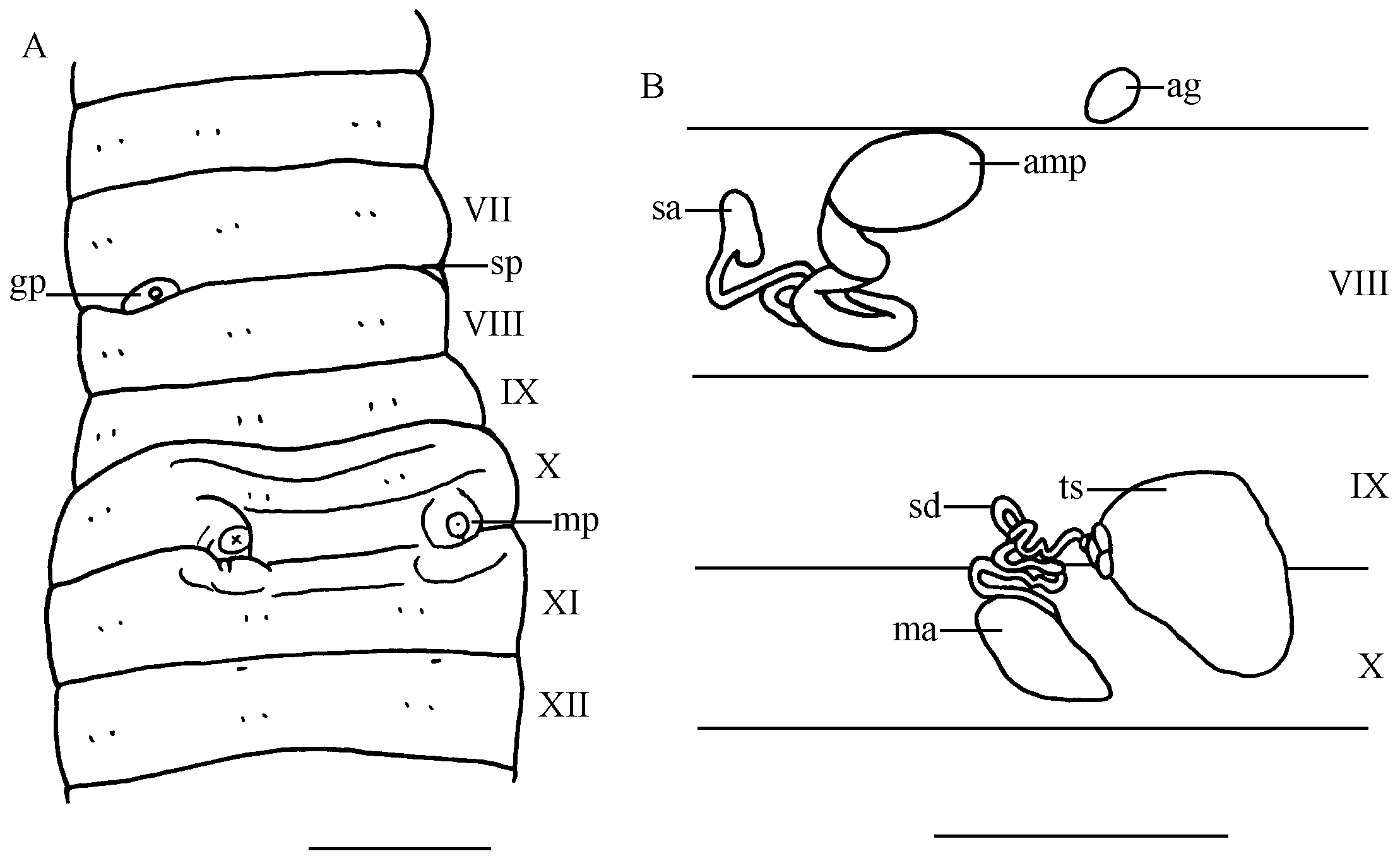
Description of MTS 45. External: Total length 54 mm, greatest diameter in clitellar region 3.6 mm, weight 0.3 g. Segments numbering 139. Prostomium prolobous. Dorsal pores absent. Setae short and closely paired, ab = cd, aa = 0.8 bc, aa> 8 ab, dd greater than 0.5 body circumference. Clitellum in X–XIII, paler, swollen. Spermathecal pores one pair in 7/8, each pore medial to seta c, sunken in the intersegmental furrow with a round genital papilla 0.47 mm in diameter anterior to it ( Fig. 6 View FIGURE 6 A). Male pores one pair, each on top of a conical protuberance formed by elevated glandular skin between b and c (much nearer to b) in postsetal X, anterior border of XI facing each protuberance also slightly elevated ( Fig. 6 View FIGURE 6 A). The elevated area surrounding each male pore pinkish. Female pores minute, one pair on anterior most border of XII, each pore in line with seta b. Preserved specimen dark bluish gray in color, pinkish around spermathecal pore and male pore areas.
Internal: Septa 5/6 thick, 6/7–8/9 thickened and muscular, 9/10 thin. Septum 10/11 fused with septum 11/12 dorsally. Gizzards three in XII–XIV, round, white, muscular and shining. Hearts VI–IX. Nephridia holoic. Testis sacs one pair, large, yellowish, elongated oval, about 1.05 mm in length and 0.9 mm in width, each suspended by septum 9/10. Sperm duct loosely spirally twisted, running in front of septum 9/10 around last heart, piercing the septum into segment X and entering into the anteromedial side of the male atrium ( Fig. 6 View FIGURE 6 B). Male atrium white, elongated oval, about 0.7 mm long and 0.4 mm wide. Ovarian chamber in XI, formed by septa 10/11 and 11/12, surrounding the esophagus in an inverted U-shape. One pair of follicular, yellowish ovisacs dorso-lateral on both sides of the digestive tract, extending posteriorly to segment XVII or XVIII, broader anteriorly and slender posteriorly, moniliform or constricted by each septum. Spermathecae one pair, both ampulla and spermathecal atrium lying on posterior face of septum 7/8. Ampulla oval-shaped, about 0.65 mm long and 0.39 mm wide, its duct at first stout and thick, and then passing into a few loose coils and becoming slender to join the spermathecal atrium ( Fig. 6 View FIGURE 6 B). Atrium small, about 0.54 mm in length, 0.2 mm in width. Accessory glands round, sessile, 0.2– 0.3 mm in diameter, corresponding to external genital papillae.
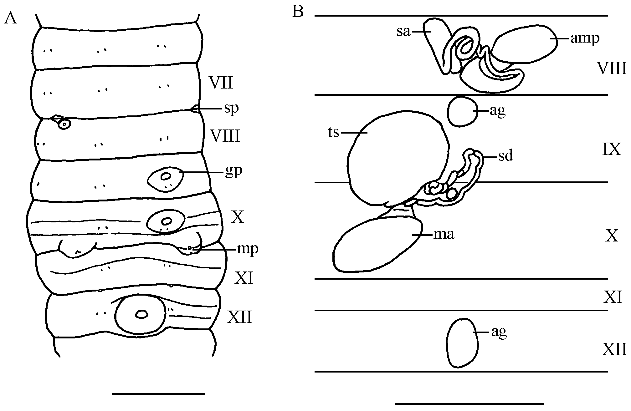
Description of MTS 87. External: Total length 46 mm, greatest diameter in clitellar region 2.85 mm, weight 0.174 g. Segments numbering 106. Prostomium prolobous. Dorsal pores absent. Setae short and closely paired, ab = cd, aa = 0.8 bc, aa = 5 ab, dd greater than 0.5 body circumference. Clitellum in X–XIII, paler, swollen. Spermathecal pores one pair in intersegmental furrow of 7/8, each pore medial to seta c, with or without a round genital papilla nearby ( Fig. 7 View FIGURE 7 A). Male pores one pair, each on an oval tumescence formed by elevated glandular skin between b and c (much nearer to b) in posterior edge of X, projecting over intersegmental furrow of 10/11 and slightly to anterior edge of XI ( Fig. 7 View FIGURE 7 A). Female pores minute, one pair on anterior most border of XII, closely adjacent to intersegmental furrow of 11/12, each pore in line with seta b. Genital papillae unpaired on IX, X and XII, each as a small, round tubercle 0.25–0.3 mm in diameter, surrounded by a large, paler, slightly elevated glandular rim 0.65–0.75 mm in diameter. Preserved specimen bluish gray on dorsum and grayish pale on ventrum.
Internal: Septa 5/6 thick, 6/7–8/9 thickened and muscular, 9/10 thin. Septum 10/11 fused with septum 11/12 dorsally. Gizzards two in XII–XIII. Hearts VI–IX. Nephridia holoic. Testis sacs one pair, large, oval, yellowish, about 1 mm in length and 0.8 mm in width, each suspended by septum 9/10. Sperm duct loosely spirally twisted, running in front of septum 9/10 around last heart, piercing the septum into segment X and entering into the anterior end of the male atrium ( Fig. 7 View FIGURE 7 B). Male atrium white, elongated oval, about 0.8 mm long and 0.6 mm wide. Ovarian chamber in XI, formed by septa 10/11 and 11/12, surrounding the esophagus in an inverted U-shape. One pair of follicular, yellowish ovisacs dorso-lateral on both sides of the digestive tract, extending posteriorly to segment XVII or XVIII. Spermathecae one pair, both ampulla and spermathecal atrium lying on posterior face of septum 7/ 8. Ampulla oval-shaped, about 0.7 mm long and 0.35 mm wide, its duct at first stout and thick, and then passing into a few loose coils and becoming slender to join the spermathecal atrium ( Fig. 7 View FIGURE 7 B). Atrium about 0.6 mm in length and about 0.2 mm in width. Accessory glands sessile, round or oval, 0.3–0.42 mm long and 0.2–0.3 mm wide, each corresponding to external genital papilla.
DNA barcodes. GenBank accession numbers KR047039 View Materials (MTS 45) and KR047040 View Materials (MTS 87).
Remarks. According to Kobayashi (1938), this species closely resembles D. japonica but differs in having larger, nipple-like male porophore and shorter, thicker spermathecal duct. In addition, Blakemore et al. (2014) indicate that D. koreana differs from D. japonica mostly in its blue color and details of the penis. However, it remains difficult to distinguish the two species due to the ambiguous morphological variation of the abovementioned characters. For example, among the three new subspecies of D. koreana published in Blakemore et al. (2014), Drawida koreana nanjiro was said to be similar to both Drawida koreana koreana Kobayashi, 1938 and D. japonica ( Michaelsen, 1892) due to its similarity to the former in its blue color and to the latter in its stubby penes, and Drawida koreana shindo was documented to comply more with D. japonica sspp. than with D. koreana sspp. as it lacks the characteristic blue coloration, has genital markings, a stubby penis and coiled spermathecal duct ( Blakemore et al. 2014). The reason for erecting these subspecies, stated by Blakemore et al. (2014), is that they are unambiguously identifiable on their DNA. In this study, both specimens MTS 45 and MTS 87 have thicker spermathecal ducts ( Table 4 View TABLE 4. A ). The former has dark blue body color and its penis structure is similar to those of above-mentioned D. koreana sspp. and thus, it should be included in D. koreana . As for the specimen MTS 87, its blue coloration is not so distinctive and its morphological characters such as the penis structure and genital markings are almost identical to those of D. japonica as described in Chen (1933). However, based on molecular analysis ( Fig. 8 View FIGURE 8 ), both specimens are conspecific to D. koreana . Apparently, morphology alone is not sufficient to distinguish D. koreana from D. japonica .
In addition to differences in body coloration and penis structure, specimen MTS 87 has higher number of genital markings than MTS 45 and two gizzards in XII–XIII while MTS 45 possesses three gizzards in XII–XIIV ( Table 4 View TABLE 4. A ). Genetically, MTS 45 is most closely related to D. koreana austri and MTS 87 to D. koreana shindo and D. koreana nanjiro ( Fig. 8 View FIGURE 8 ). There is almost no difference of the COI sequences between austri and MTS 45 and also, they are morphologically similar ( Table 4 View TABLE 4. A ). However, the spermathecal ducts of austri are quite short whereas MTS 45 has coiled spermathecal ducts. As for MTS 87, its penis structure which is nearly identical to those of D. japonica distinguishes it from other D. koreana sspp.
Drawida koreana seems to be a species with intraspecific variation. This species has been found in Korea, Japan and northern China ( Kobayashi 1938; 1940; Easton 1981). Its occurrence in Hsiju, Matsu constitutes the island as the southernmost range of this species to date.

No known copyright restrictions apply. See Agosti, D., Egloff, W., 2009. Taxonomic information exchange and copyright: the Plazi approach. BMC Research Notes 2009, 2:53 for further explanation.
|
Kingdom |
|
|
Phylum |
|
|
Class |
|
|
Order |
|
|
Family |
|
|
Genus |
Drawida koreana Kobayashi, 1938
| Shen, Huei-Ping, Chang, Chih-Han & Chih, Wen-Jay 2015 |
Drawida
| Easton 1981: 39 |
| Kobayashi 1940: 268 |
Drawida koreana
| Kobayashi 1938: 102 |