
Bosmina (Liederobosmina) korineki, Garibian & Juračka & Kotov, 2021
|
publication ID |
https://doi.org/ 10.11646/zootaxa.4990.2.4 |
|
publication LSID |
lsid:zoobank.org:pub:4E6FE456-A7EA-4922-AF0C-D7EF4C6C6BAE |
|
DOI |
https://doi.org/10.5281/zenodo.5096971 |
|
persistent identifier |
https://treatment.plazi.org/id/DC1EB339-FF93-FFEA-0AC5-F9169E04B0B5 |
|
treatment provided by |
Plazi |
|
scientific name |
Bosmina (Liederobosmina) korineki |
| status |
sp. nov. |
Bosmina (Liederobosmina) korineki sp.nov.
( Figures 1–4 View FIGURE 1 View FIGURE 2 View FIGURE 3 View FIGURE 4 )
Etymology. The taxon is named after Professor Vladimír Kořínek (1934–2019) (see Fott 2019), who detected this taxon in the Andean samples and send the samples to AAK for their detailed study.
Type locality. Laguna Chisaca (N 4.28°, W 74.21°; 3709 m.a.s.l.), Distrito Capital de Bogotá, Colombia GoogleMaps .
Type material. Holotype. A parthenogenetic female in 96% alcohol, MGU Ml 240 in the Collection of Moscow State University, Moscow, Russia. Label of holotype: “ Bosmina korineki sp.nov., 1 parth. ♀ from Laguna Chisaca, Colombia, HOLOTYPE ”.
Paratypes. 4 parthenogenetic females, MGU Ml 241. 4 parthenogenetic females, AAK 2020-017 in the personal collection of A.A. Kotov, Moscow, Russia .
The type series was collected in 10.01.1990 by S. Gaviria.
Other material studied. 16 females from Embalse de Neusa (N 5.16°, W 73.95°; 2967 m.a.s.l.), Cundimarca Province, Colombia GoogleMaps , collected in 15.03.1999 by P. Alvarez in the personal collection of V. Kořínek , sent to the collection of the Natural History Museum, London, United Kingdom .
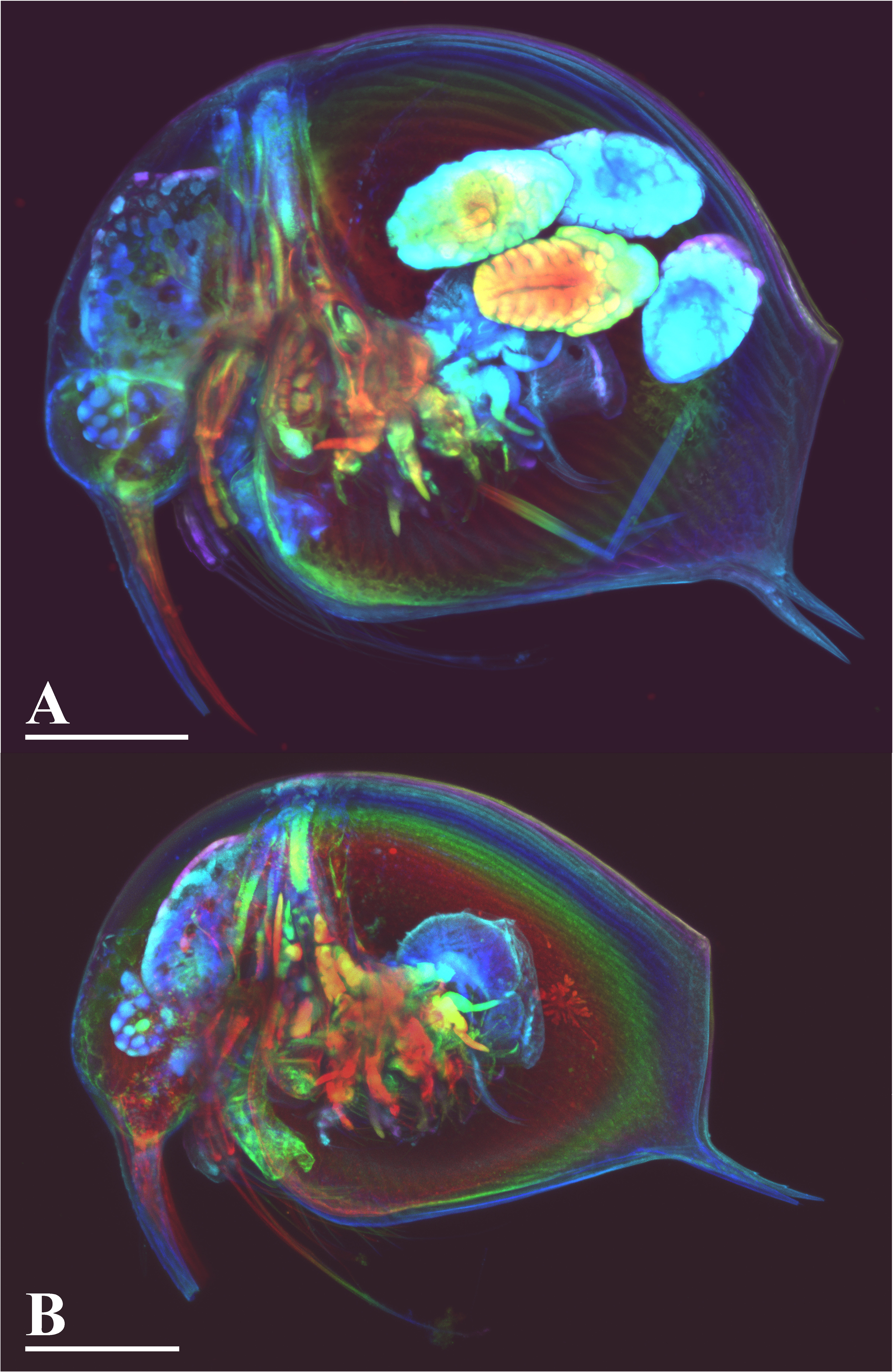
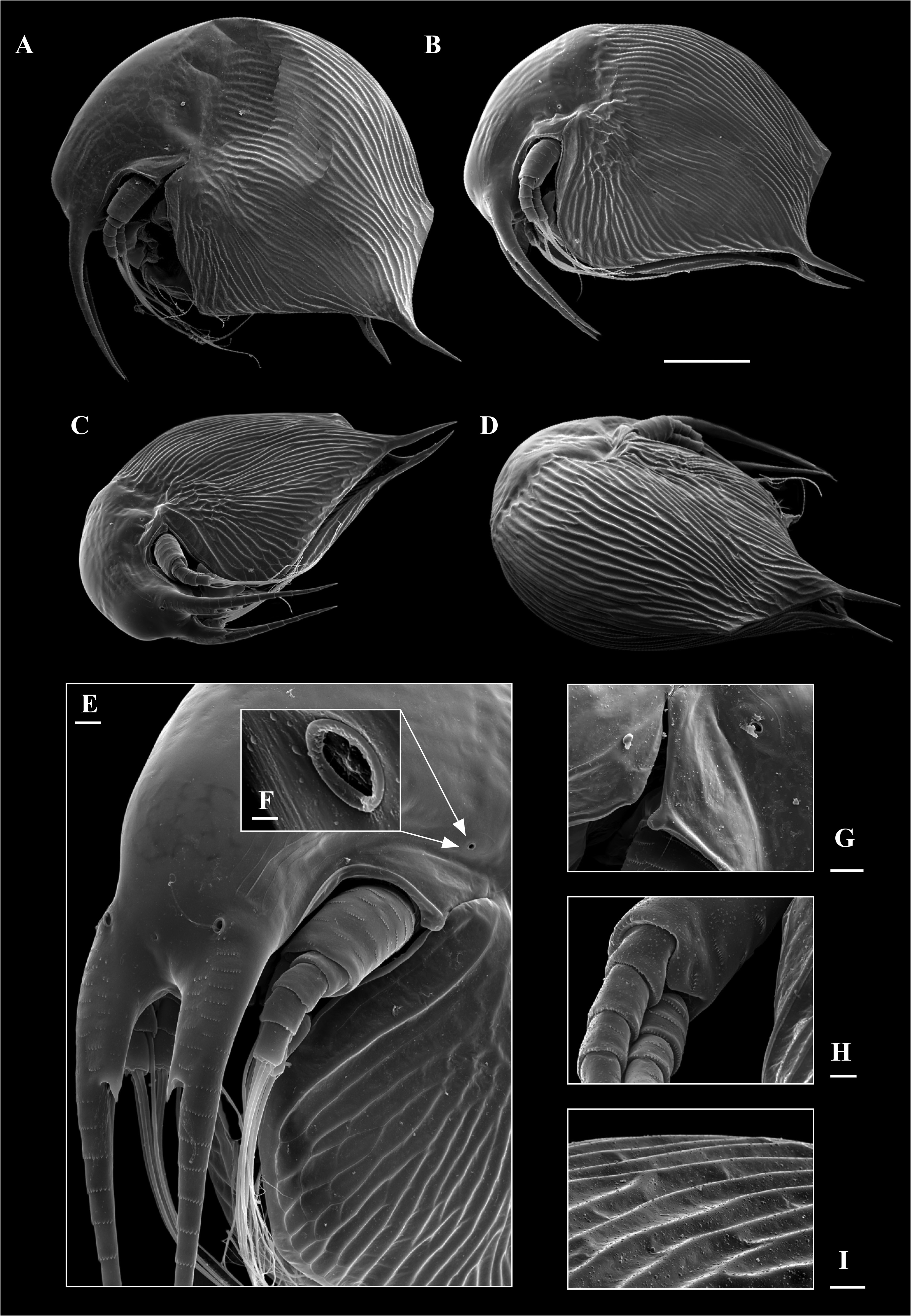
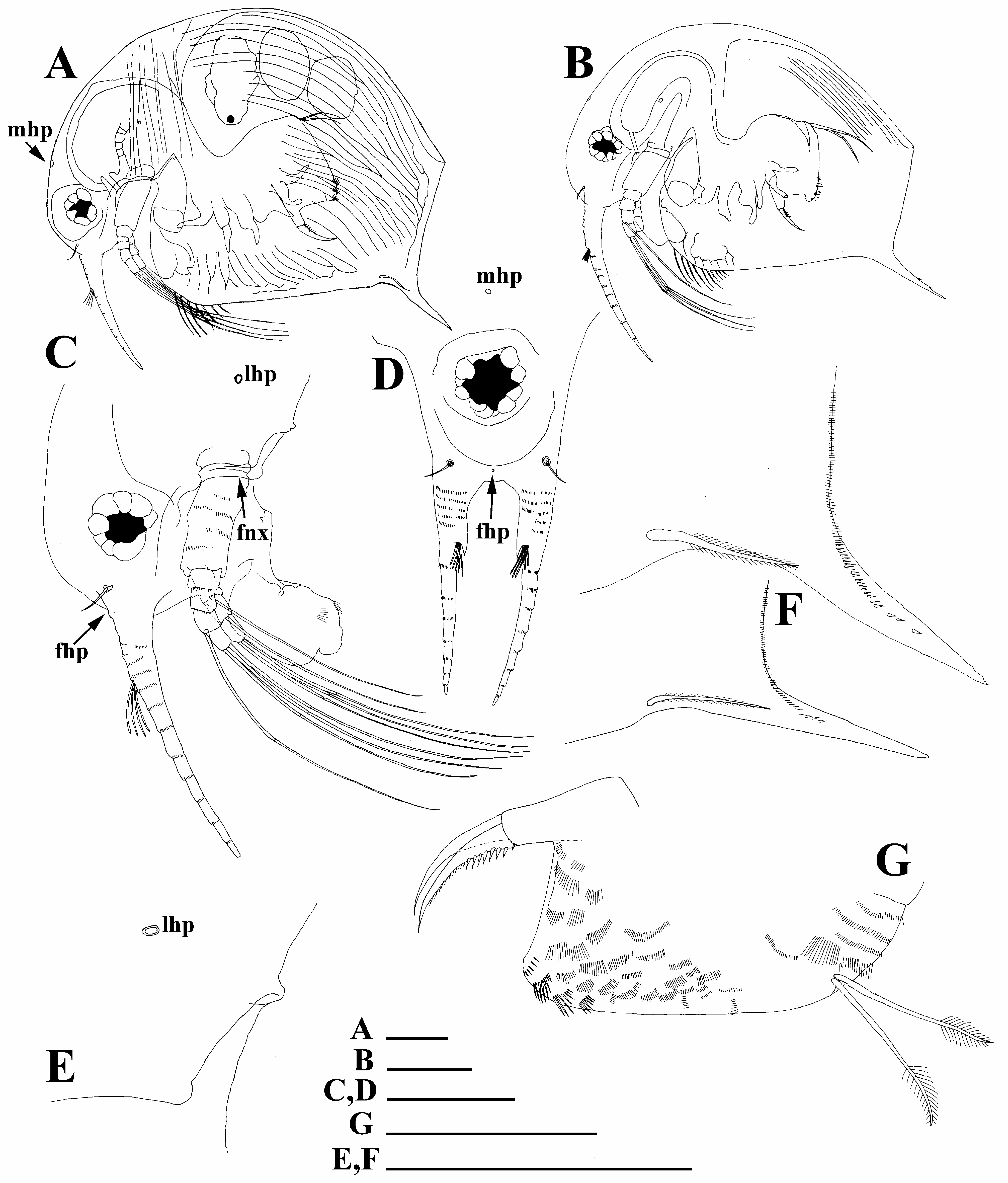
Description. Parthenogenetic female. Body short and wide in lateral view in adults ( Figs 1A View FIGURE 1 , 2A–B View FIGURE 2 , 3A View FIGURE 3 ) and more elongated in juveniles ( Figs 1B View FIGURE 1 , 3B View FIGURE 3 ); dorsal margin regularly curved from distalmost extremity to posterodorsal angle; posterior margin straight, its height about half of body height; ventral margin almost straight, with a shallow depression anterior to mucro. Sculpture as a strong striation on head and on valves ( Fig. 2A–E, I View FIGURE 2 ), but not on head shield. Head without ocular dome and without a pre-ocular depression; rostrum very short, rounded. Frontal head pore small, located slightly dorsally to the level of antennae I ( Fig. 3C–D View FIGURE 3 : fhp). Median head pore minute ( Fig. 3A, D View FIGURE 3 : mhp), located posteriorly to ocular capsule. Fornix well-developed, covering coxal part of antenna II. Lateral head pore small ( Figs 2E–G View FIGURE 2 . 3C, E View FIGURE 3 : lhp), with a raised ring-like margin, located at a great distance from ventral margin of head shield (fornix, Fig. 3C View FIGURE 3 : fnx), dorsally to the level of mandibular articulation. Compound eye of moderate size. Labrum as a fleshy appendage lacking significant projections, distal labral plate small. Anterior portion of ventral valve margin with a series of stout setae. Seta kurzi with small setulae, located on internal side of valve anterior to abovementioned depression near mucro, which is strong and long even in large adults; no incisions on ventral side of mucro ( Fig. 3F View FIGURE 3 ), while some incisions present on dorsal side of mucro in some juveniles ( Fig. 3B View FIGURE 3 ). On inner side of mucro, there are several relatively strong denticles, as a continuation of a series of setulae at posterior valve margin.
Thorax relatively long, with 6 limb pairs, abdomen short, with transverse rows of setulae ( Fig. 3G View FIGURE 3 ). Postabdomen strongly compressed laterally, with width approximately equal along all its length, with ventral (functionally dorsal!) margin straight. Preanal margin with groups of setulae; sides of postabdomen supplied with series of finer setulae. Distal (anal) margin nearly directly truncated. Postanal portion as a cylindrical projection bearing paired postabdominal claws. Each claw regularly bent, with two pectens on concave (dorsal) margin, distal pecten consists of fine setulae, while proximal pecten consists of 7–9 rather strong, sparsely located teeth. Postabdominal seta shorter than preanal margin; its distal segment about two times shorter than proximal one, supplied with fine, long setulae.
Antenna I fused with rostrum; its body relatively short; its length from tip to tip of rostrum about 0.3–0.4 body lengths ( Fig. 3A–B View FIGURE 3 ). Antennular (frontal) sensory seta located on head surface. Free part of antenna I consists of a pre-aesthetasc portion, fused with head, and post-aesthetasc portion, presence of which is a unique synapomorphy of Bosmina ( Kotov et al., 2009) . Nine aesthetascs delicate, slightly differing in size. Post-aesthetasc portion directed ventrally and somewhat posteriorly, almost straight. Both portions supplied with crossing series of fine denticles. Antenna II typical for the genus ( Figs 2H View FIGURE 2 , 3C View FIGURE 3 ).
Six pairs of thoracic limbs with morphology indistinguishable from that in other species ( Kotov, 1995).
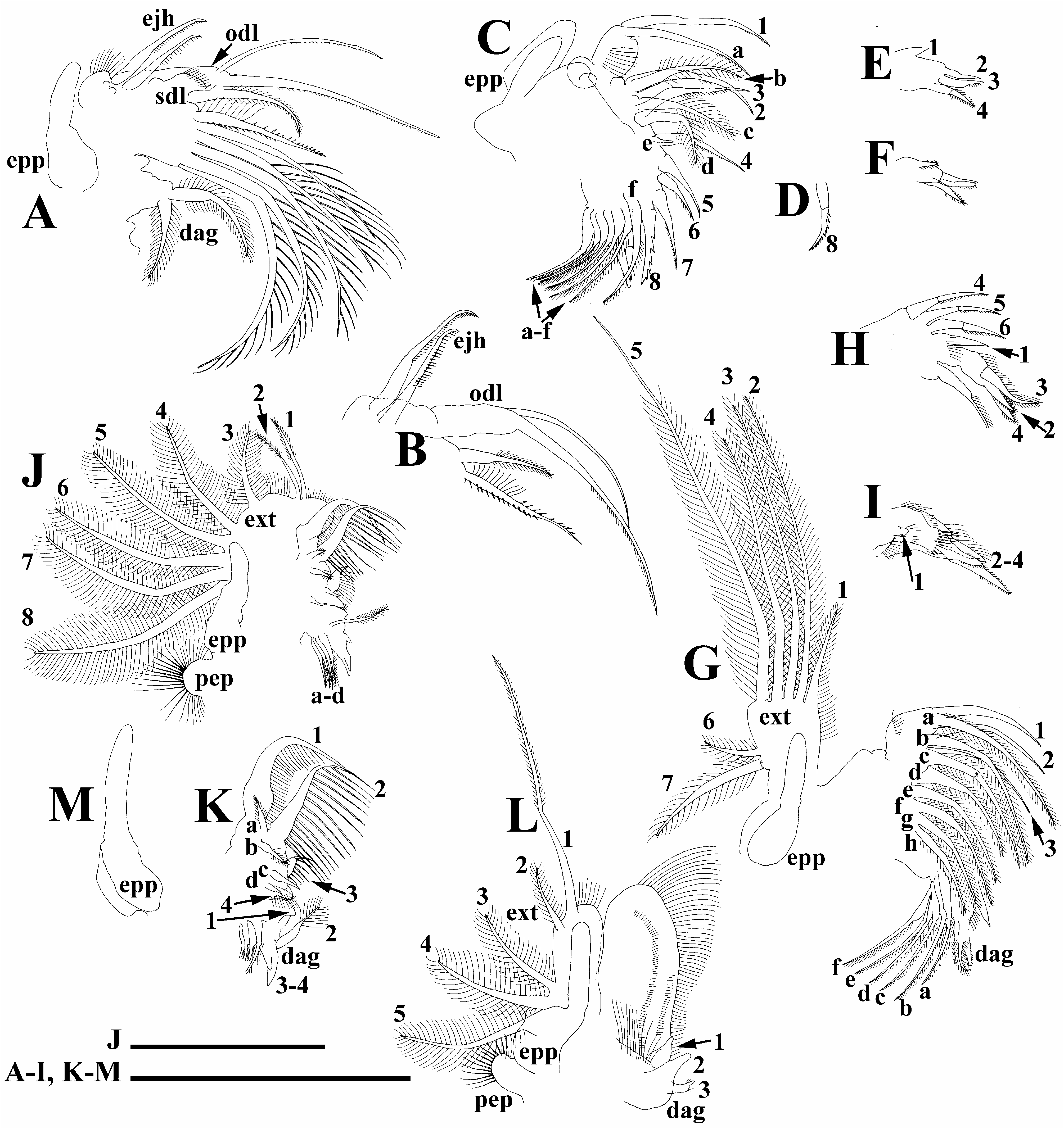
Limb I with epipodite bearing a finger-like projection ( Fig. 4A View FIGURE 4 : epp). Outer distal lobe ( Fig. 4A–B View FIGURE 4 : odl) cylindrical, bearing a long distal seta and a long lateral seta. Subdistal lobe with a single seta of unclear homology; limb corm with six setae of unclear homology and a pair of ejector hooks ( Fig. 4A–B View FIGURE 4 : ejh). Distal armature of gnathobase with two setulated setae ( Fig. 4A View FIGURE 4 : dag).
Limb II with epipodite bearing a long finger-like projection ( Fig. 4C View FIGURE 4 : epp). Exopodite fully reduced. Limb corm with an inner-distal lobe bearing a single anterior (1) and single posterior (a) setae; the rest of inner-distal limb portion with seven anterior (2–8) and five posterior (b–f) setae; seta 8 armed by strong denticles varying in size among specimens from same population ( Fig. 4C–D View FIGURE 4 ). Gnathobase with distal armature of four setae ( Fig. 4E–F View FIGURE 4 : 1–4); seta 1 represents a sensillum. Filter plate with six setae ( Fig. 4C View FIGURE 4 : a–f).
Limb III with epipodite having a finger-like projection ( Fig. 4G View FIGURE 4 : epp). Exopodite subrectangular, with five distal (1–5) and two lateral (6–7) setulated setae. Distal endite (in terms of Kotov 2013) with three anterior setae ( Fig. 4G View FIGURE 4 : 1–3); proximal endite with three setae ( Fig. 4H View FIGURE 4 : 4–6). Eight setae (a–h) on posterior limb face. Distal armature of gnathobase with four elements ( Fig. 4H–I View FIGURE 4 : 1–4), including a sensillum (1). Filter plates with six setae (a–f).
Limb IV with ovoid densely setulated preepipodite ( Fig. 4J View FIGURE 4 : pep) and epipodite ( Fig. 4J View FIGURE 4 : epp) supplied by a finger-like projection. Exopodite ( Figure 4J View FIGURE 4 : ext) rounded, bearing two distal (1–2) and six lateral (3–8) setae. Inner distal portion of limb IV with four anterior setae strongly decreasing in size proximally ( Figure 4K View FIGURE 4 : 1–4) and four small posterior setae (a–d). Distal armature of gnathobase with four setae ( Fig. 4K View FIGURE 4 : 1–4). Filter plate with four setae (a–d).
Limb V with densely setulated preepipodite ( Fig. 4L View FIGURE 4 : pep) and ovoid epipodite (epp) supplied by a long fingerlike projection. Exopodite with two distal setae (1–2) and three lateral setae (3–5). Inner limb portion as elongated, flat lobe, with setulated margin. Distal armature of gnathobase with three setae ( Fig. 4L View FIGURE 4 : 1–3).
Limb VI represented by epipodite only ( Fig. 4M View FIGURE 4 : epp).
Ephippial female, male. Unknown.
Size. Total length (without mucro) 0.45–0.62 mm.
Differential diagnosis. Our new species belongs to the subgenus B. (Liederobosmina) having: (1) lateral head pore located dorsally to the level of mandibular articulation; (2) incisions on the dorsal side of mucro in juveniles (see Lieder 1983b; Kotov et al. 2009). This taxon is unique among all the taxa of Bosmina (Liederobosmina) having heavily striated valves. Paggi (1979) recognized three species from this subgenus in South America. B. (L.) korineki sp. nov. has no elongated flattened rostrum as B. (L.) huaronensis Delachaux , and has no pre-ocular depression as B. chilense Daday. To date, we can only say that it differs from B. hagmanni Stingelin only in striated valves in the females as the males are unknown, but they possess most diagnostic characters in the bosminids ( Kotov et al. 2009).
Distribution. To date the taxon is known only from two large, deep high mountain lakes in Colombia. Embalse de Neusa is an artificial reservoir with surface of over 955 ha, maximum depth of 50 m and mean depth of 10 m. Secchi depth ranges from 3.6 m to 1.5 m; the lake is considered as oligo mesotrophic ( Carrillo et al. 2006).
We did not find in previous literature any descriptions which could be attributed to B. (L.) korineki sp.nov., although different bosminids were noted in many papers on the Neotropical fauna. Most probably, this taxon has a narrow distribution area in the Andean high mountains.
| V |
Royal British Columbia Museum - Herbarium |
No known copyright restrictions apply. See Agosti, D., Egloff, W., 2009. Taxonomic information exchange and copyright: the Plazi approach. BMC Research Notes 2009, 2:53 for further explanation.
|
Kingdom |
|
|
Phylum |
|
|
Class |
|
|
Order |
|
|
SubOrder |
Radopoda |
|
Family |
|
|
Genus |
|
|
SubGenus |
Bosmina |