
Philmontoides, Ingrisch, 2022
|
publication ID |
https://doi.org/ 10.11646/zootaxa.5182.2.1 |
|
publication LSID |
lsid:zoobank.org:pub:8920DE84-2BE6-4A68-A7F7-AC987F1F894E |
|
DOI |
https://doi.org/10.5281/zenodo.7053839 |
|
persistent identifier |
https://treatment.plazi.org/id/DB181868-FF83-FFD0-FF67-D2E72AE4F435 |
|
treatment provided by |
Plazi |
|
scientific name |
Philmontoides |
| status |
gen. nov. |
Philmontoides gen. nov.
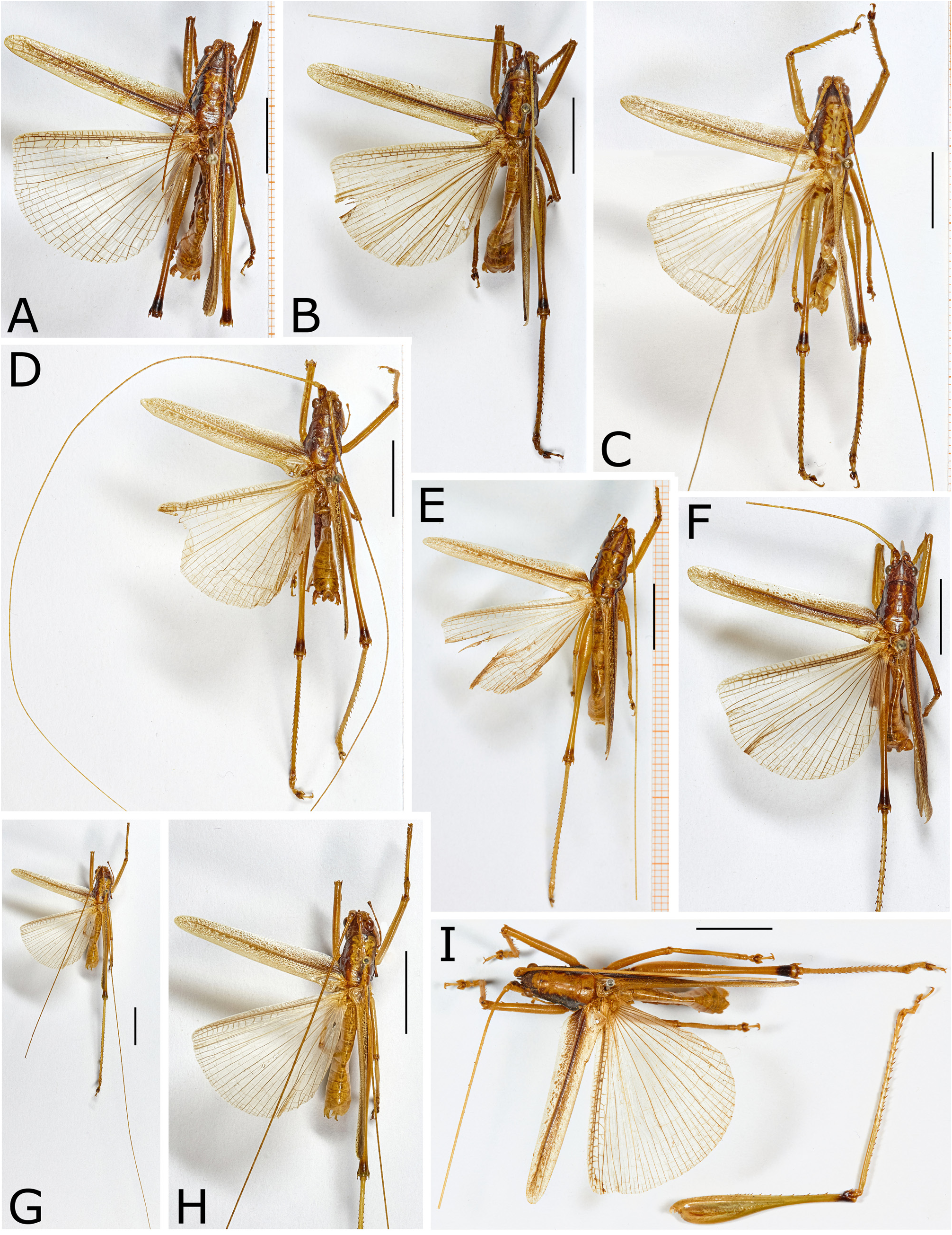
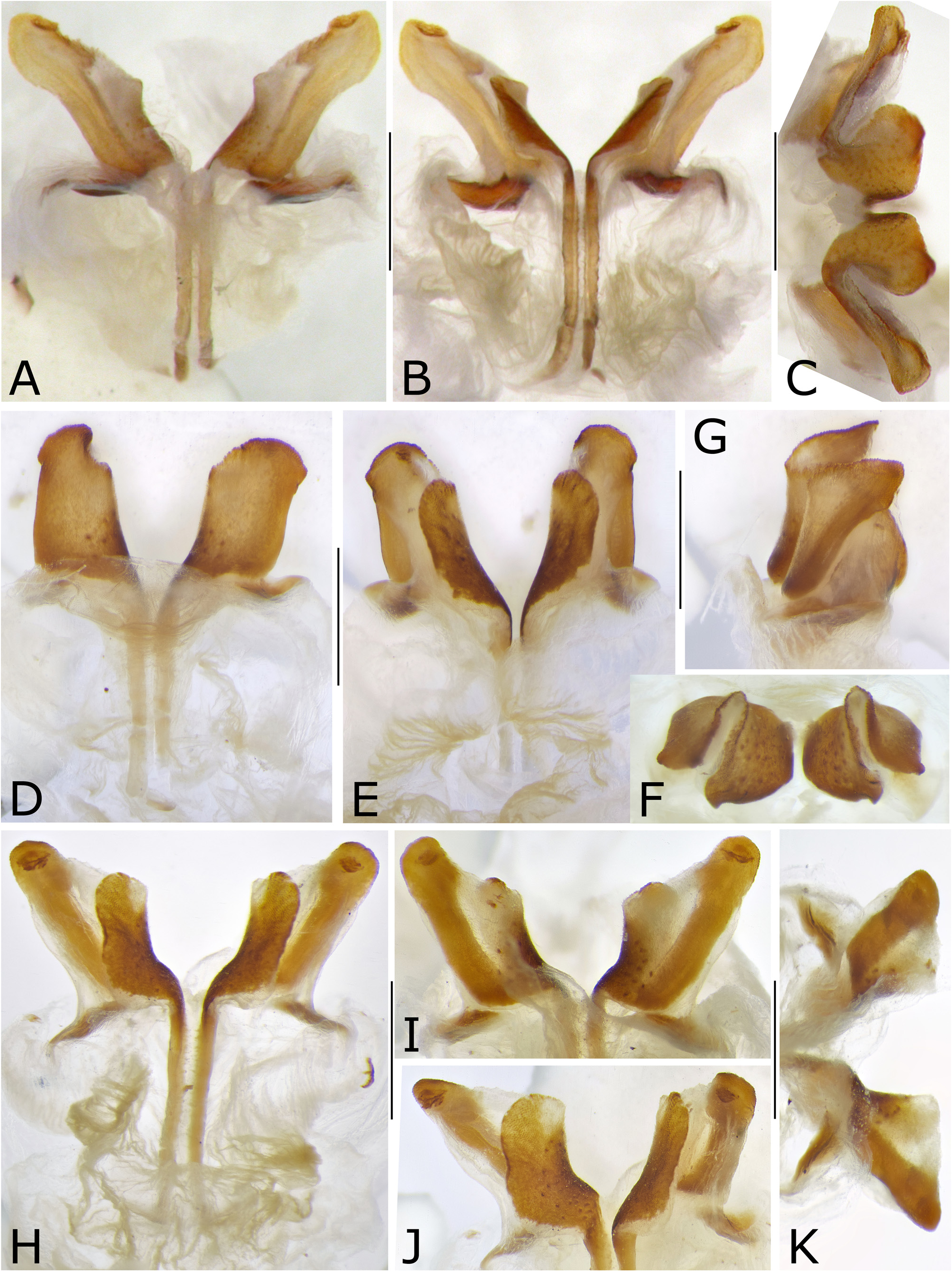
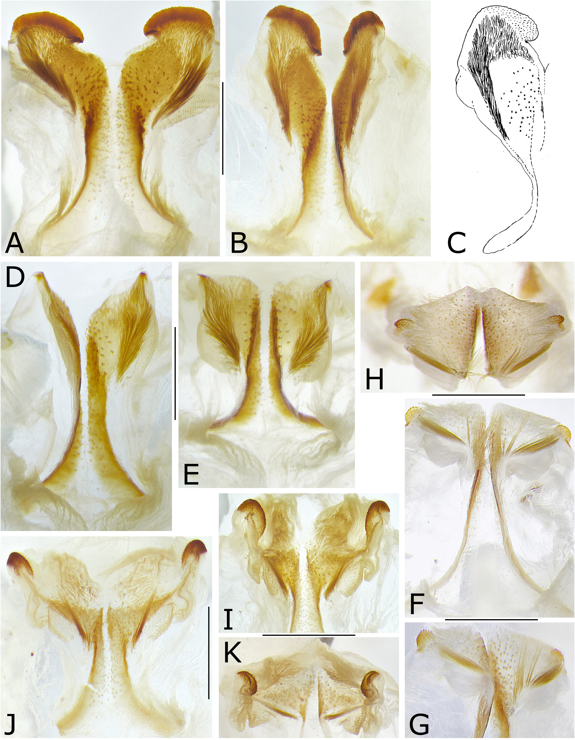
Figures 9–17 View FIGURE 9 View FIGURE 10 View FIGURE 11 View FIGURE 12 View FIGURE 13 View FIGURE 14 View FIGURE 15 View FIGURE 16 View FIGURE 17 , map 2 View MAP 2
Type species: Lobaspis hageni Dohrn, 1905 here designated
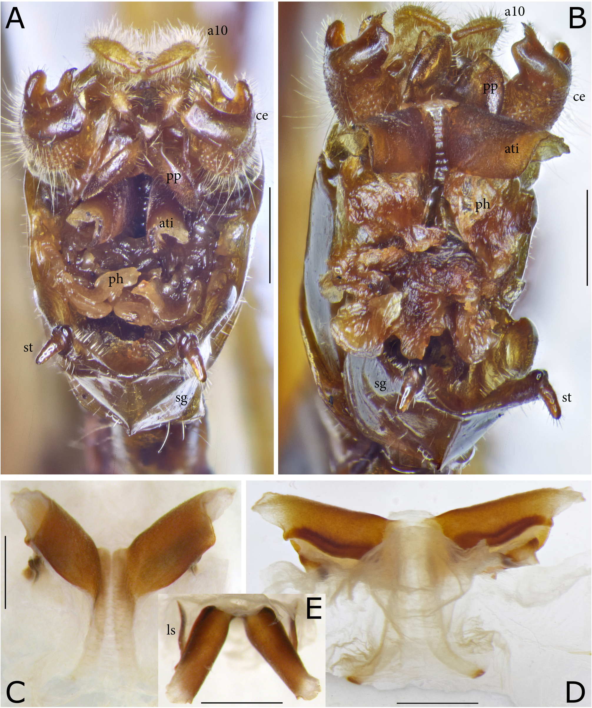
Generic diagnosis and discussion. The new genus is similar to Philmontis Willemse, 1966 in general shape and coloration. The dark band from lateral lobes of pronotum to and along midline of tegmen is often more conspicuous than in the latter genus. The lateral lobes of pronotum are narrow as in Philmontis but less strongly prolonged behind, leaving the male stridulatory apparatus largely free. The prosternal spines are long instead of short, minute or absent in Philmontis ; the hind femur is provided with spines on both ventral margins instead of only at anterior margin and the hind knees are provided with two spines on both sides instead of only one spine. The male titillators of Philmontoides gen. nov. vary in shape between species but are never provided with a small sclerotized disc at end which is typical for the genus Philmontis . Females of Philmontoides can easily be separated from those of the latter genus by the shape of the dorsal ovipositor valves that are dorso-ventrally expanded around mid-length while in Philmontis they are regularly narrowed toward end.
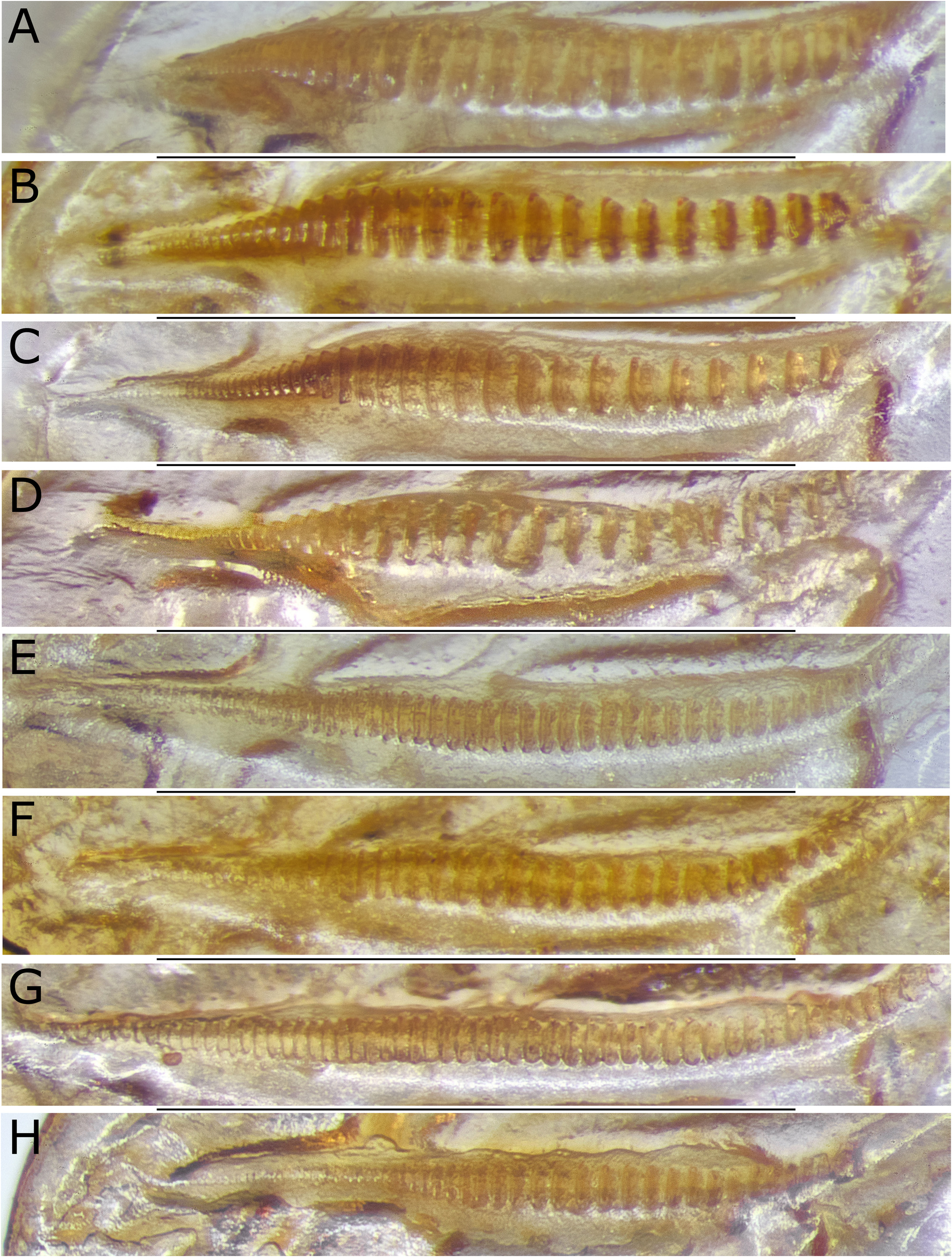
All species of Philmontoides gen. nov. are of similar general appearance, coloration, length and width of tegmen but differ in the shapes of the male tenth abdominal tergite, cerci, and titillators as well as in the shape of the female subgenital plate. Regarding the male stridulatory file on the underside of the male left tegmen, there are four species, P. hageni ( Dohrn, 1905) , P. lobatus ( Naskrecki & Rentz, 2010) , P. globosus sp. nov., and P. disjunctus sp. nov. that have large and stout teeth in the basal two thirds of the file, while small to minute teeth in the apical area. Males of the other four species studied have smaller and narrower teeth but differ in the length of the area useable for stridulation. Regarding the male titillators of the phallus ( Figs 13–15 View FIGURE 13 View FIGURE 14 View FIGURE 15 ), there is a gradual changeover from shapes with simple, sub-flat but widened apical areas (e.g., in P. affinis (Willlemse, 1966) or P. commodus sp. nov.) similar to those found in some other genera of Agraeciini , via species in which the apical area is bent in an about rectangle against the basal stem (e.g., P. striatus sp. nov.), to species, in which this bent apical area becomes vaulted (e.g., P. wau sp. nov.) and in a further step strengthened to form a strong tunnel-shaped structure (e.g., P. hageni Dohrn ). That structure became even further modified in three other species. In two species, both sides of the vaulted apical area differ in size and shape ( P. lobatus ( Naskrecki & Rentz, 2010) and P. globosus sp. nov.), while in one species ( P. disjunctus sp. nov.), the apical area is completely divided into two separate branches, connected only at very base where they arise from the basal stem of the titillator.
The general habitus of the genus Philmontoides gen. nov. is also similar to that of the genus Habetia Kirby 1906 . Shape and length of pronotum with narrow lateral lobes and the apical area covering only the base of the stridulatory area in males are similar in both genera. Coloration of the pronotum differs however between both genera: the lateral lobes are black in Philmontoides while of light general color in Habetia . Also, the spination of legs is similar in both genera especially on the hind femur having spines on both ventral margins and the hind knee lobes are provided with two spines on both sides. The fore wings of Philmontoides are narrow throughout and provided with a dark medial band while in Habetia they are of uniform general color, often wider in more than basal half and have the apical area narrow and parallel-sided. Differences between both genera are also found in the male tenth abdominal tergite that is usually provided with a pair of projections from central apical area in Philmontoides while simply excised at end in Habetia ; the male cerci are divided into apical lobes or are provided with two projections from various areas in Philmontoides while with an apical internal projection and often with a dorsal apical crest in Habetia . The basic shape of the male titillators consists mainly of soft structures in Philmontoides but is based on a basic sclerite in Habetia ; but these structures are strongly modified between species in both genera. Females differ strikingly between both genera by the shape of the ovipositor which is curved and has the dorsal valves widened in Philmontoides but strongly prolonged and except at base substraight in Habetia .
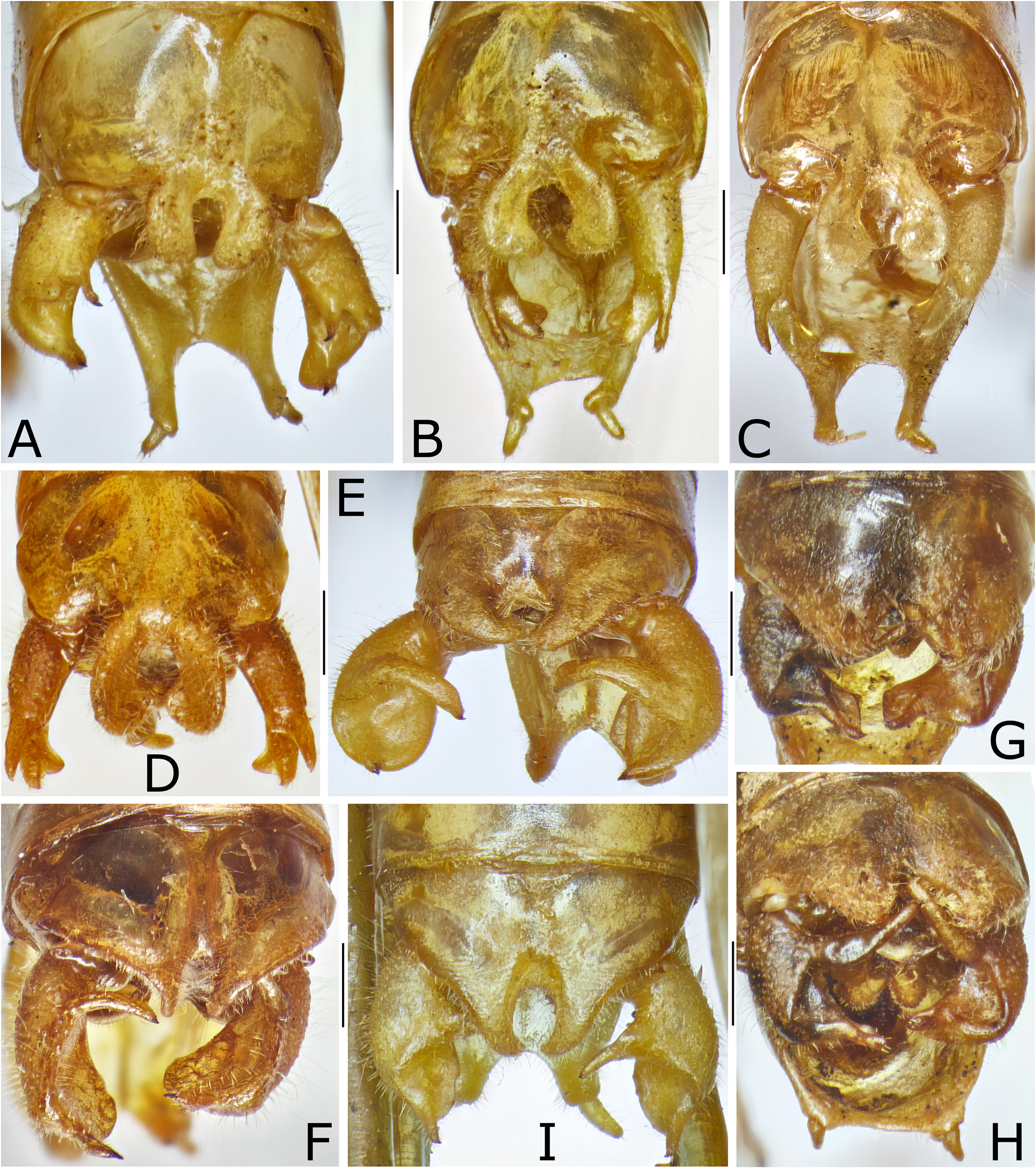
Description. Elongate species with narrow tegmen reaching about to end of hind femur, little shorter or longer. Pronotum in males not or only little prolonged behind, leaving the stridulatory area largely uncovered, ventroposterior angle of lateral lobes distinctly behind middle of pronotum length; in females lateral lobes not prolonged; apical margin of disc slightly convex or subtruncate in both sexes; sulci and furrows of pronotum distinct, interrupted in middle; lateral lobes not very deep, ventral margin slightly concave, at end forming a rounded angle with lateral margin of posterior area that is projecting laterad and clearly visible from above; auditory swelling small. Tegmen rather narrow, reaching or slightly surpassing hind knees, little narrowing from basal area to rounded or subtruncate end. Prosternal spines of medium size or long, but shorter than coxa. Postfemur with spines on both ventral margins, on ventro-internal margin usually with small spinules in basal half and a few large spines before apex as in genus Habetia . Hind knee lobes always bi-spinose.
Male. Stridulatory file often with large and spaced teeth from near base to behind mid-length, narrow and dense teeth at apical end; in other species differences in tooth size between areas less pronounced but with largest teeth also before or around mid-length, while in apical area with small or indistinct teeth; in few species (e.g., P. commodus sp. nov.) all teeth distinctly visible from base to tip despite of differences in size. Tenth abdominal tergite little globular and apex with two projections in middle. Subgenital plate with variable end; styli small. Titillators variable between species but always with a simple titillator stem that is extended at end into a large, complex, apical structure.
Female. Cerci rather short, conical, slightly curved, apex pointing. Subgenital plate with a medial carina, often compressed; with baso-lateral angles projecting and modified, but not as strongly as in Philmontis , sometimes with separate lateral sclerites. Ovipositor moderately curved, with dorsal valves widened and highest little behind midlength, apex acute.
Coloration. Flagellum of antennae white or yellowish. Tegmen along anterior and posterior margins of light color or whitish, partly with dark spots, along central area with a broad, dark brown or black longitudinal band with dark brown or blackish main veins but often light veinlets even within the dark band. Hind femur with a black pregenicular ring. Ovipositor rufous; dorsal valves yellowish along ventral margin.
Tentative key to the known species of the genus Philmontoides gen. nov.
Key to known species—Males
1. Cerci elongate, only little curved, in apical area dividing into an elongate dorsal branch with a spine at tip and a stout ventral branch widening at end and there with stiffened rims connected by a thin septum with concave apical margin; the dorsal, stiffened rim with a spinule at tip ( Figs 10 E–J View FIGURE 10 , 11B–D View FIGURE 11 ). Titillators, behind thin and long basal areas, strongly widened and compressed, with the thin surface curved around for nearly 180° thus that they almost touching each other when seen in apical view; lateral sclerites sinuate and rather large ( Fig. 14 View FIGURE 14 )........................................................2.
- Cerci dorso-ventrally widened, with concave internal surface at least in apical area, and with projecting spines and lobes ( Figs 10A–D, K–M View FIGURE 10 , 11A, E–I View FIGURE 11 )...............................................................................4.
2. Titillators, behind thin and long basal areas, divided into two lobes each that are connected only at very base; lateral sclerites simple ( Figs 14H–K View FIGURE 14 ).................................................................. P. disjunctus sp. nov.
- Titillators, behind thin and long basal areas, with widened apical area curved in a nearly 180°-angle around the longitudinal axis but not divided into separate branches.....................................................................3.
3. Titillators with main branches elongate, with re-curved area markedly shorter than elongate main area; in apical view, main area long and narrow, recurved area oval ( Figs 14A–C View FIGURE 14 ).............................. P. lobatus ( Naskrecki & Rentz, 2010) View in CoL
– Titillators with main branches wider but shorter, with re-curved area only little shorter than main area; in apical view titillators appear like rounded globes furrowed in middle ( Figs 14D–G View FIGURE 14 )................................... P. globosus sp. nov.
4. Tenth abdominal tergite terminating into a pair of wide, about oval lobes forming together nearly a circle with open end ( Fig. 11A View FIGURE 11 ). Cerci with convex external and concave internal surface; dorsal-internal surface terminating into a short, curved and narrow projection with spine at tip, dorsal-external surface terminating into a short, rounded lobe; lateral surface prolonged and compressed with substraight to slightly convex dorsal and faintly concave ventral margins, end of lateral surface truncate with rounded ventral angle and from dorsal angle on internal surface with a narrow, curved projection with a spine at tip ( Figs 10K–L View FIGURE 10 , 11A View FIGURE 11 ). Titillators behind thin and long basal areas with large, vaulted projections ( Figs 13C–E View FIGURE 13 )................................................................................................... P. hageni ( Dohrn, 1905)
- Tenth abdominal tergite of different shape. Cerci lobular with convex external and concave internal surface, carrying a long spined projection on dorsal and a short spined projection on apical margin. Titillators of different shape ( Fig. 15 View FIGURE 15 ).........5.
5. Tenth abdominal tergite in middle of apical margin divided into a pair of short obtuse lobes, not forming a distinct projection. Cerci with dorsal projection very long and upcurved ( Figs 10B View FIGURE 10 , 11G–H View FIGURE 11 )............................ P. striatus sp. nov.
- Tenth abdominal tergite of different shape, cerci with dorsal projection down-curved................................6.
6. Tenth abdominal tergite from hind margin with a pair of short and straight, styliform projections, margin angularly excised in between. Basal projection of cercus strongly curved; apical projection long ( Figs 10A View FIGURE 10 , 11F View FIGURE 11 )...... P. affinis ( Willemse, 1966) View in CoL
- Tenth abdominal tergite terminating into a pair of long, curved projections........................................7.
7. Apical projections of tenth abdominal tergite dorso-ventrally compressed, curved mediad, narrowing toward tip and overlapping each other for a short distance at end; dorsal projection of cercus long, curved, and little compressed ( Figs 10C, 10M View FIGURE 10 , 11E View FIGURE 11 ). P. commodus sp. nov.
- Apical projections of tenth abdominal tergite rather stout, curved ventrad and little approaching each other; dorsal projection of cercus rather short and thin ( Figs 10D View FIGURE 10 , 11I View FIGURE 11 )...................................................... P. wau sp. nov.
Key to known species—Females
1. Subgenital plate with stout medial carina and on both sides in subbasal area with a wide transverse groove with ribbed bottom ( Figs 17I–J View FIGURE 17 )............................................................................ P. striatus sp. nov.
- Subgenital plate of different shape....................................................................... 2.
2. Subgenital plate in basal area with a pair of duplicate plates interrupted along midline by a membranous zone and the medial carina ( Figs 17G–H View FIGURE 17 ).................................................................... P. geminus sp. nov.
- Subgenital plate of different shape........................................................................3.
3. Subgenital plate in about basal half with a pair of auricular expansions that are from dorso-proximal side extended into conical basal extensions ( Figs 17K–L View FIGURE 17 )....................................................... P. affinis ( Willemse, 1966) View in CoL
- Subgenital plate of different shape........................................................................4.
4. Subgenital plate with moderately converging lateral margin (in specimens that had been collected when still soft they may appear parallel-sided), with a pair of grooves in apical area; very basal area of plate elevated and laterally extended ( Figs 17O–P View FIGURE 17 ).................................................................................. P. wau sp. nov.
- Subgenital plate with a medial carina; grooves if present in more basal area.......................................5.
5. Subgenital plate with a pair of grooves in basal half and with a pair of elongate basal, lateral projections, separated by a gap from main area of the plate..............................................................................6.
- Subgenital plate without grooves; basal, lateral projections rounded or oval, not separated by a gap from main area of the plate.............................................................................................. 7.
6. Subgenital plate little widening toward base, with a strong medial carina and with semi-globular lateral projections from basal lateral margins ( Fig. 17C–D View FIGURE 17 ); some specimens that were probably not fully hardened when collected with a pair of groves and the lateral projections prolonged ( Figs 17E–F View FIGURE 17 )................................. .. P. lobatus ( Naskrecki & Rentz, 2010) View in CoL
- Subgenital plate markedly widening toward base and with a pair of groves in subbasal area ( Figs 17M–N View FIGURE 17 )................................................................................................... P. commodus sp. nov.
7. Sclerotized basal, lateral projections arising from very base of the subgenital plate and are moveable against the plate ( Figs 17A–B View FIGURE 17 ).......................................................................... P. hageni ( Dohrn, 1905)
- Sclerotized basal, lateral projections arising from the basal lateral margin of the subgenital plate and are fixed ( Figs 16K View FIGURE 16 , 17C–D View FIGURE 17 ).................................................................................................. P. globosus sp. nov. and P. lobatus ( Naskrecki & Rentz, 2010) View in CoL fully hardened specimens at time of collecting cannot be reliably separated without corresponding males.


No known copyright restrictions apply. See Agosti, D., Egloff, W., 2009. Taxonomic information exchange and copyright: the Plazi approach. BMC Research Notes 2009, 2:53 for further explanation.
|
Kingdom |
|
|
Phylum |
|
|
Class |
|
|
Order |
|
|
Family |
|
|
SubFamily |
Conocephalinae |
|
Tribe |
Agraeciini |