
Blepharisma steini, Kahl, 1932
|
publication ID |
https://doi.org/ 10.1111/zoj.12369 |
|
DOI |
https://doi.org/10.5281/zenodo.10543810 |
|
persistent identifier |
https://treatment.plazi.org/id/DA7BBB25-1F2E-A130-FF1F-00444DA4FD36 |
|
treatment provided by |
Marcus |
|
scientific name |
Blepharisma steini |
| status |
|
BLEPHARISMA STEINI View in CoL FORMA PENARDI KAHL, 1932
Diagnosis Body about 80–180 × 45–55 μm in vivo; cells colourless to dark brownish; peristome extending to mid-body; 36–63 adoral membranelles; 24–34 somatic kineties; single macronucleus; three to five micronuclei nodules; granules pale pink to colourless; habitat freshwater or moss.
Type locality A freshwater pond in Baihuayuan Garden (36°4′N, 120°20′E), Qingdao , China GoogleMaps .
Type material A protargol slide containing the holotype specimen marked with an ink circle is deposited in the Laboratory of Protozoology , Ocean University of China ( OUC) , China (slide number YY2013102901 ) . A paratype slide is deposited in the Natural History Museum , London, UK (registration number NHMUK 2015.4.23.1) .
Etymology The species name penardi is named in honour of Dr Eugene Penard who first described this organism.
Gene sequence The SSU rDNA sequence, derived from a single cell isolated from the same sample as the holotype, is deposited in GenBank (accession number KR815913 View Materials ) .
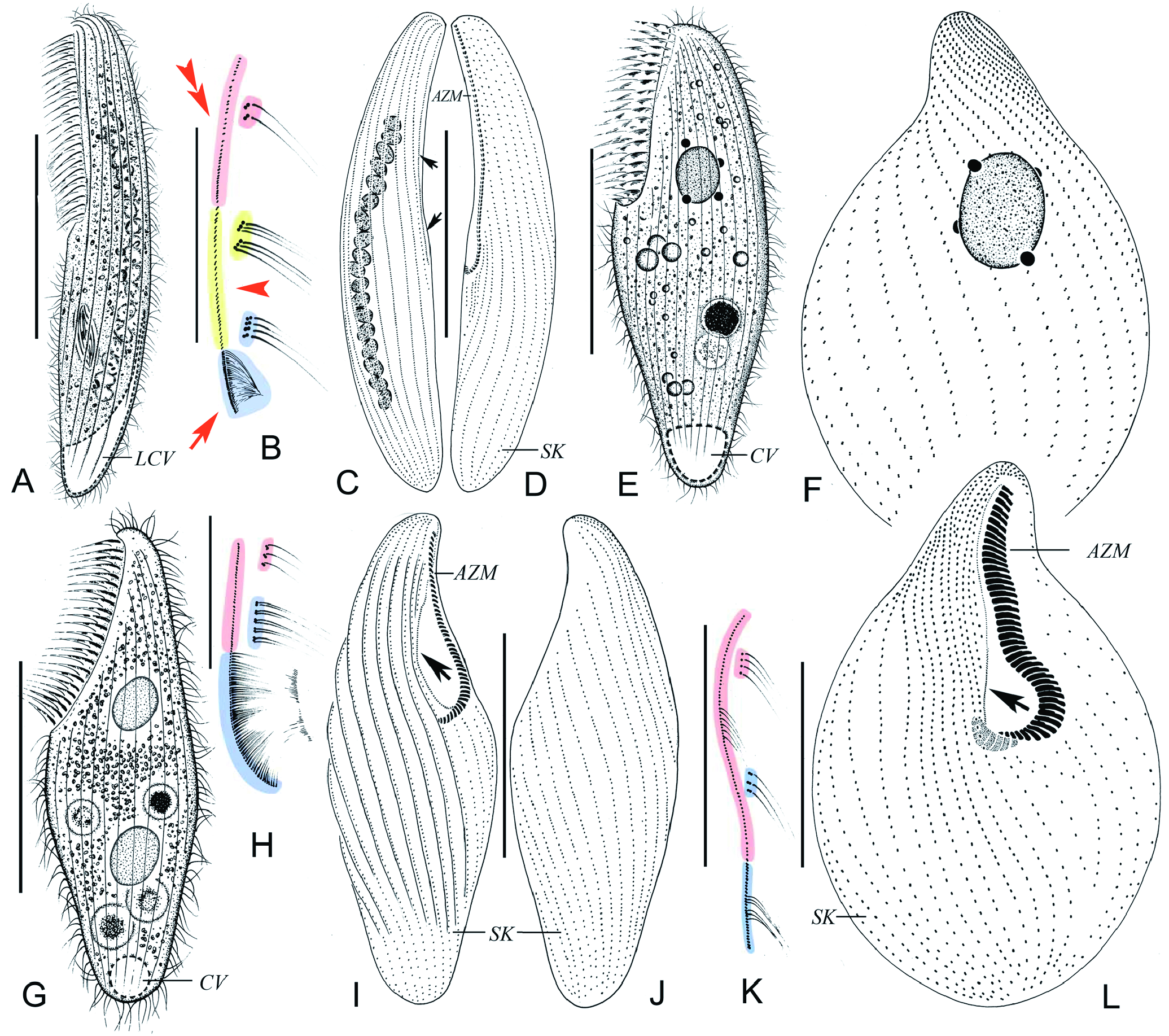
Description Body about 150–180 × 45–55 μm in vivo, slender and irregularly sigmoid, flexible and slightly
All measurements in μm. Abbreviations: CV, coefficient of variation in %; M, median; Ma, macronucleus; Max., maximum; Mean, arithmetic mean; Min., minimum; N, number of specimens; –, data not available.
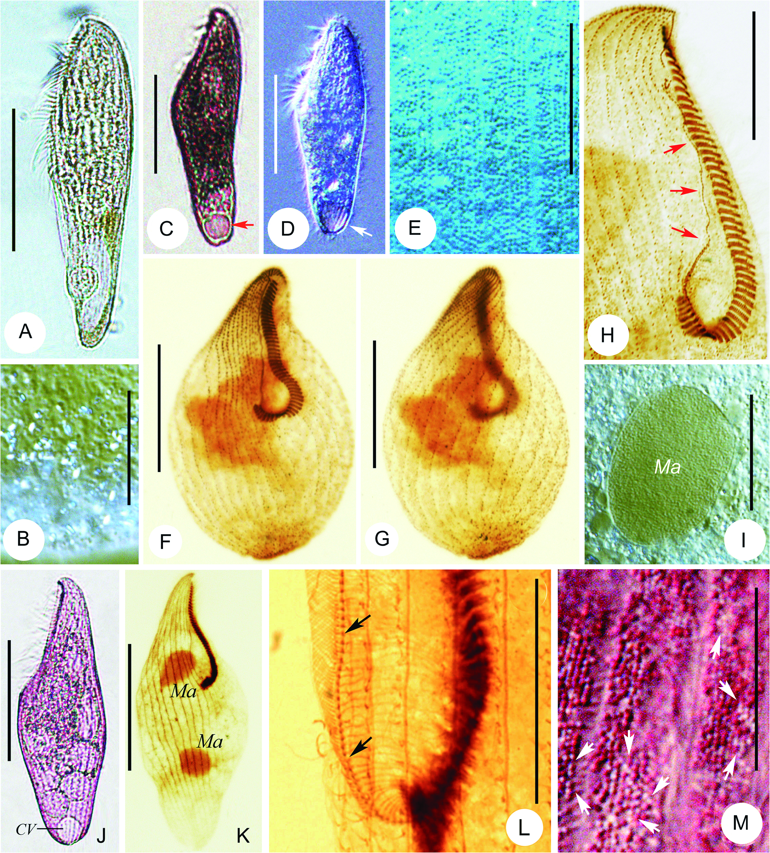
bilaterally flattened ( Figs 1E View Figure 1 , 4A, C, D View Figure 4 ). Buccal area about 50% body length. Cell coloration in both freshly isolated and cultivated (for about 1 to 2 weeks) samples rather variable from almost colourless to darkbrownish at low magnification using bright-field microscopy ( Fig. 4A, C, D View Figure 4 ) although most individuals are slightly brownish or pale pink. It remains unclear what causes the cell coloration as neither pigments nor coloured food vacuoles were detected. Cortical granules colourless, round, c. 0.2–0.3 μm in diameter, densely arranged in rows located between kineties ( Fig. 4E View Figure 4 ). Paroral membrane inconspicuous, difficult to detect in vivo ( Fig. 4A, C, D View Figure 4 ). Macronucleus located slightly above mid-body region, spherical to ovoid, about 20 μm in diameter, with three to five closely associated globular micronuclei ( Fig. 1F View Figure 1 ). Contractile vacuole conspicuous, c. 20 μm in diameter, terminally located ( Figs 1E View Figure 1 , 4C, D View Figure 4 ). Locomotion mainly by gliding slowly on bottom of Petri dish.
Infraciliature consists entirely of dikinetids except for anterior two-thirds of paroral membrane, which is composed of monokinetids ( Fig. 1F, K, L View Figure 1 ). In posterior third of paroral, only left basal body of each dikinetid is ciliated ( Fig. 1K View Figure 1 ). Adoral zone composed of 36–63 membranelles, each of which consists one short and two long rows of basal bodies ( Figs 1L View Figure 1 , 4H View Figure 4 ). Twenty-four to 34 longitudinal somatic kineties including two to five shortened postoral (right) rows ( Figs 1F, L View Figure 1 , 4F, G View Figure 4 ), with cilia about 12–13 μm long in vivo.
Abbreviations: BL, body length; Ma, macronucleus; SK, somatic kineties; –, data not available.
*data from the drawings.
No known copyright restrictions apply. See Agosti, D., Egloff, W., 2009. Taxonomic information exchange and copyright: the Plazi approach. BMC Research Notes 2009, 2:53 for further explanation.
|
Kingdom |
|
|
Phylum |
|
|
Class |
|
|
Order |
|
|
Family |
|
|
Genus |