
Geitogonalia buccina, Cavichioli & Rendón-Mera & Domahovski & Mejdalani, 2018
|
publication ID |
https://doi.org/ 10.11646/zootaxa.4531.4.8 |
|
publication LSID |
lsid:zoobank.org:pub:FABAD4FF-BDAD-419B-A807-1DF309FE1F3F |
|
DOI |
https://doi.org/10.5281/zenodo.6489562 |
|
persistent identifier |
https://treatment.plazi.org/id/D4395124-FFDC-FFE4-FF01-ABAF86A5FBF3 |
|
treatment provided by |
Plazi |
|
scientific name |
Geitogonalia buccina |
| status |
sp. nov. |
Geitogonalia buccina View in CoL sp. nov.
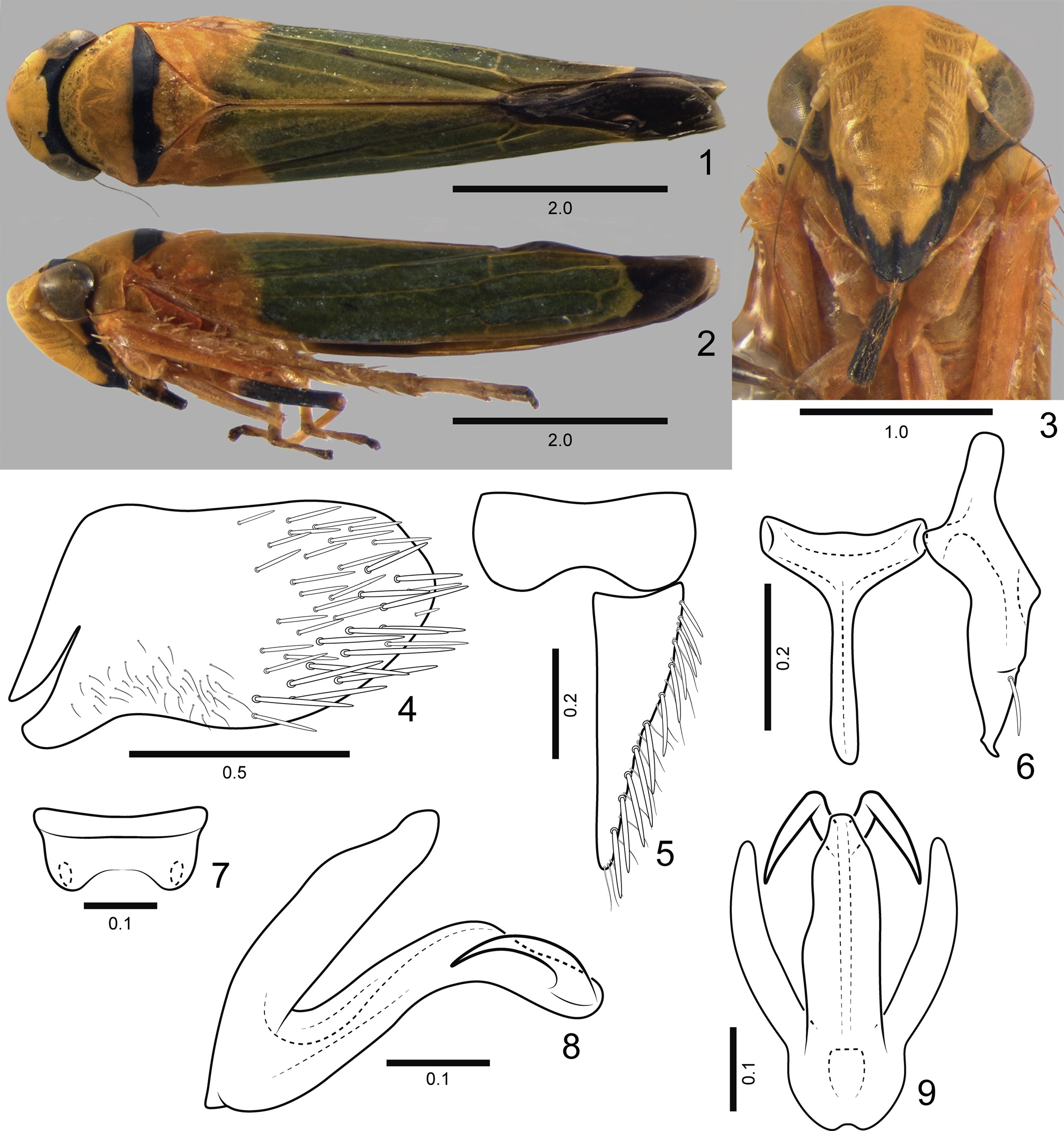
( Figs 1–14 View FIGURES 1–9 View FIGURES 10–14 )
Diagnosis. Aedeagus ( Figs 8, 9 View FIGURES 1–9 ) shaft elongate, apical third strongly curved ventrally, apex with pair of strong horn-shaped processes directed anteriorly, shorter than shaft apical third.
Measurements (in mm; 2 ♂, 2 ♀). Length of body ♂ 6.3–6.7, ♀ 6.9–7.2.
Male. Coloration. Anterior dorsum (crown, pronotum, and mesonotum) yellow ( Figs 1, 2 View FIGURES 1–9 ). Crown with black transverse stripe posteriorly, broadening on each side of midline between ocellus and eye. Pronotum with black transverse stripe on posterior margin. Mesonotum with black transverse narrow anterior stripe continuous with posterior stripe of pronotum. Mesoscutellum entirely yellow. Forewing ( Figs 1, 2 View FIGURES 1–9 ) yellow near base; corium and clavus green and yellowish-green along anal margin; apex and appendix smoky; veins yellow, but R brown along most of its length. Face ( Fig. 3 View FIGURES 1–9 ) yellow; gena black on its posterior half (also posterior to eye), continuing black stripe on crown; lorum black; clypeus with irregular black boundary; rostrum black. Thorax lateral and ventral portions yellow. Legs yellowish-orange, protibia brownish, mesofemur black. Abdomen black.
Structure. Head ( Figs 1–3 View FIGURES 1–9 ) moderately produced, median length of crown approximately 6/10 of interocular width and 4/10 of transocular width; ocelli located slightly behind imaginary line between anterior eye angles; coronal suture slightly visible. Other features as in the descriptions of Young (1977: 524) and of G. viridis Mejdalani & Cavichioli (2014: 374) .
Male genitalia. Pygofer ( Fig. 4 View FIGURES 1–9 ), in lateral view, well produced posteriorly; posterior margin broadly rounded, slightly angulated subapically; without processes; macrosetae distributed on posterior half, thicker and longer macrosetae on ventral half; thin setae present ventrally on anterior half. Valve ( Fig. 5 View FIGURES 1–9 ), in ventral view, with lateral margins subrounded, constricted medially. Subgenital plate ( Fig. 5 View FIGURES 1–9 ), in ventral view, triangular, not fused to its counterpart basally, outer lateral margin sinuous, gradually narrowing towards apex, with uniseriate row of macrosetae intercalated with thin setae; in lateral view, not extending posteriorly as far as pygofer apex. Connective ( Fig. 6 View FIGURES 1–9 ), in dorsal view, T-shaped, stalk long and narrow, longer than arms, keeled dorsally. Style ( Fig. 6 View FIGURES 1–9 ), in dorsal view, with apophysis elongate, distinctly longer than apodeme, extending approximately as far posteriorly as connective; with lobe on outer median portion bearing single elongate seta, apex narrow, obliquely truncate. Paraphyses ( Fig. 7 View FIGURES 1–9 ) present but very small, consisting of well sclerotized basal transverse plate and pair of weakly sclerotized rami with sclerotized apices, each curved inwards and directed upwards. Aedeagus ( Figs 8, 9 View FIGURES 1–9 ) without preatrial process; dorsal apodeme elongate, about 2/3 as long as shaft and about as wide in lateral view, apex sinuous and narrow; shaft elongate, apical third strongly curved ventrally, apex with pair of strong hornshaped processes directed anteriorly, less than one-third shaft length.
Female. Coloration. As in male, with additional black macula medially at base of frons, visible from dorsal and anterior view, and may present transverse black stripe anteriorly on pronotum, continuous with that of crown.
Structure. As in male.
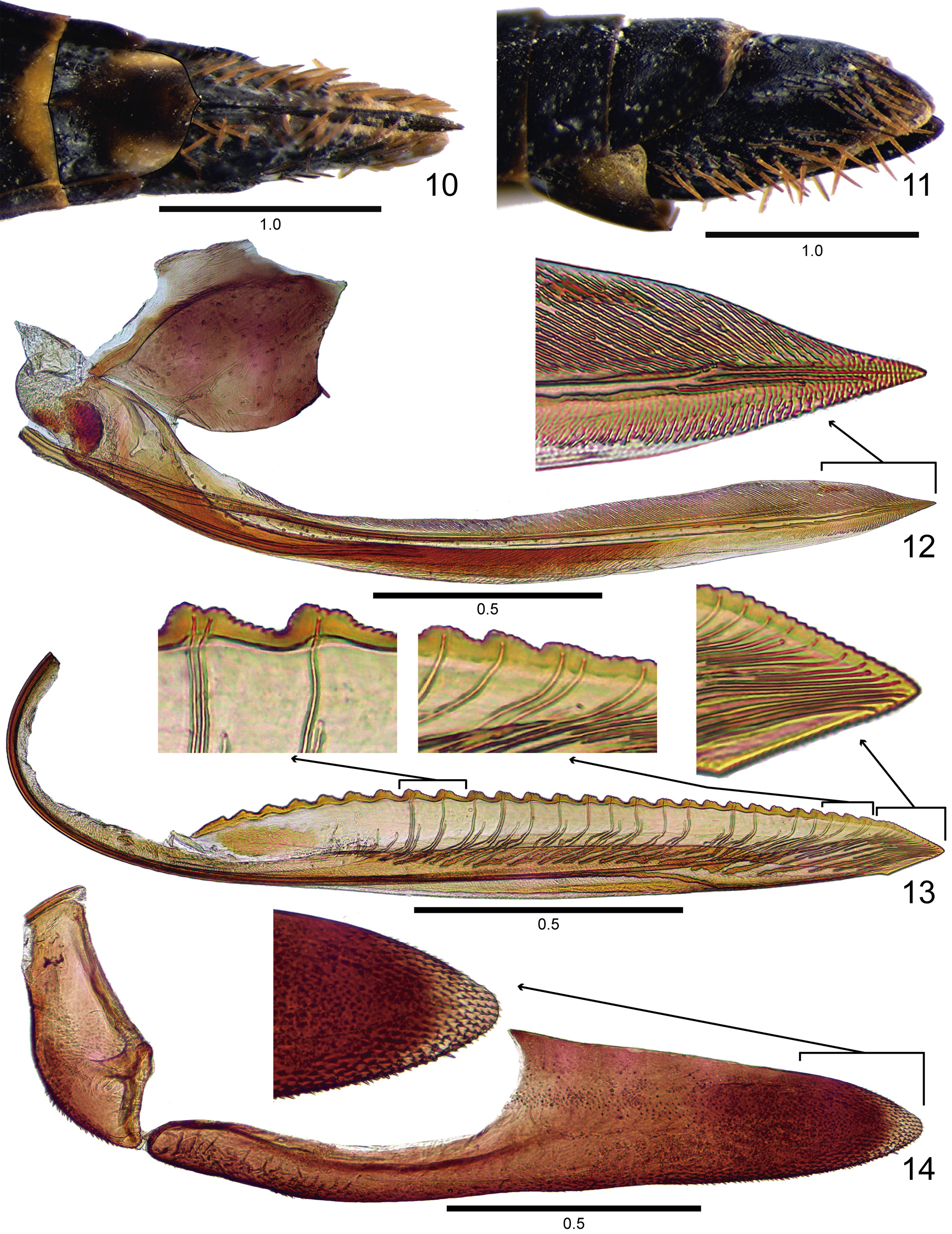
Female genitalia. Abdominal sternite VII ( Figs 10, 11 View FIGURES 10–14 ), in ventral view, slightly longer than wide, posterior margin with slight medial dentiform projection. “Internal” sternite VIII, in dorsal view, without distinct sclerotized areas. Pygofer ( Fig. 11 View FIGURES 10–14 ), in lateral view, strongly produced posteriorly, posterior margin narrowly rounded, macrosetae located mostly on posterior portion and ventral margin. First valvifer ( Fig. 12 View FIGURES 10–14 ) approximately as long as wide. First valvula ( Fig. 12 View FIGURES 10–14 ) slightly curved dorsally, with apex acute; dorsal sculptured area strigate, extending from basal portion of blade to apex; ventral sculptured area scale-like, restricted to apical portion of blade; ventral interlocking device restricted to basal half of blade, located along ventral margin with distal portion directed dorsally. Second valvula ( Fig. 13 View FIGURES 10–14 ) moderately expanded beyond basal curvature; dorsal margin moderately convex; ventral margin with slight preapical prominence; apex subacute; blade with about 27 subtriangular continuous teeth; denticles distributed on teeth and on dorsal and ventral apical portions of blade; ducts extending to apical portion of blade and to teeth or terminating below them (about seven basal-most teeth do not receive ducts). Gonoplac ( Fig. 14 View FIGURES 10–14 ) with basal half narrow and apical half distinctly expanded; apex obtuse; blade with many tiny spiniform processes on apical portion and extending anteriorly along ventral margin.
Etymology. The specific epithet, buccina (noun in apposition), is Latin for “horn.” It refers to the aedeagus with long horn-shaped apical processes ( Figs 8, 9 View FIGURES 1–9 ).
Material examined. Holotype Ƌ: GoogleMaps Southeastern Brazil GoogleMaps , state of Rio de Janeiro: “P. [Parque] N. [Nacional] Itatiaia, RJ [Rio de Janeiro], Brasil \ 22°27’16”S 44°36’29”W \ 800–1300m 23/IX/2011 luz \ R.R.Cavichioli leg.” (DZUP). Paratypes: 1 ♀: “P. N. Itatiaia, RJ, Brasil \ Trilha do Hotel Simon \ 22°27’16”S 44°36’29”W \ 800– 1300m 24/IX/2011 \ R.R.Cavichioli leg.” ( DZUP) GoogleMaps ; 1 ♀: “ BRAZIL: \ Minas Gerais \ Delfim Moreira , \ 1100m. ii.1972.; F.M.Oliveira \ B.M.1972–541” ( DZUP) ; 2 ♂: “ Brasil, RJ, P. N. Itatiaia, \ Travessia Rui Braga \ 22°25’50.4”S 40°37’12.6”W \ 1100m 12/X/2013 sweep \ D.M. Takiya, C. Moraes \ C. Gonçalves” (1 ♂ DZRJ and 1 ♂ MNRJ) GoogleMaps ; 1♀: “ Brasil, RJ, P. N. Itatiaia,\ Hotel Simon \ 22°26’14.4”S 44°36’28.2”W \ 804m 7/ VI /2013 sweep \ D.M. Takiya, C. Moraes \ C. Gonçalves, A. Silva” ( DZRJ) GoogleMaps ; 4♀: “ Biota FAPERJ \ 2.JUL [julho]— 3 AGO [agosto]. 2015 \ COLETA 1”; “ BRASIL: RJ Itatiaia, PNI [ Parque Nacional do Itatiaia ] \ Complexo do Maromba , \ Travessia Ruy Braga , PNI-M2B \ 22°26’07.50”S \ 44°37’33.20”W, 1234m a.s.l. ” (2 ♀ DZRJ and 2 ♀ MNRJ) GoogleMaps .
Remarks. This species is similar in coloration to G. tetracorni sp. nov., whereas its aedeagus is similar to that of G. viridis , except for the shape of the apical processes ( Figs 8, 9 View FIGURES 1–9 ).
No known copyright restrictions apply. See Agosti, D., Egloff, W., 2009. Taxonomic information exchange and copyright: the Plazi approach. BMC Research Notes 2009, 2:53 for further explanation.
|
Kingdom |
|
|
Phylum |
|
|
Class |
|
|
Order |
|
|
Family |
|
|
Genus |