
Acostatrichia simulans Mosely, 1939
|
publication ID |
https://doi.org/ 10.11646/zootaxa.4755.2.1 |
|
publication LSID |
urn:lsid:zoobank.org:pub:CAD4295B-2456-48EE-98F6-723FDEF5C0EB |
|
DOI |
https://doi.org/10.5281/zenodo.3812817 |
|
persistent identifier |
https://treatment.plazi.org/id/D40B8780-CA4D-FFEA-D7F1-FB78FCB2FD23 |
|
treatment provided by |
Carolina |
|
scientific name |
Acostatrichia simulans Mosely, 1939 |
| status |
|
Acostatrichia simulans Mosely, 1939 View in CoL
Figs. 6 View FIGURE 6 , 16 View FIGURE 16
Acostatrichia simulans Mosely 1939: 229 View in CoL , figs. 179–182, male; type locality: Brazil, Santa Catarina, Nova Teutonia; type depository: BMNH.
Angrisano (1995), reported from Uruguay.
Angrisano & Sganga (2010) View Cited Treatment , larva, pupa, case, biology, reported from Argentina.
Santos et al. (2016), phylogenetic placement.
Redescription. Length from front of head to tips of folded forewings 2.0–3.0 mm (n = 9). General color, in alcohol, light brown. Head unmodified. Ocelli 3. Antenna 19-articulated; scape cylindrical, twice as long as wide, inner margin not produced; pedicel cylindrical; flagellomeres cylindrical, unmodified. Forewings each with costal vein bearing short basal bulla. Abdominal segment VII bearing two acute ventromesal processes, basal one shorter ( Figs. 6A, 6C View FIGURE 6 ).
Male genitalia. Segment VIII shorter dorsally than ventrally ( Fig. 6C View FIGURE 6 ); in ventral view, posterior margin of sternum with shallow median V-shaped incision ( Fig. 6A View FIGURE 6 ), without lateral processes; tergum with scattered setae. Segment IX mostly within segment VIII, ventrally open; with pair of elongate dorsolateral processes, gradually curved inwards in dorsal and ventral views ( Figs. 6A, 6B View FIGURE 6 ), almost straight in lateral view ( Fig. 6C View FIGURE 6 ), each one with sinuous subapical spine ( Figs. 6A, 6B, 6C View FIGURE 6 ). Preanal processes digitate and each bearing very long seta ( Fig. 6C View FIGURE 6 ). Inferior appendages paired, short and triangular in ventral view, without apical or basal processes ( Fig. 6A View FIGURE 6 ). Subgenital plate, in ventral view broad and concave at apex ( Fig. 6A View FIGURE 6 ); in lateral view, triangular ( Fig. 6C View FIGURE 6 ). Tergum X membranous, posterior margin convex in dorsal view ( Fig. 6B View FIGURE 6 ), slightly bilobed in lateral view ( Fig. 6C View FIGURE 6 ). Phallus tubular basally, bearing midlength complex with dorsal window and basal loop shorter than basal portion of phallus; apical portion with Y-shaped sclerite and internal spine ( Figs. 6D, 6E View FIGURE 6 ).
Material examined. Brazil, Santa Catarina, Nova Teutonia, ix.1963, F Plaumann leg., Flint 1975 det., 1 male ( NMNH); Nova Teutonia, i.1963, F. Plaumann, Flint det., 6 males ( NMNH). Argentina, Província Misiones, Salto Encantado Provincial Park, Arroyo Azul , 23-28.i.2008, Angrisano & Sganga leg., Sganga 2012 det., 1 male ( DZRJ). Uruguay, Artigas, Rio Cuareim, Sepulturas , 15.xii.1952, CS Carbonell , Flint det., 1 male ( NMNH).
Remarks. Acostatrichia simulans shows typical features of the A. plaumanni Species Group, such as the following: (1) a costal bulla in each forewing, (2) a double ventromesal process on segment VII, (3) the inferior appendages short and not fused to each other, and (4) a pair of long dorsolateral processes on segment IX. In the A. plaumanni Group, A. simulans shares more similarities in male genitalia with A. plaumanni and A. fluminensis , but it can be distinguished from those by a shallow median incision on the posterior margin of segment VIII in ventral view ( Fig. 6A View FIGURE 6 ); and by the dorsolateral processes of segment IX curved inwards in ventral and dorsal views, each one with a short sinuous subapical spine ( Figs. 6A, 6B View FIGURE 6 ). This is the only Acostatrichia species with immature stages described ( Angrisano & Sganga 2010). According to Angrisano & Sganga (2010), the larva is similar to those described for other Leucotrichiini genera, in particular to larvae of Zumatrichia Mosely 1937 , but the A. simulans larva differs by having the frontoclypeus with a reticulate sculpturing and each tarsal claw with an enlarged basal seta.
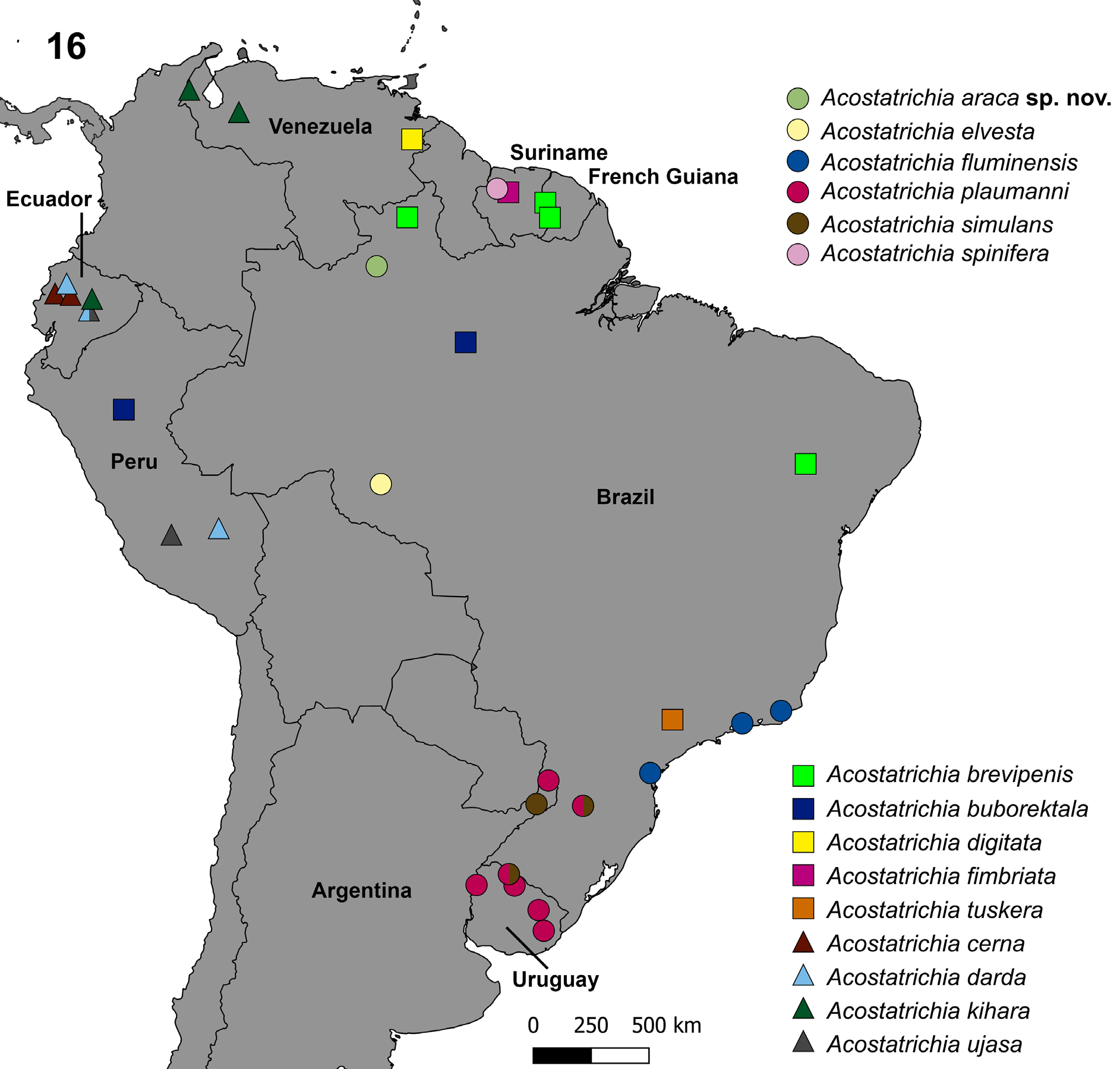
Distribution. Argentina, Brazil, and Uruguay ( Fig. 16 View FIGURE 16 ).

No known copyright restrictions apply. See Agosti, D., Egloff, W., 2009. Taxonomic information exchange and copyright: the Plazi approach. BMC Research Notes 2009, 2:53 for further explanation.
|
Kingdom |
|
|
Phylum |
|
|
Class |
|
|
Order |
|
|
Family |
|
|
Genus |
Acostatrichia simulans Mosely, 1939
| Santos, Allan Paulo Moreira 2020 |
Acostatrichia simulans
| Santos, A. P. M. & Nessimian, J. L. & Takiya, D. M. 2016: 209 |
| Angrisano & Sganga 2010: 209 |
| Angrisano 1995: 209 |
| Mosely, M. E. 1939: 229 |