
Tupiniquim Linzmeier, Oliveira & Konstantinov, 2021
|
publication ID |
https://doi.org/ 10.11646/zootaxa.5068.1.4 |
|
publication LSID |
lsid:zoobank.org:pub:4DAB2CFA-B192-434D-A697-8B1067BBFDED |
|
DOI |
https://doi.org/10.5281/zenodo.5706167 |
|
persistent identifier |
https://treatment.plazi.org/id/D26D412E-FFF3-8F50-FF14-F2D28450FF5A |
|
treatment provided by |
Plazi |
|
scientific name |
Tupiniquim Linzmeier, Oliveira & Konstantinov |
| status |
gen. nov. |
Tupiniquim Linzmeier, Oliveira & Konstantinov , new genus
( Figs 14–28 View FIGURES 14–16 View FIGURES 17–22 View FIGURE 23 View FIGURES 24–26 View FIGURES 27–28 )
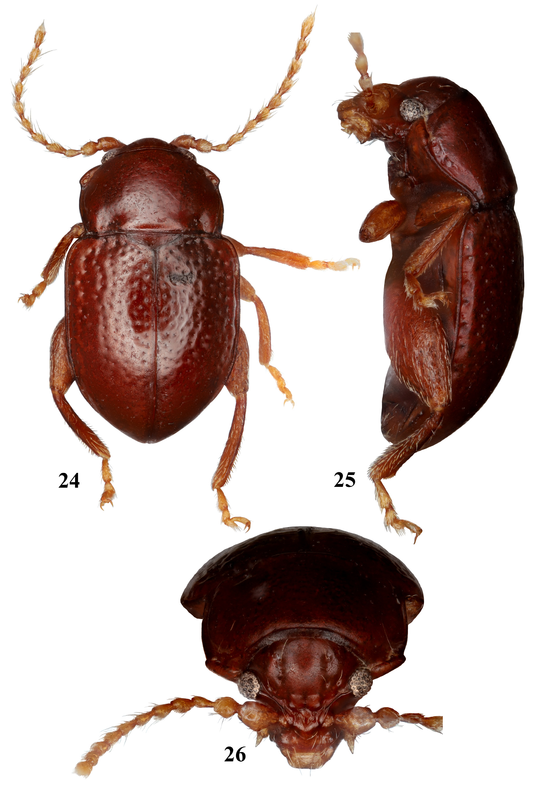
Description. Body 1.62–2.05 mm long and 0.91–1.13 mm wide, oval, glabrous, shiny, nearly flat in lateral view ( Figs 14, 15 View FIGURES 14–16 , 23 View FIGURE 23 , 24, 25 View FIGURES 24–26 ). Color yellow, reddish brown to dark brown. Head hypognathous; frons and vertex in same plane or forming about 135º angle, in lateral view. Vertex glabrous. Supraorbital pore distinct to almost indistinguishable. Two or four setiferous pores form vertical line or small cluster on each side of vertex ( Figs 16 View FIGURES 14–16 , 26 View FIGURES 24–26 ). Antennal calli somewhat rounded (pentagonal), smooth, placed between the line of the inner margin of antennal sockets, in same plane as vertex surface. Midfrontal and suprafrontal sulci shallow to absent. Supracallual sulcus absent. Suprantennal and supraorbital sulci deep, bordered by anterior ridge, which reaches almost to middle of eye. Orbital sulcus close to eye. Orbit as wide as diameter of eye, slightly concave. Interantennal space slightly wider than orbit. Antennal socket laterally directed. Frontal ridge wider posteriorly, between antennal sockets, than anteriorly, on clypeus, raised higher than surface of antennal calli. Anterofrontal ridge low throughout. Eye small, rounded, slightly projected laterally. Labrum slightly notched in middle, with anterior angles rounded and six setiferous pores, four with long setae and two with short setae. First maxillary palpomere cylindrical, thin, slightly wider at apex; second maxillary palpomere widely inflated, as long as first; third palpomere thinnest, slightly shorter than second, conical, with acute apex. Labial palpomeres minute, first and second labial palpomeres slightly longer than wide; third conical. Antenna with 11 antennomeres, filiform, pubescent; antennomeres IV to XI with length twice width, obliquely widened at apex. Antennomeres I and II much wider than remainder. Some antennomeres with long setae, at times as long as antennomeres themselves ( Figs 14 View FIGURES 14–16 , 24 View FIGURES 24–26 ).
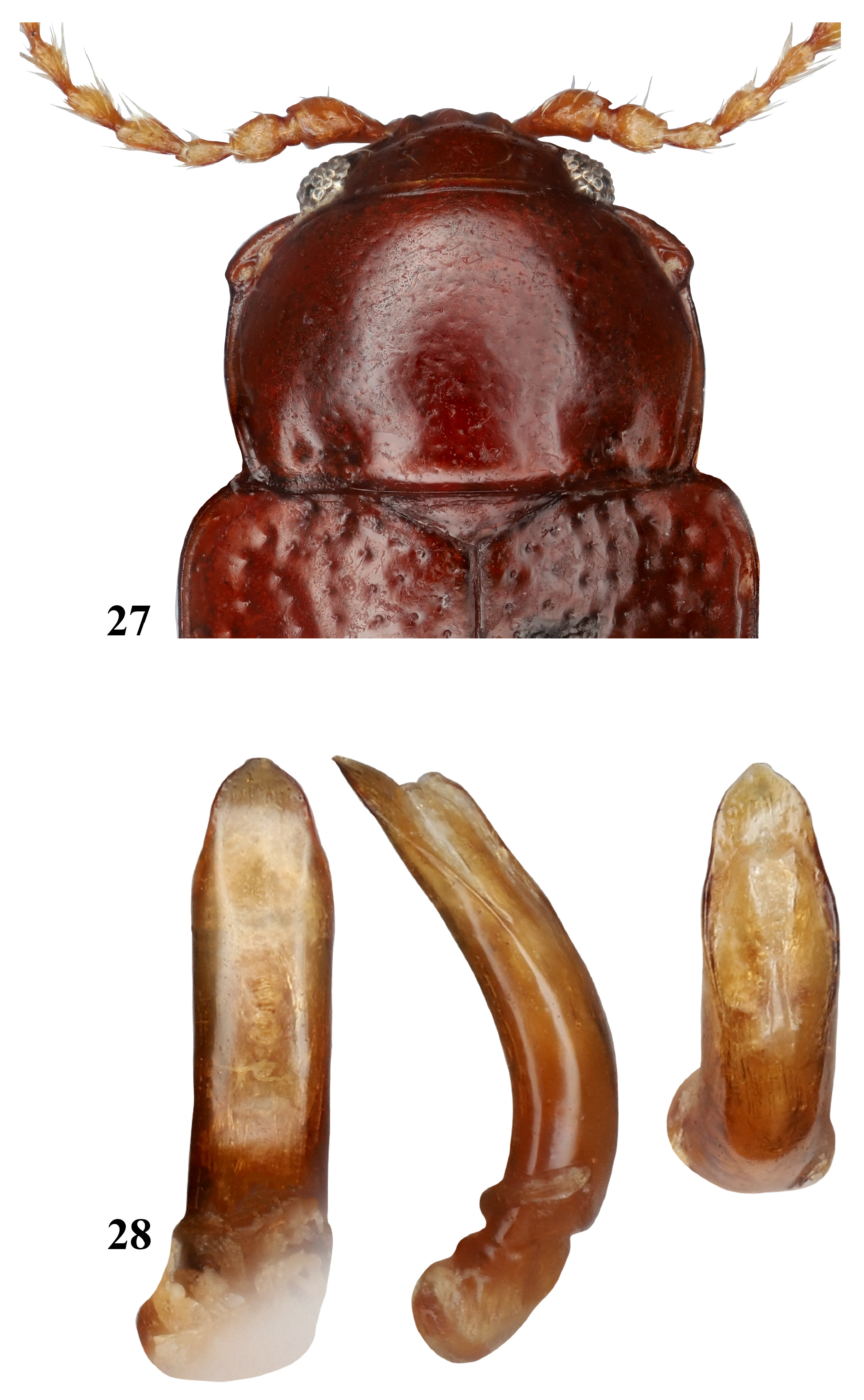
Pronotum rectangular, large, about ¼ length of body, wider than long, margined laterally. Anterior margin widening laterally towards anterolateral callosity near its setiferous pore; posterior margin slightly convex in middle, concave laterally across impressions; sides slightly rounded. Anterior angle beveled, with setiferous pore on posterior corner, facing dorsally, bearing a long seta; posterior angle setiferous pore smaller, facing laterally. Surface shiny, almost smooth, finely punctured to microreticulate, pilosity short, almost imperceptible. Basal margin with or without antebasal impressions ( Figs 14 View FIGURES 14–16 , 24, 26 View FIGURES 24–26 ). Scutellum triangular, short and broad, four times wider than long, smooth, glabrous. Elytra oval, apical half curved towards venter from peak at middle in lateral view. Elytral surface shiny, punctured with small and shallow to large and deep punctures, organized in confused rows, bearing short, thin, almost imperceptible pilosity. Basal and humeral calli absent. Epipleura wide, sinuous, oblique, almost horizontal, narrowing at elytral apex, nearly reaching it. Elytral apex rounded. Membranous wings absent.
Prosternal surface smooth, margined with long setae. Prosternal intercoxal process thin, extended posteriorly beyond procoxae. Procoxal cavities opened posteriorly. Procoxae elongate. Pro- and mesofemora slightly dilated medially; pro- and mesotibiae subcylindrical, with a median dorsal ridge, pubescence sparsely distributed. First pro- and mesotarsomeres enlarged in males ( Fig. 14 View FIGURES 14–16 ), as long as third and fourth tarsomeres together; in females as long as second and third, similar in size, as wide as long; third not bilobed; fourth tarsomere thinnest. Metafemora about 1.5 times longer than wide, sparsely pilose, with sharply margined inner ventral groove, for receiving tibia ( Fig. 19 View FIGURES 17–22 ). Metatibia longer than metafemur, straight in lateral view, nearly straight and slightly wider at apex, in dorsal view; denticles along apical half of outer dorsal margin and at apex of the inner dorsal margin. Dorsal surface of metatibia canaliculate from about basal third to apex. Metatibial spur short. Metatarsomeres inserted apically; first tarsomere slightly longer than rest; second and third similar in size, as wide as long; third not bilobed; fourth as long as second and third together, thinnest. Claws appendiculate.
Abdomen sparsely pubescent, with five visible ventrites, each convex in lateral view. Intercoxal process of ventrite I punctured. Ventrite V distinctly sexually dimorphic: males with concavity medially on posterior margin, females conical at apex ( Fig. 17 View FIGURES 17–22 ). Median lobe simple, curved, with apical third slightly flat in lateral view; lateral margins slightly expanded above middle, apex triangular with poorly defined denticle varying in length in ventral view ( Figs 21 View FIGURES 17–22 , 28 View FIGURES 27–28 ).
Female genitalia lacking vaginal palpi. Spermatheca with ovoid receptacle clearly separated from pump, shorter than it; pump widening apically; canal short ( Figs 18, 22 View FIGURES 17–22 ). Tignum relatively short, with distal area triangular, with wide posterior sclerotization ( Figs 18, 20 View FIGURES 17–22 ).
Type species. Tupiniquim confusa Linzmeier, Oliveira & Konstantinov , new species.
Etymology. The generic name refers to the indigenous tribe Tupiniquim that inhabited the Brazilian coast when the Portuguese arrived in state of Bahia ( Zorzetto 2020), near the genus type locality. In addition, “ Tupiniquim ” is used popularly as a synonym for “Brazilian”. The name is feminine.
Remarks. Tupiniquim gen. nov. is remarkably different from all known New World alticine genera, including those inhabiting leaf litter or moss. It can be immediately differentiated from known flea beetles based on the following characters: head with two or four setiferous pores forming vertical line or a small cluster on each side of vertex (the vertical line of punctures has not been observed in flea beetles before); midfrontal and suprafrontal sulci shallow to absent; supracalinal sulcus absent; suprantennal and supraorbital sulci deep, bordered by ridge on lower side, this ridge comes close to middle of eye; antennomeres I and II much wider than the rest, with antennomere II nearly globose and misshaped; some antennomeres with long setae, at times as long as antennomeres themselves; pronotum rectangular, large, about ¼ of length of body; with anterior margin widening laterally approaching anterolateral callosity near its setiferous pore; scutellum triangular, four times wider than long, pro- and mesotibiae subcylindrical, with a dorsal ridges running medially; vaginal palpi absent.
In Scherer’s key to Neotropical alticines ( Scherer, 1983), Tupiniquim could be placed close to Gioia Bechyné, 1955 due to anterior angles of pronotum beveled and elytra confusedly punctate. However, Tupiniquim is easily separated from Gioia (in parenthesis) by small eyes (large eyes); presence of a ridge from suprantennal sulcus to midline of inner margin of eye (ridge absent); pronotum almost smooth, minutely punctured to finely textured (pronotum distinctly punctured); scutellum short and broad (triangular, nearly as long as wide); humeral calli not developed (developed).
Two recently described moss or leaf little inhabiting alticines that are somewhat similar are Nicaltica and Stevenaltica . Tupiniquim can be immediately separated from Nicaltica based on the small and flat body (in Nicaltica - much larger and convex) absence of supracallual sulci (in Nicaltica supracallual sulci present), frontal and anterofrontal ridges forming a T-shaped structure (in Nicaltica - Y-shaped); spermatheca with distinct border between receptacle and pump (in Nicaltica spermatheca with border absent). Tupiniquim is separated from Stevenaltica by the following features: absence of supracallual sulci (in Stevenaltica supracallual sulci present), absence of transverse antebasal impression (transverse antebasal impression present in Stevenaltica ), absence of ridges near base of elytra (in Stevenaltica ridges present); spermatheca with round receptacle and short canal without coils (in Stevenaltica spermatheca with elongate receptacle and long duct with coils).
Absence of vaginal palpi is an infrequent feature in flea beetles, but it is not possible to say that because of it Tupiniquim is somehow related to Monomacra or other genera of the group, which also lack vaginal palpi. Regardless, it can be easily differentiated from them based on the unique combination of features mentioned at the beginning of Remarks.
Interestingly, Tupiniquim shares some features with Amydus Chen, 1935 a ground living genus found in Himalaya ( Scherer 1989). Among these features are large pronotum, with anterior angles oblique and without prebasal impression; extremely short and broad scutellum; winglessness; puncturation on elytra confused to barely organized in rows and procoxal cavities opened posteriorly. They can be differentiated mainly by the smaller size of eyes in Tupiniquim , the ridge that reaching the midpoint of inner margin of eye (absent in Amydus ); absence of orbital sulcus (present in Amydus ).
The two known species of Tupiniquim are substantially different. However, they are clearly congeneric sharing the main characters of the body, head, pronotum and male genitalia, including relatively unusual shape of the latter in ventral view having median lobe widening above the middle.


No known copyright restrictions apply. See Agosti, D., Egloff, W., 2009. Taxonomic information exchange and copyright: the Plazi approach. BMC Research Notes 2009, 2:53 for further explanation.