
Delavalia reducta, Gómez, 2021
|
publication ID |
https://doi.org/10.11646/zootaxa.5051.1.12 |
|
publication LSID |
lsid:zoobank.org:pub:A99E653A-EBDF-48B1-BF24-0194136E03F9 |
|
DOI |
https://doi.org/10.5281/zenodo.5570777 |
|
persistent identifier |
https://treatment.plazi.org/id/4490AD3C-AB74-4025-B975-2D89DD7CB309 |
|
taxon LSID |
lsid:zoobank.org:act:4490AD3C-AB74-4025-B975-2D89DD7CB309 |
|
treatment provided by |
Plazi |
|
scientific name |
Delavalia reducta |
| status |
sp. nov. |
Delavalia reducta sp. nov.
( Figs. 19–23 View FIGURE 19 View FIGURE 20 View FIGURE 21 View FIGURE 22 View FIGURE 23 )
urn:lsid:zoobank.org:act:
Type locality. Off Todos Santos Bay , Baja California ( Eastern Tropical Pacific), Mexico; Talud XVIB cruise, sampling station 25 ( 31.805°N, 116.925°W); depth 825 m; organic carbon content, 4.17%; organic matter content, 7.17%; sand 2.13%; clay, 10.36%; silt, 87.51 GoogleMaps %.
Specimens examined. Adult female holotype dissected and mounted onto ten slides (EMUCOP-260514-01); May 26, 2014; coll. S. Gómez.
Etymology. The specific epithet from the Latin reducta , reduced, makes reference to the loss of the inner seta of P1 ENP1, and to the reduced armature complement of P1 ENP2 from three to two. It is in the nominative singular. Gender feminine.
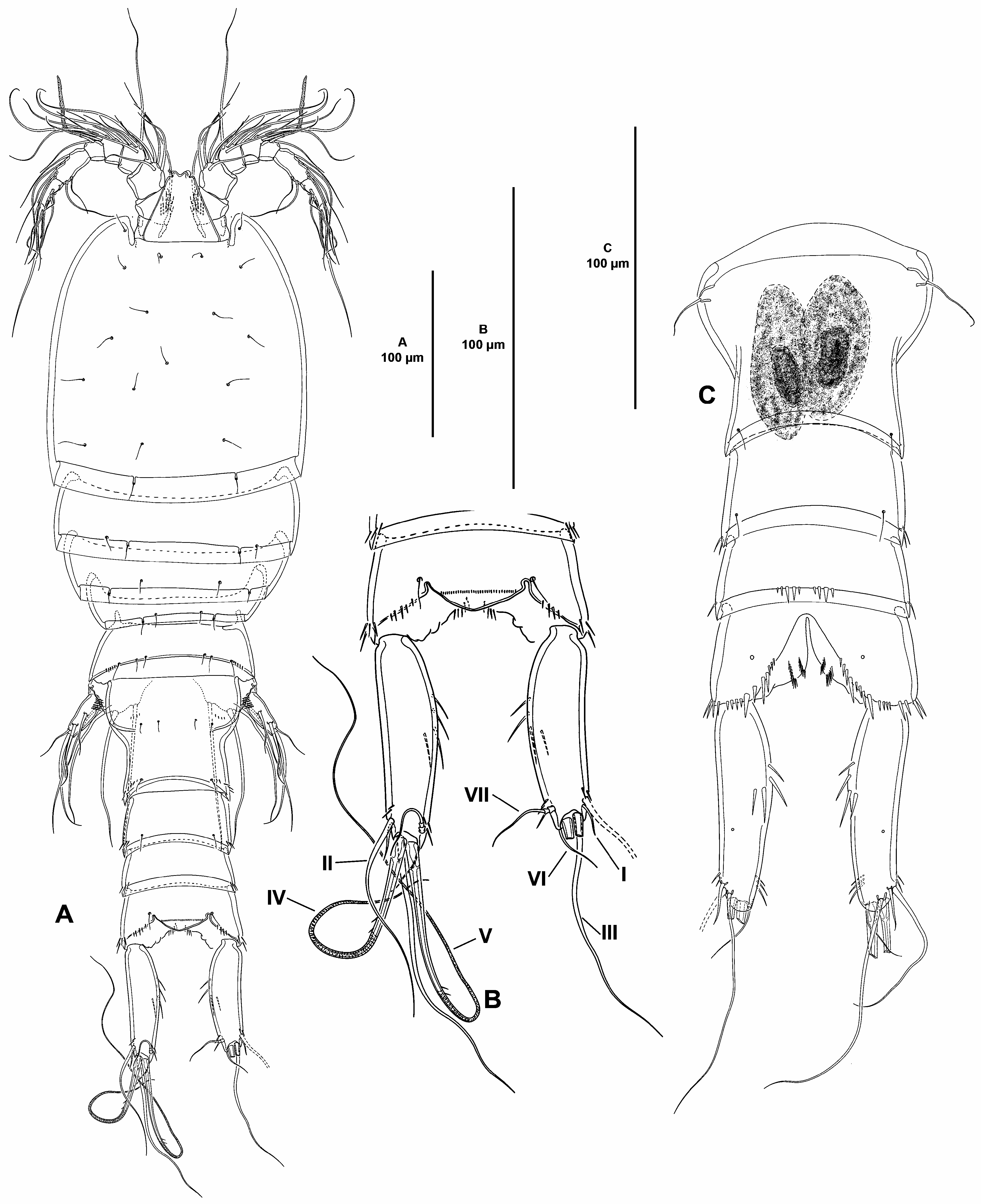
Description of female. Total body length measured from tip of rostrum to posterior margin of caudal rami, 525 µm; habitus pyriform, widest at posterior end of cephalothorax, tapering posteriad ( Fig. 19A View FIGURE 19 ); cephalothorax/body length ratio, 0.35.
Prosome and pedigerous somites ( Fig. 19A View FIGURE 19 ) largely as in previous species.
Urosome ( Fig. 19A–C View FIGURE 19 ) consisting of fifth pedigerous somite (first urosomite), genital double-somite (genital— second urosomite—and third urosomites fused), two free urosomites, and anal somite. Urosomites without expansions laterally nor dorsally; integument weakly sclerotized.
Fifth pedigerous somite ( Fig. 19A View FIGURE 19 ) narrower than preceding somites; with two dorsolateral sets of small spinules.
Second and third urosomites completely fused dorsally and ventrally forming genital double-somite ( Fig. 19C View FIGURE 19 ), with dorsolateral trace of division ( Fig. 19A View FIGURE 19 ); genital double-somite as long as wide, widest part measured in proximal third close to P6; proximal half of genital double-somite with sensilla and two sets of small spinules dorsally ( Fig. 19A View FIGURE 19 ), ventrally without sensilla nor spinules ( Fig. 19C View FIGURE 19 ); distal half of genital double-somite with posterior sensilla and dorsolateral spinular rows ( Fig. 19A View FIGURE 19 ), ventrally with few sensilla and without spinules ( Fig. 19C View FIGURE 19 ); posterior hyaline fringe broad and smooth; genital complex hardly distinguishable, copulatory pores not exposed, paired genital apertures located ventrolaterally and covered by P6 ( Fig. 19C View FIGURE 19 ).
Fourth urosomite ( Fig. 19A, C View FIGURE 19 ) as distal half of genital double-somite; no ventral pores detected.
Fifth urosomite with dorsolateral spinules and without dorsal sensilla ( Fig. 19A View FIGURE 19 ); ventrally without sensilla but with two medial sets of spinules ( Fig. 19C View FIGURE 19 ); no ventral pores detected.
Anal somite about 2.2. times as wide as long ( Fig. 19A–B View FIGURE 19 ); with dorsal, ventrolateral and ventral spinules around joint of caudal rami ( Fig. 19A–C View FIGURE 19 ); with spinules along medial cleft ventrally ( Fig. 19C View FIGURE 19 ); with one ventral pore on each side ( Fig. 19C View FIGURE 19 ); anal operculum with transverse row of minute spinules, semicircular, flanked by one sensilla on each side ( Fig. 19B View FIGURE 19 ).
Caudal rami elongate, about 3.6 times as long as wide ( Fig. 19A–C View FIGURE 19 ) and about as long as fifth and anal somites combined; outer margin nearly straight, inner margin convex; with spinules at base of setae I and II, and VII, and with inner medial spinules ( Fig. 19A–C View FIGURE 19 ), with one medial pore ventrally ( Fig. 19C View FIGURE 19 ); with seven elements ( Fig. 19B View FIGURE 19 ); setae I and II subdistal, lateral, the former a small seta ventral to seta II, the latter long; seta III subdistal, arising ventrally ( Fig. 19B–C View FIGURE 19 ); setae IV and V distal, rat-tail like in distal half, with fracture plane; seta VI small, issuing at inner distal corner; dorsal seta VII triarticulate at base, situated subdistally close to inner margin.
Rostrum ( Fig. 20A View FIGURE 20 ) trapezoidal, not fused to cephalothorax, bifid, with two subdistal sensilla, without dorsal pore.
Antennule ( Fig. 20A View FIGURE 20 ) seven-segmented; all segments smooth, except for first segment with proximal spinular row; first segment without pore. All setae smooth, except for one pinnate seta on second, third and last segments; second and third segments with one seta with fracture plane each; sixth segment with two, seventh segment with three articulated setae. Armature formula: 1(1); 2(10); 3(9); 4(5 + (1 + ae)), 5(3); 6(8); 7(5 + acro). Acrothek consisting of two setae and one minute aesthetasc fused basally.
Antenna ( Fig. 20B View FIGURE 20 ). Coxa short, with some outer spinules. Allobasis as long as free endopodal segment; with some slender spinules at base of exopod, and with few long slender inner spinules at proximal third; with one abexopodal seta arising slightly above the middle of segment. Free endopodal segment elongate; proximal half with longitudinal row of strong inner spinules, distal half with subdistal outer strong spinules, with two outer subdistal frills; armature composed of two lateral spines and two setae, distally with one inner apical element, three apical geniculate setae and one slender element, and one outer distal pinnate element fused basally to slender seta. Exopod three-segmented; first and third segments longest; first and middle segment without, third segment with spinules as shown; first and second segment with one distal seta each, third segment with one proximal and three apical setae, two of which seemingly fused basally.
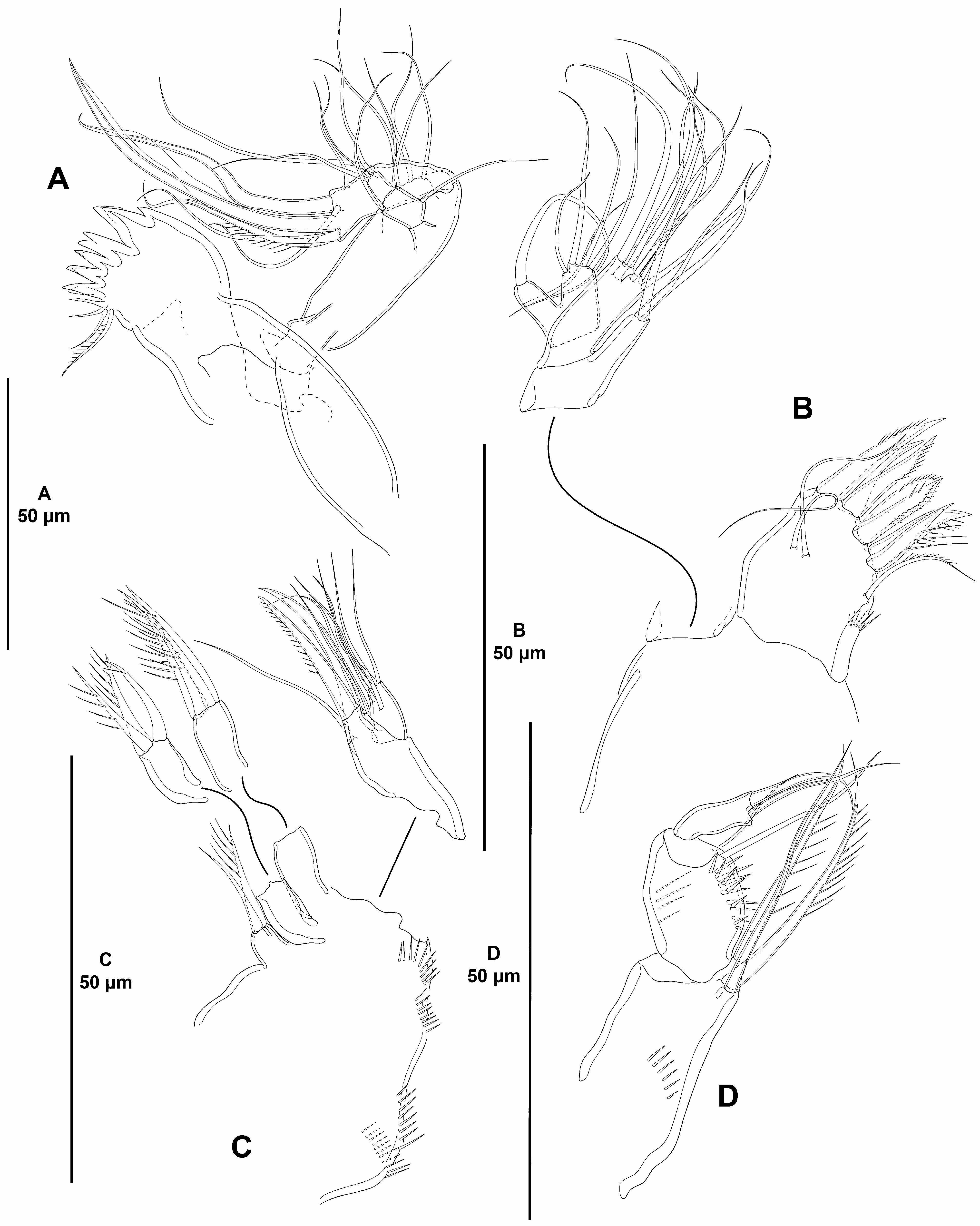
Mandible ( Fig. 21A View FIGURE 21 ). Coxa relatively short. Gnathobase wide; ventral distal corner produced into small sharp semi-hyaline process; with two strong and several smaller teeth, two spines and two setae, one of which pinnate. Basis elongate, spinular ornamentation difficult to see, not detected, with three subdistal outer setae. Exopod arising from short pedestal, one-segmented, elongate, about 2.2 times as long as wide, and 0.3 times as long as basis, with three lateral and three apical setae, none of which fused basally. Endopod recurved, twisted over exopod, with three lateral setae, and five distal elements (three slender setae, one of which spinulose and one strong element, and longest element fused to endopod basally and with hyaline flange in middle part).
Maxillule ( Fig. 21B View FIGURE 21 ). Arthrite of praecoxa with two surface setae and some dorsal spinules; distal armature composed of seven strong spines as shown —no setiform elements detected—, and one lateral pinnate seta. Coxal endite with three setae, spinular ornamentation not detected. Basis with two endites; proximal endite with four, distal endite with three slender setae. Exopod and endopod fused basally, separated from basis, one-segmented; endopod larger than exopod, with four setae; exopod with two setae.
Maxilla ( Fig. 21C View FIGURE 21 ). Large syncoxa with outer spinules as shown; with three endites; proximal endite bilobed, each lobe with one seta; middle and distal endites elongate, the latter slightly longer, with three spinulose setae each. Basis drawn out into strong claw, with strong spinulose spine and two slender setae, one of which arising from elongate setophore. Endopod one-segmented, with six slender setae (one arising basally, two medially, and three apically).
Maxilliped ( Fig. 21D View FIGURE 21 ) subchelate. Syncoxa rectangular; about two times as long as wide; with medial spinules as shown; with one bare and two spinulose strong elements, of which bare seta and one spinulose element at the same level, the other arising distally from long pedestal. Basis shorter than syncoxa, oval, with some outer spinules, with one anterior and one posterior inner spinular row, with two slender distal setae subequal in length. Endopod one-segmented, with one claw-like element and one seta.
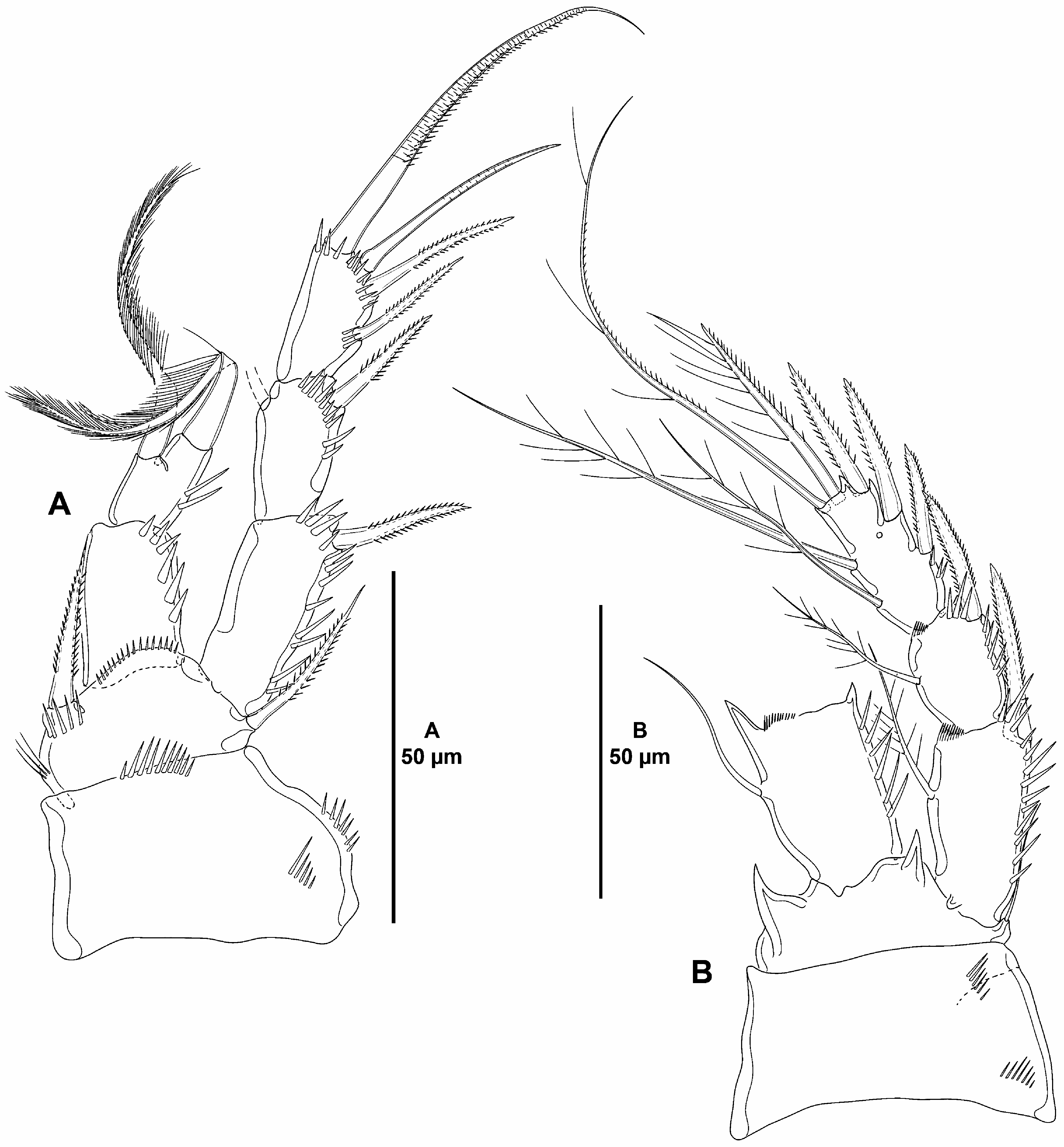
P1 ( Fig. 22A View FIGURE 22 ). Coxa massive, 1.6 times as wide as long; with two outer, and one medial subdistal row of spinules. Basis with outer and inner pinnate spines, with spinules at base of endopod and at base of inner spine, and with few inner setules. Exopod three-segmented, longer than endopod; no pores detected on exopodal segments; EXP1 longest, EXP3 shortest; all segments without outer nor inner acute distal processes; EXP1 and EXP2 with longitudinal row of outer spinules, EXP1 without, EXP2 with inner seta; EXP3 with spinules at the base of setae/ spines, with two outer spines and two apical elements, of which outermost apical element spine-like, innermost apical element pinnate and rat-tail like in distal half. Endopod two-segmented, reaching middle of EXP2, segments without inner nor outer acute distal processes; no pores detected on endopodal segments; ENP1 barely reaching tip of EXP1, 1.5 times as long as wide, visibly longer than ENP2, with outer and distal spinules, without armature; ENP2 small, rectangular, about 1.4 times as long as wide, and 0.7 times as long as ENP1, with few outer spinules, with one inner and one apical seta, both strong and plumose.
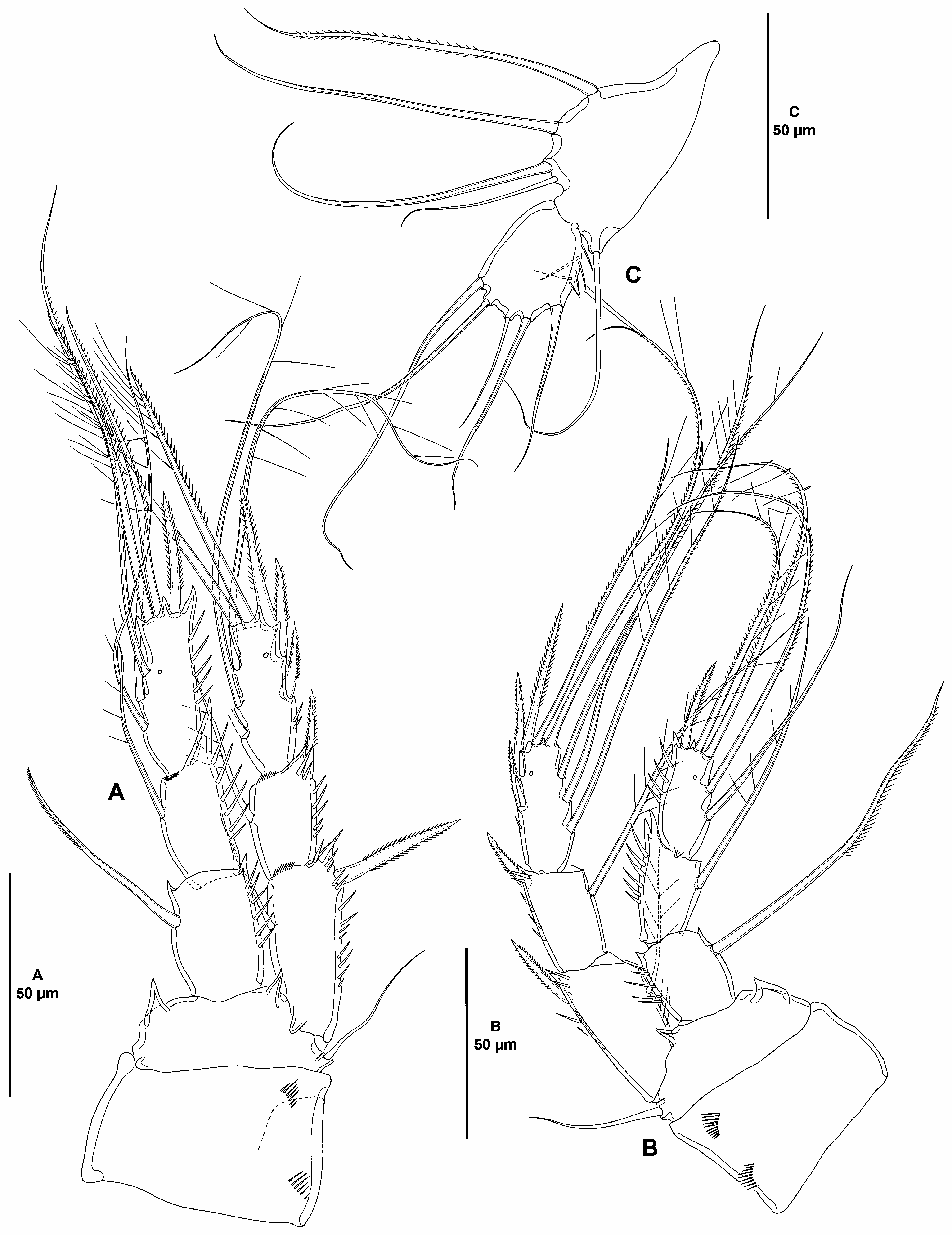
P2–P4 ( Figs. 22B View FIGURE 22 , 23A–B View FIGURE 23 ). Intercoxal sclerite (not shown) not transversely elongate, trapezoidal, with strong pointed process on distal outer corners, without surface ornamentation. Coxa with outer spinules proximally and subdistally. Basis with strong acute process between rami and at inner distal corner, the latter larger, of P2 largest, of P4 smallest; seemingly without spinular ornamentation. Exopod three-segmented, of P3 slightly shorter than, of P4 visibly longer than ENP, relative length of P2 EXP uncertain due to bad condition of endopod; pores detected on last exopodal segments only; EXP1 and EXP2 with outer distal process (of P4 less developed), with longitudinal row of outer spinules, of P2 and P3 with, of P4 seemingly without inner distal frill, with inner seta; EXP3 with distal processes as shown, with few spinules proximally, with three outer spines and two apical setae, of P2 with two, of P3 and P4 with three inner setae. Endopod three-segmented (P2 badly damaged and second and third endopodal segments lost during dissection), of P2 most probably longer than, of P3 slightly longer than EXP, of P4 reaching proximal third of EXP3; pores present on the last endopodal segments of P3 and P4 (P2 ENP2–3 unknown); P3–P4 ENP1 shortest, ENP3 longest; ENP1 and ENP2 (of P2 unknown) with outer acute and inner small distal process, outer process of ENP2 (at least of P3 and P4) visibly longer, outer process of P4 ENP2 bifurcate, most probably aberrant, distal processes of ENP3 as shown; ENP1 with inner element, of P2 a slender seta, of P3 and P4 a stiff element with inner margin pinnate, of P4 very long; armature of P2 ENP2 unknown (most probably two setae), P3 and P4 ENP2 with one inner seta; P2 ENP3 unknown, P3–P4 ENP3 with one apical outer spine, two apical elements, and three (P3), or two (P4) inner setae.
Setal formula of swimming legs as follows:
P5 ( Fig. 23C View FIGURE 23 ). Baseoendopod pentagonal, transversely elongate; endopodal lobe poorly-developed, with four setae, of which outermost shortest and set closely to adjacent seta, all setae naked, except for innermost seta pinnate. Exopod oval, with some outer spinules proximally, with five setae as shown.
P6 ( Fig. 19C View FIGURE 19 ) represented by a minute flap covering ventrolateral genital aperture, fused to somite, without surface ornamentation, with one slender seta.
Male. Unknown.
Variability. No variability was detected in the single female found in the sediment samples.
Remarks. The genus Delavalia is the most diverse genus within Stenheliinae both in number of species and morphologically. Due to the latter, the monophyly of the genus has been questioned repeatedly ( e.g. Karanovic & Kim 2014; Mu & Huys 2002; Willen 2000, 2002, 2003). Several studies have focused their efforts towards the monophyly of the genus and several authors have contributed importantly (see Gómez & Cruz-Barraza 2021). The disparity in maxilliped structure ( Gómez & Cruz-Barraza 2021), the different positions of caudal setae I and II (either subdistally or more proximally) ( Gómez & Cruz-Barraza 2021), the disparity in structure and armature of the male P5 ( Mu & Huys 2002; Willen 2003), the different shapes of the anal operculum ( Mu & Huys 2002; Willen 2003), the different shapes of caudal seta I (either a spine or a small seta) ( Gómez & Cruz-Barraza 2021), the different shapes of the 2-segmented endopod of P1, the disparity in swimming leg pattern, and morphology of the dimorphic endopod of the male P2 ( Mu & Huys 2002) in different species of Delavalia support the di- or polyphyletic status of the genus ( Mu & Huys 2002). Some authors have subdivided the genus Delavalia with diagnostic purposes only. Willen (2003) subdivided the genus Delavalia into a number of groups and subgroups based on the shape of the anal operculum, on the combination of a specialized setation pattern on the female P5 and presence/reduction/absence of the distal inner setae on P2–P4 EXP3, shape of the male and female P5, and reduction of the setation of swimming legs. In their key to the genera of Stenheliinae , Huys & Mu (2008) subdivided the genus Delavalia into four groups based on the segmentation pattern of the antennary exopod and number of outer spines on P2–P4 EXP3.
The species described above were attributed to Willen’s (2003) longicaudata -group based on the apomorphic endopod of P 1 in which ENP1 is longer than ENP2, the latter being short and with one multiplumose and flagellate apical seta. To the longicaudata -group belong De. noodti, De. islandica, De. lima, De. diegensis, De. longipilosa, De. longicaudata, De. coineauae, De. intermedia, De. mastigochaeta and De. nuwukensis ( Willen 2003) . Some of these are marine shallow water species ( e.g. De. longipilosa, De. longicaudata, De. coineauae, De. intermedia, De. mastigochaeta and De. nuwukensis ), but some have been described from the deep sea. Delavalia noodti and De. islandica were described from the Iceland-Faroe Ridge at 500 m depth ( Schriever 1982), De. lima was described from the Peru Trench at 920 m depth ( Becker & Schriever 1979), De. diegensis was found in the San Diego Trough at 1,200 m depth ( Thistle & Coull 1979), and an undescribed species ( Stenhelia spec. 6 in Willen (2003)), probably related to De. lima , was found in the Angola Basin at 5,389 m depth ( Willen 2003).Also, in their appendix A, George et al. (2014) reported four unidentified species of Delavalia found in deep-sea sediment samples ( 5,389 m depth) from the Angola Basin; it is conceivable that one of George’s et al. (2014) species of Delavalia could be conspecific to Willen’s (2003) Stenhelia “spec 6”. The depth distribution of this and some other lineages (see Gómez & Cruz- Barraza 2021; Gómez 2021) support the view that deep-sea harpacticoids may have originated from shallow-water ancestral stocks as suggested earlier by Mu & Huys (2002). The shallow-water species De. longicaudata and De. nuwukensis are assumed here to be the most primitive species of the longicaudata -group based on the retention of four setae on P1 ENP2.
The deep-sea De. reducta sp. nov. is unique within the genus by the autapomorphic P1 ENP2 with reduced armature from three to two well-developed and densely setulose setae. Amongst the species of the longicaudata - group, the loss of the inner seta of P1 ENP1 is shared with the deep-sea De. noodti only, but a close relationship between these two species seems unlikely given the remarkable differences in the general structure and setation of the antennary exopod (one-segmented and with two setae in De. noodti , and typical for the genus in De. reducta (three-segmented with one, one, and four setae on the first, second and third segments, respectively), armature formula of the swimming legs (P1 ENP2 with three setae in De. noodti , but two setae only in De. reducta sp. nov., loss of the inner seta on P2 ENP 1 in De. noodti —De. noodti also lacks the inner seta on P2 ENP2 and possesses two inner setae on P2 ENP3, but these segments are unknown for De. reducta sp. nov. —, presence of two inner setae on P3 and P4 EXP 3 in De. noodti , but three in De. reducta sp. nov., and loss of the inner seta on P3 ENP 1 in De. noodti ).
The deep-sea Delavalia asetosa sp. nov. and the shallow-water De. mastigochaeta are the only species within the longicaudata -group without inner armature on P1 EXP2. Amongst the species of the longicaudata -group, the deep-sea species Delavalia asetosa sp. nov., De. noodti , D, islandica , and the shallow water species De. longipilosa share the reduction of the inner armature of P3 EXP3 from three to two setae. However, De. asetosa sp. nov. seems to be unique within the longicaudata -group by the loss of inner armature on P3–P4 EXP1.
Amongst the species of the longicaudata -group, only four species, the shallow-water De. coineauae , and the deep-sea De. californiensis sp. nov., De. profunda sp. nov., and De. lima share exactly the same armature formula of P1–P4 (P1 EXP/ENP: 0,1,022/1,111; P2 EXP/ENP: 1,1,223/1,2,121; P3 EXP/ENP: 1,1,323/1,1,321; P4 EXP/ ENP: 1,1,323/1,1,221). All the other species display some reduction in the armature complement of one or more swimming legs. Within this core of species, De. californiensis sp. nov. and De. coineauae display a combination of six and five setae on the exopod and endopodal lobe, respectively, of the female P5. Delavalia profunda sp. nov. retained the plesiomorphic armature complement of six setae on the exopod of the female P5, but the armature of the endopodal lobe is reduced from five to four setae, while the armature complement of the female P5 EXP and baseoendopod is reduced from six to five and from five to four setae, respectively, in De. lima .
The polyphyletic status of Delavalia is evident from the disparity in the structure and armature complement of a number of appendages both in the female and male (see above). The monophyly of the longicaudata -group is not clear yet. Willen (2003) commented on the apomorphic status of the P1 ENP, and noted that in the groundpattern of this taxon the male P5 EXP is discrete, the armature complement of the female P5 is complete and that the setae on the third exopodal segment of swimming legs are not reduced. Although the shape and structure of the maxilliped seem to be constant within this group, other appendages need some attention. For example, the mandible of De. coineauae seems to possess a two-segmented exopod and four setae on the basis, and the exopod and endopod of the maxillule appear as discrete in Soyer (1971), the maxillulary rami of De. lima and De. diegensis appear also as discrete in Becker & Schriever (1979) and Thistle & Coull (1979), respectively, and the female antenna of De. noodti seems to bear a basis with a one-segmented exopod with two setae and a two-segmented endopod with two inner setae on the first segment ( Schriever 1982: 28, Fig. 1 View FIGURE 1 (A2)). The female of all the species of the longicaudata - group have been described, but the male is known only for the shallow-water De. longipilosa, De. coineauae , and De. mastigochaeta , and for the deep-sea De. noodti and De. californiensis sp. nov. Although the disparity in the structure and shape of the male P2 ENP in the species of Delavalia is indicative of the polyphyletic status of the genus ( Mu & Huys 2002), the male P2 ENP seems to be similar in those species of the longicaudata -group for which the male has been described.


No known copyright restrictions apply. See Agosti, D., Egloff, W., 2009. Taxonomic information exchange and copyright: the Plazi approach. BMC Research Notes 2009, 2:53 for further explanation.
|
Kingdom |
|
|
Phylum |
|
|
Class |
|
|
Order |
|
|
Family |
|
|
SubFamily |
Stenheliinae |
|
Genus |