
Fonsecaiulus unciformis, Felix & Mejdalani & Domahovski & Cavichioli, 2022
|
publication ID |
https://doi.org/ 10.11646/zootaxa.5195.2.1 |
|
publication LSID |
lsid:zoobank.org:pub:5807272E-D0C5-46D8-BC37-B86A035D5B24 |
|
DOI |
https://doi.org/10.5281/zenodo.7184637 |
|
persistent identifier |
https://treatment.plazi.org/id/83DF800C-F1D8-4079-83A9-971F122E73EC |
|
taxon LSID |
lsid:zoobank.org:act:83DF800C-F1D8-4079-83A9-971F122E73EC |
|
treatment provided by |
Plazi |
|
scientific name |
Fonsecaiulus unciformis |
| status |
sp. nov. |
Fonsecaiulus unciformis View in CoL sp. nov.
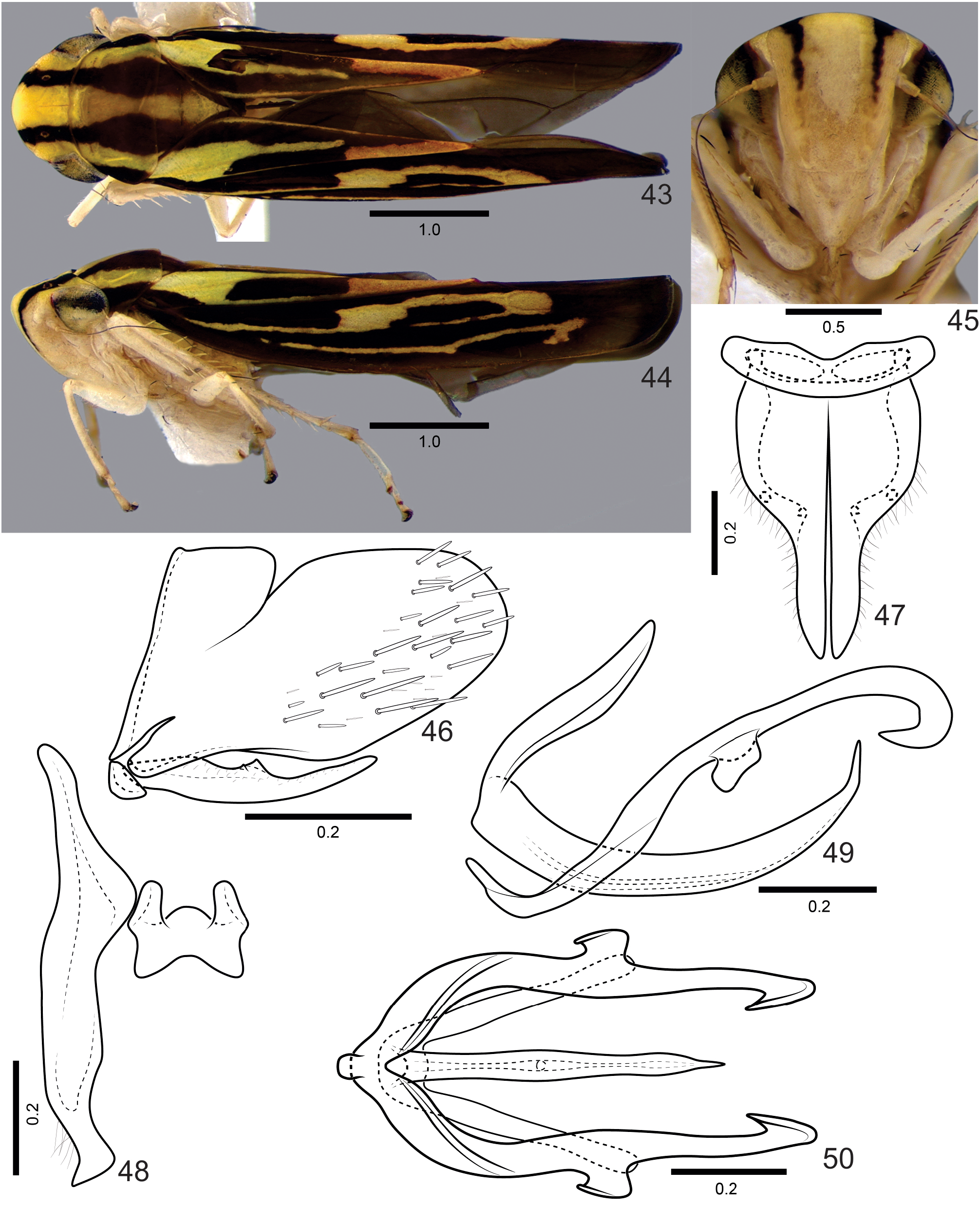
( Figs 43–60 View FIGURES 43–50 View FIGURES 51–60 )
Etymology. The specific epithet, unciformis , refers to the hook-shaped apical portion of the paraphyses rami in lateral view ( Fig. 49 View FIGURES 43–50 ).
Total length (mm). Male holotype 5.63; female paratype 5.69.
Color ( Figs 43–45 View FIGURES 43–50 ). Dorsum brown with three longitudinal yellow stripes extending from anterior margin of crown to apex of clavus; median stripe narrowed posteriorly from median portion of pronotum, continuing as narrow line along commissural margins; lateral stripes strongly narrowed on median portion of clavus. Corium with irregular yellow stripe extending from anterior portion of brachial cell to inner anteapical cell, strongly narrowed along anterior third; very narrow yellow stripe extending longitudinally near costal margin, posteriorly connected to yellow macula on median and outer anteapical cells.
Male terminalia. Pygofer ( Fig. 46 View FIGURES 43–50 ), in lateral view, moderately produced posteriorly; posterior margin broadly rounded; without processes; sparse macrosetae of distinct sizes distributed mostly on posterior portion and extending anteriorly over ventral portion. Valve ( Fig. 47 View FIGURES 43–50 ), in ventral view, short and broad, subrectangular; anterior margin concave. Subgenital plate ( Figs 46, 47 View FIGURES 43–50 ), in ventral view, with basal half broad and apical half narrow, outer margin rounded at basal half; plate fused along basal third to its counterpart; without macrosetae; in dorsal view, with two tiny dentiform processes at apical portion of basal half, not located close to each other, anterior process associated with style apex; in lateral view, plate not extending as far posteriorly as pygofer apex. Connective ( Fig. 48 View FIGURES 43–50 ), in dorsal view, subquadrate, without stalk, with slight median keel. Style ( Fig. 48 View FIGURES 43–50 ), in dorsal view, extending farther posteriorly than connective; apophysis without preapical lobe; apical portion strongly narrowed, bearing setae; apex truncate, foot-shaped. Aedeagus ( Figs 49, 50 View FIGURES 43–50 ) symmetrical; shaft, in lateral view, elongate, subcylindrical, curved dorsally, with acute apex; without processes; gonopore located ventrally at apical third. Paraphyses ( Figs 49, 50 View FIGURES 43–50 ), in lateral view, with elongate rami, each one with ventral subquadrate lobe at median portion and with apex curved ventrally, hook-shaped.
Female terminalia. Sternite VII ( Figs 51–53 View FIGURES 51–60 ), in ventral view, with posterior margin trilobed; median lobe broad, subquadrate. “Internal” sternite VIII without distinct sclerites. First valvifer ( Figs 54, 55 View FIGURES 51–60 ), in lateral view, somewhat trapezoidal; anterior portion with sclerotized bifurcated structure with short dorsal and long ventral branch associated with first valvula (indicated by an arrow in Fig. 54 View FIGURES 51–60 and magnified in Fig. 55 View FIGURES 51–60 ); surface of this structure distinctly covered by tegumentary processes. First valvula ( Figs 54, 56 View FIGURES 51–60 ), second valvula ( Figs 57–59 View FIGURES 51–60 ), and gonoplac ( Fig. 60 View FIGURES 51–60 ) much as described for F. spinosus sp. nov. Second valvula with about 22 teeth.
Type material. Holotype: male, “ Brasil, Minas Gerais, \ São Roque P. N. [Parque Nacional] Serra \ da Canastra \ 14–19.xii.2013 Malaise \ Melo & Rosa legs.” ( DZUP) . Paratypes: one female, same data as the holotype ( DZUP) ; one female, “BRA [ Brazil], MG [Minas Gerais], Serra do Salitre, \ RPPN [Reserva Particular do Patrimônio Natural] Cachoeira do Campo \ (19º09′45,7”S / 46º34′01,9”W, \ alt. 1063m) 11–15.X.2012 \ Lima & Kumagai col.” ( DZUP) GoogleMaps .
Remarks. Males of F. unciformis sp. nov. can be recognized by the subquadrate lobe at the median portion and the hook-shaped apex of the paraphyses rami ( Figs 49, 50 View FIGURES 43–50 ). In females, the posterior margin of the sternite VII is characteristically trilobed ( Fig. 53 View FIGURES 51–60 ) and, as aforementioned, a peculiar bifurcate structure is associated with the first valvifer and valvula of the ovipositor ( Figs 54, 55 View FIGURES 51–60 ).

| DZUP |
Universidade Federal do Parana, Colecao de Entomologia Pe. Jesus Santiago Moure |
No known copyright restrictions apply. See Agosti, D., Egloff, W., 2009. Taxonomic information exchange and copyright: the Plazi approach. BMC Research Notes 2009, 2:53 for further explanation.
|
Kingdom |
|
|
Phylum |
|
|
Class |
|
|
Order |
|
|
Family |
|
|
Genus |