
Characodoma latisinuatum, Harmer, 1957
|
publication ID |
https://doi.org/ 10.11646/zootaxa.4419.1.1 |
|
publication LSID |
lsid:zoobank.org:pub:03CAFD21-185F-4C86-ACC3-8CEB61E7F7DD |
|
DOI |
https://doi.org/10.5281/zenodo.3799568 |
|
persistent identifier |
https://treatment.plazi.org/id/CF6D87AA-E860-D27E-FF7D-F9430EA9FB9C |
|
treatment provided by |
Plazi |
|
scientific name |
Characodoma latisinuatum |
| status |
|
‘Characodoma’ latisinuatum Harmer, 1957 View in CoL
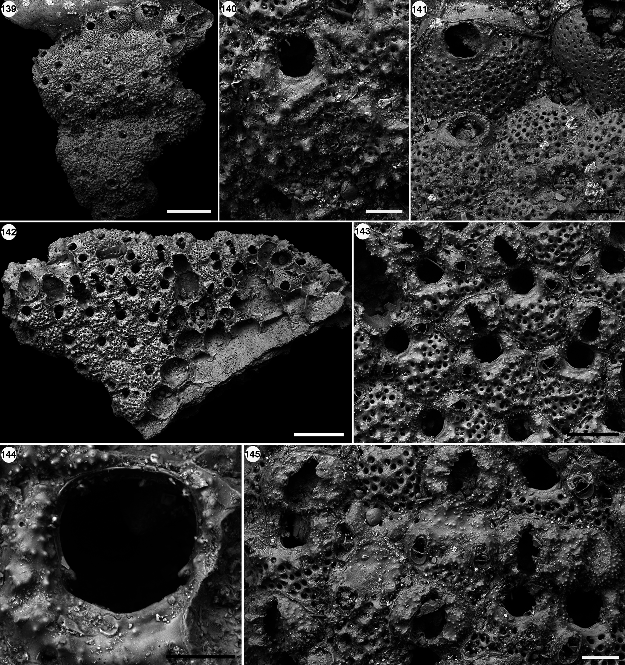
( Figs 139–145 View FIGURES 139–145 ; Table 31)
Characodoma latisinuatum Harmer, 1957: 1007 View in CoL , pl. 68, figs 28–30.
“ Characodoma View in CoL ” latisinuatum: Cook & Bock, 1996: p. 85 View in CoL , figs 18, 19.
Figured material. RGM.1350582, RGM.1350583, RGM.1350584, Holocene, UPGG 041, off South Sulawesi.
Description. Colony characterized by an encrusting phase covering minute fragments of shells, sometimes on both sides, and subsequently developing an erect phase. Autozooids arranged quincuncially, distinct by shallow
interzooidal furrows, hexagonal to irregularly polygonal, slightly longer than wide (mean L/W = 1.14); porechamber windows small (about 20 µm long), transversely oval, visible on the inner vertical walls of broken or incomplete zooids. Frontal shield convex, evenly pseudoporous except around the orifice, at first smooth becoming finely tuberculate and almost spinous with ontogeny; pseudopores numerous, small (12–18 µm in diameter), circular; marginal areolar pores undistinguishable. Primary orifice cleithridiate, almost equidimensional, a rounded anter separated from a widely arcuate sinus by two small, triangular, proximomedially directed condyles. Oral spines absent. Avicularia adventitious, commonly single, sometimes paired, monomorphic, triangular, placed lateral and pointing the orifice; rostrum raised and crossbar complete. Ooecia prominent, globular, broader than long, finely tuberculate, with an uncalcified frontal slit, almost as long as the entire ooecium (about 150 µm long) and usually broadest at its mid-length (60–80 µm wide), becoming closed only by the development of secondary calcification, which may also seal the orifice.
Remarks. Fourteen specimens of ‘Characodoma’ latisinuatum were found in our samples, mostly as encrusting phases on bivalve and gastropod shell fragments. Harmer (1957) described ‘ C.’ latisinuatum from the Makassar Strait at 59 m depth. Cook & Bock (1996, p. 86) pointed out that the generic attribution of this species requires further investigation. ‘Characodoma’ latisinuatum differs from the type species of Characodoma , as well as other species assigned to the genus, in having a pseudoporous frontal shield and mature ovicells that are uncalcified frontally. The Miocene Australian Characodoma rotundum ( Waters, 1881) is, at first sight, very similar to ‘C.’ latisinuatum in having a tuberculate frontal shield, though imperforate, and oral, unilateral avicularium pointing the orifice. The specimens figured by Cook & Bock (1996) also have incompletely calcified ovicells, accentuating the similarity. Anchicleidochasma mirabile ( Harmer, 1957) is also superficially very similar because of its marginal areolar pores migrating distally and the oral unilateral avicularium, but differs in having a completely calcified hood-like ovicell, overhanging a subtriangular opening ( Soule et al. 1991).
N, Number of colonies and number of zooids measured; SD, standard deviation.
| RGM |
National Museum of Natural History, Naturalis |
No known copyright restrictions apply. See Agosti, D., Egloff, W., 2009. Taxonomic information exchange and copyright: the Plazi approach. BMC Research Notes 2009, 2:53 for further explanation.
|
Kingdom |
|
|
Phylum |
|
|
Class |
|
|
Order |
|
|
SubOrder |
Neocheilostomina |
|
InfraOrder |
Ascophorina |
|
SuperFamily |
Mamilloporoidea |
|
Family |
|
|
Genus |
Characodoma latisinuatum
| Martino, Emanuela Di & Taylor, Paul D. 2018 |
Characodoma latisinuatum
| Harmer, S. F. 1957: 1007 |