
Kokkocynips Pujade-Villar & Melika, 2013
|
publication ID |
https://doi.org/ 10.11646/zootaxa.4938.2.3 |
|
publication LSID |
lsid:zoobank.org:pub:D16922EF-8508-49DB-8238-1D836B598749 |
|
DOI |
https://doi.org/10.5281/zenodo.4579773 |
|
persistent identifier |
https://treatment.plazi.org/id/C82E87E2-FF82-FFDB-42A0-C4245E84F7F7 |
|
treatment provided by |
Plazi |
|
scientific name |
Kokkocynips Pujade-Villar & Melika, 2013 |
| status |
|
Kokkocynips Pujade-Villar & Melika, 2013
Type species: Kokkocynips doctorrosae Pujade-Villar, 2013 .
Diagnosis. Kokkocynips differs from all other known Cynipini genera by the following combination of characters: Malar (subocular) sulcus absent or sometimes visible in the form of a short and shallow impressed area marked by some irregular striae; rarely complete, but weakly impressed. Facial striae radiating from clypeus indistinct or very short to absent. Anterior and posterior tentorial pits large and very deeply impressed and distinctive. Epistomal line strongly curved. Gular sulci widely separated at hypostomata. Female antennae 14-segmented, F1 1.4 to 2x as long as F2. Notauli percurrent, well-marked. Median mesoscutal impression either absent or if present only short and weakly impressed. Scutellar foveae rounded or ovate, usually indistinctly margined posteriorly; widely separated in K. panamensis n. sp. but narrowly in other species. Mesoscutum with delicate and shiny coriaceous sculpture, almost smooth in some species. Mesopleuron medially with a longitudinal band of coriaceous sculpture, sometimes also with some longitudinal rugae or carina; upper and lower parts of mesopleuron reticulate in the type species K. doctorrosae , but more usually without visible sculpture. Lateral propodeal carinae distinct, more often subparallel but slightly divergent dorsally and curved ventrally in K. panamensis n. sp.; central and lateral propodeal areas dull rugose. Forewings with open radial cell; distal margin of forewing with fringe of short setae. Metatarsal claws simple, without acute basal lobe or tooth. Metasoma with a group of sparse setae latero-basally. Terga entirely and regularly reticulate in the type species; with conspicuous micropunctures to almost smooth in other species. Projecting part of hypopygial spine long, 4.5x to 6x as long as wide in ventral view, laterally with some setae, basal setae longer while the apical ones are short and not reaching apex of the spine.
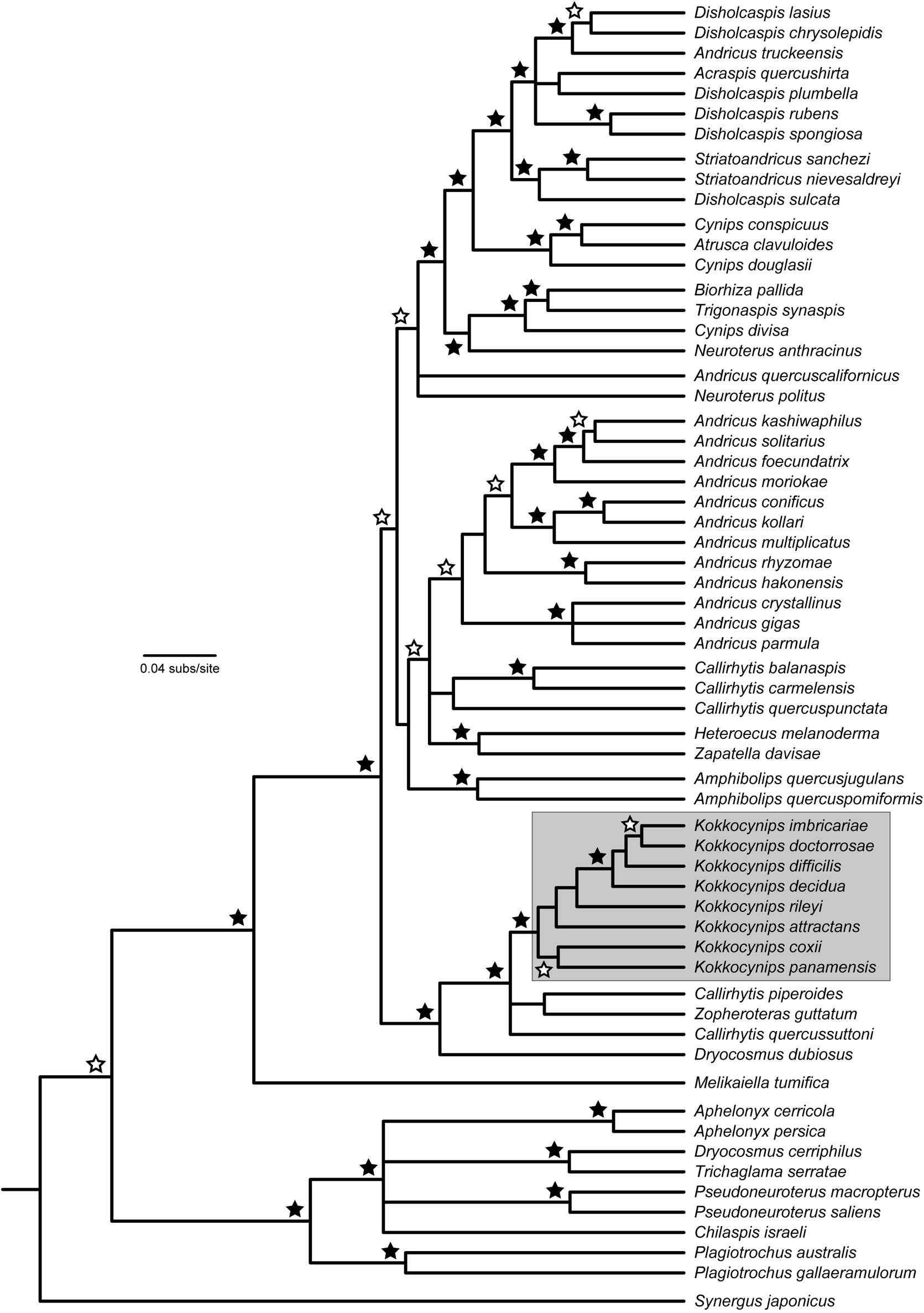
One of the diagnostic morphological characters mentioned in the original description in support of a monotypic Kokkocynips was the presence of a striking feature, namely the reticulate sculpture on the metasomal terga ( Pujade-Villar et al. 2013). A similar morphological feature is present in a group of Andricus species ( A. georgei Pujade-Villar, 2011 and A. nievesaldreyi Pujade-Villar, 2011 ), leading to comparison of Kokkocynips with this group. However, as mentioned in Pujade-Villar et al. (2013), these species have toothed claws, are associated with white oaks and have recently been moved into their own new genus Striatoandricus ( Cuesta-Porta et al. 2020) . In fact, Kokkocynips is placed in a very different part of the phylogeny to Striatoandricus (see Fig. 1 View FIG ), implying that the distinct sculpturing on the metasomal tergites shared by these genera has been independently derived. Hence it can be concluded that within the Cynipini this character of reticulate sculpturing of the metasoma is not reliable for assessing relationships among genera but only for determining relatedness within genera.
In the original description of Kokkocynips the new genus and species was not compared with some Nearctic species that were originally classified within Dryocosmus and Callirhytis , which we show here share other important diagnostic characters such as the simple tarsal claw, similar antennae, particular sculpturing of the mesoscutum and mesopleuron, their gall morphology and an association with red oaks. Hence, we provide the following more detailed diagnoses.
Within the group of the Cynipini genera with a transscutal fissure and fully winged asexual females, Kokkocynips is most similar to Dryocosmus , Callirhytis , Loxaulus and Plagiotrochus . Kokkocynips differs from Dryocosmus (sensu Giraud 1859, not Weld 1926b), in the long spine of the hypopygium without long subapical setae, absence of radiating facial striae, presence of a band of sculpture on the mesopleuron, and the absence of a median carina in the propodeal sculpture. It differs from Plagiotrochus in lacking radiating facial striae from the clypeus, the relatively longer hypopygial spine and difference in sculpture of the mesoscutum. The indistinct malar sulcus, distal forewing margin with setae, the sculpture of propodeum, and the structure of the hypopygial spine, readily differentiate Kokkocynips from Callirhytis and the allied genera Melikaiella and Zapatella . Kokkocynips differs from Andricus by having simple metatarsal claws, not having a whitish band of dense setae on metasomal terga, nor radiating striae from the clypeus reaching or almost reaching the ventral margin of the eye, and having a weak but still discernible malar sulcus (if the metasoma is not reticulated). It differs from Belonocnema by the apex of the fore tibia, which in Belonocnema extends far behind the base of fore tarsomere I, and the 2nd abscissa of Rs is strongly curved. Loxaulus differs from Kokkocynips by the finely transversely coriaceous sculpture of the mesoscutum, the presence of the median propodeal carina and the short prominent part of the ventral spine of the hypopygium, which is always less than 3.5 times as long as broad while in Kokkocynips it is always longer. Finally, Erythres differs from Kokkocynips by the sculpture of the head, mesosoma and dorsal surface of legs, which are uniformly and finely punctate-rugose; the notauli are also incomplete in Erythres .
Re-description
Asexual female largely glabrous, hardly pubescent. Head in anterior view wider than high, genae slightly expanded behind eyes. Facial striae radiating from clypeus very short, barely distinctive. Anterior and posterior tentorial pits large and very deeply impressed and distinctive. Epistomal line strongly curved. Clypeus trapezoidal with ventral margin sinuate, moderately projecting over mandibles. Malar sulcus absent or only present in the form of a shallow impressed area; rarely complete but weak impressed. Head, in posterior view, without occipital carina. Gular sulci widely separated at hypostomata. Antenna 14-segmented, F1 1.4 to 2x as long as F2. Pronotum short medially, pronotal plate indistinct dorsally. Mesoscutum reticulate or delicately coriaceous to almost smooth and shiny in the middle lobe, overall weakly pubescent. Notauli percurrent, deeply impressed. Median mesoscutal impression present, usually short and weakly impressed. Transscutal fissure present. Scutellar foveae rounded or oval, more or less widely separated medially and usually with indistinct posterior margins. Mesopleuron medially with a longitudinal band of fine coriaceous sculpture variably extended, some weak longitudinal rugae also present in some species; the upper and lower parts of the mesopleuron smooth except in K. doctorrosae . Lateral propodeal carinae distinct, slightly divergent dorsally and curved ventrally. Metatarsal claws simple, without acute basal lobe or tooth. Forewing with open radial cell along anterior margin; areolet triangular; apical margin of wing with short hair fringe.
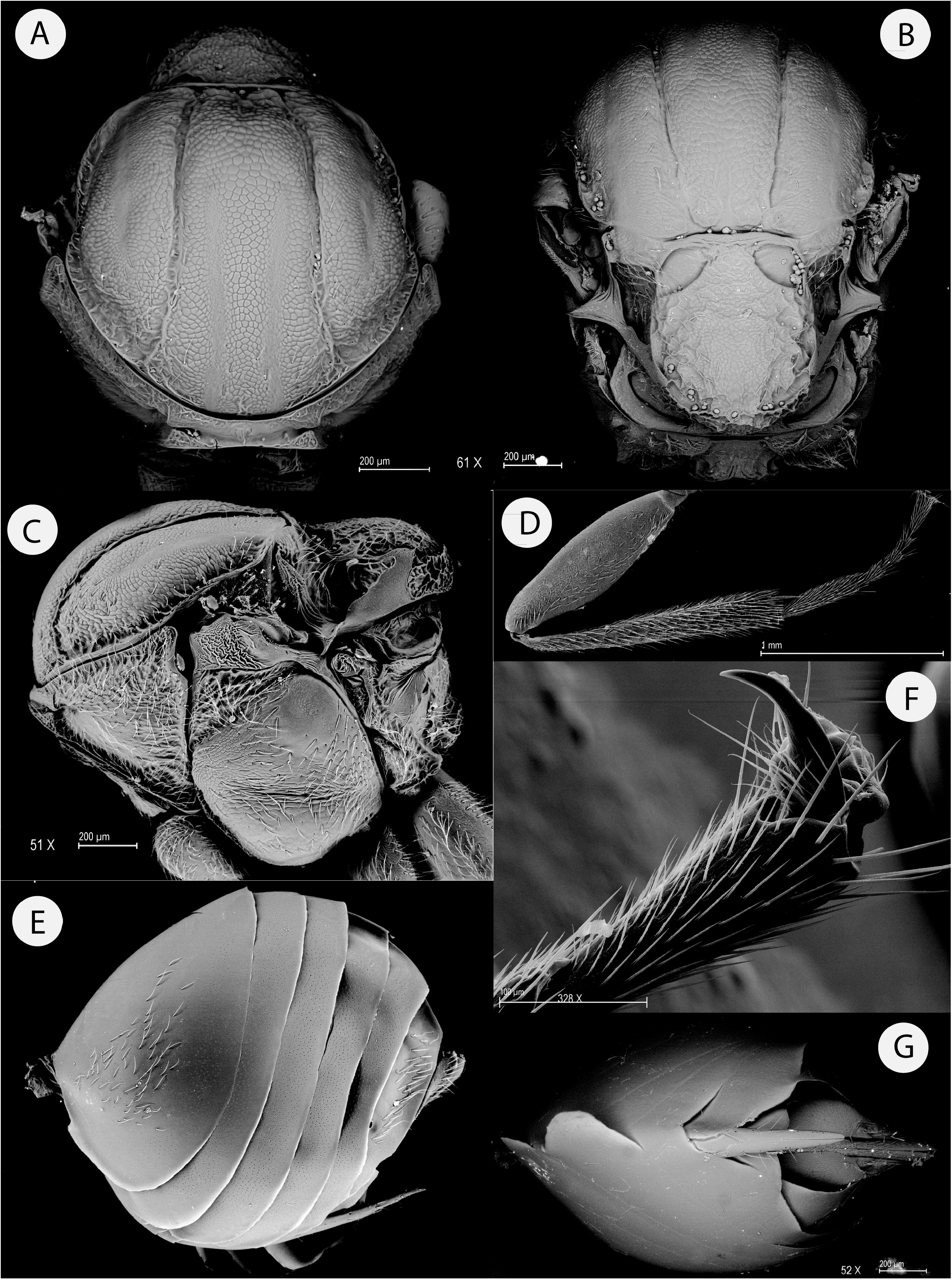
Metasoma with a group of sparse setae latero-basally. Projecting part of hypopygial spine long, about 4.5 to 6 times as long as wide in ventral view, laterally with some setae, basal setae longer, the apical ones short and not reaching apex of the spine ( Figs. 3 View FIG E-G).
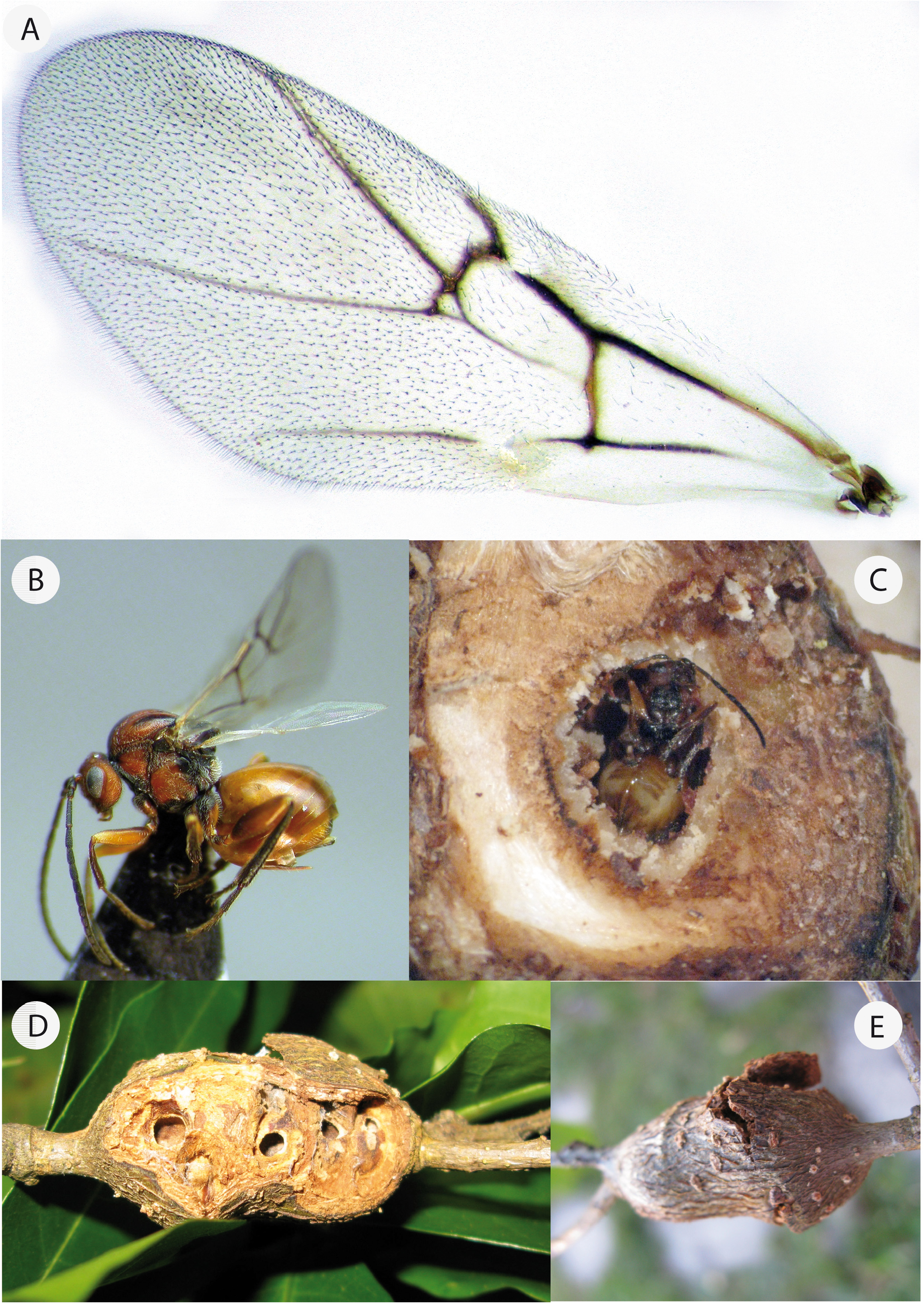
Gall structures: the galls induced by asexual forms of Kokkocynips species range from multilocular (manychambered) stem swellings to unilocular (single-chambered) solitary or aggregated galls on stems or leaves ( Figs. 4 View FIG D-F and 11).
Host plant association: Kokkocynips species are associated only with red oaks, in Quercus section Lobatae.
Biology: all but one Kokkocynips species are known only from their asexual generations. However, Callirhytis attractans (Kinsey, 1922) (= Callirhytis uvellae Weld, 1944 ), that our phylogenetic analysis shows is included in the genus Kokkocynips , is known only from its sexual generation.
Distribution: Nearctic and Neotropical. Currently known from Panama, Mexico, the United States and Canada.
Key to asexual females of Kokkocynips
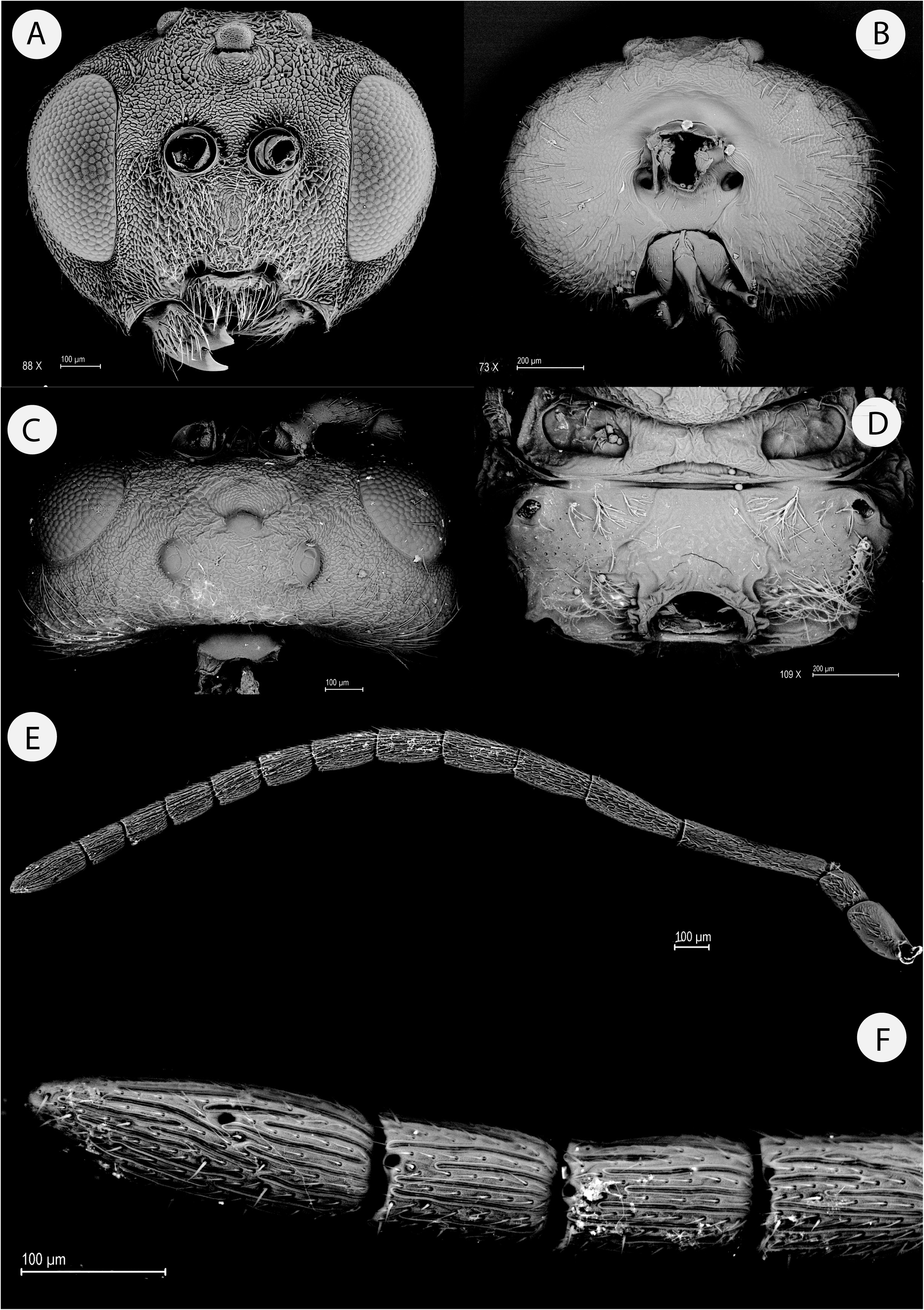
1. Scutellar foveae with inner margins widely separated by a broad septum; with distinct posterior margins ( Fig. 3B View FIG ). Lateral carinae of propodeum narrow and clearly divergent in anterior 2/3 of propodeum ( Fig. 2D View FIG ). Head and body contrasting in colour with antennae and metatibia ( Fig. 4B View FIG )..................................................... K. panamensis n. sp.
• Scutellar foveae with inner margins in contact anteriorly or narrowly separated by a carina or narrow septum; posterior margins usually indistinct ( Figs. 5B View FIG , 6C View FIG , 7C View FIG , 8C View FIG ). Lateral carinae of propodeum usually broader, subparallel or only slightly divergent ( Figs. 7E View FIG , 8E View FIG , 9E View FIG ). Head and body usually not contrasting in colour with legs and antennae ( Figs. 10A, 10C, 10D, 10E, 10F View FIG ) 2
2. Metasomal terga with regular coriaceous-reticulate sculpture ( Figs. 5 View FIG F-G). Sculpture of mesopleuron extended beyond the medial area towards upper and lower areas of mesopleuron ( Fig. 5C View FIG ). Scutellar foveae with rugose sculpture ( Fig. 5B View FIG )................................................................................. K. doctorrosae Pujade-Villar
• Metasomal terga punctate to almost smooth ( Figs. 6E View FIG , 7F View FIG , 8F View FIG , 9F View FIG ). Sculpture of mesopleuron usually present only in the medial area; upper and lower areas of mesopleuron smooth ( Fig. 6D View FIG , 7D View FIG , 8D View FIG , 9D View FIG ). Scutellar foveae smooth or only with some longitudinal rugae ( Figs. 6C View FIG , 7C View FIG , 8C View FIG , 9C View FIG )...................................................................... 3
3. Head, mesosoma and metasoma dark brown to black ( Fig. 10A View FIG ). Notauli subparallel or only slightly converging towards the transscutal fissure; mid lobe of mesoscutum almost smooth ( Fig. 10B View FIG )..................... K. decidua (Beutenmueller)
• Head, mesosoma and metasoma amber to reddish ( Figs. 10C, 10D, 10E, 10F View FIG ). Notauli more converging towards transscutal fissure ( Figs. 6C View FIG , 7C View FIG , 9C View FIG ); mid lobe of mesoscutum with fine coriaceous sculpture ( Figs. 7C View FIG , 8C View FIG , 9C View FIG ).................. 4
4. Malar sulcus well defined but weakly impressed ( Fig. 7B View FIG ). Projection of ventral spine of hypopygium relatively short, less than 4x as long as broad in lateral view ( Fig. 7H View FIG ). Scutellum not marginated and coriaceus at its centre ( Fig. 7E View FIG ); scutellar foveae smooth, not margined posteriorly. Metasomal punctures absent ( Fig. 7F View FIG ). Mesosoma, metasoma and coxae darker than rest of body in lateral view ( Fig. 10C View FIG ).............................................................. K. coxii (Basset)
• Malar sulcus usually shallowly impressed, sometimes indistinct ( Figs. 6A View FIG , 8B View FIG , 9B View FIG ). Projection of ventral spine of hypopygium longer, more than 4 times as long as broad in lateral view ( Figs. 6E View FIG , 8H View FIG , 9H View FIG ). Scutellum marginated and surface rugose ( Figs. 6 View FIG C-D, 8C-D, 9C-D); scutellar foveae smooth or with some longitudinal rugae, margined posteriorly in K. difficilis ( Figs. 9C, 9E View FIG ). Metasomal punctures sometimes weak but present ( Figs. 6E View FIG , 8F View FIG , 9F View FIG ). Mesosoma, metasoma and coxae concolorous with the rest of body in lateral view ( Figs. 10D, 10E, 10F View FIG )........................................................ 5
5. Notauli strongly converging posteriorly, only slightly separated at the junction with the transscutal fissure ( Fig. 6C View FIG ); scutellar foveae with indistinct posterior margins, separated medially by a longitudinal carina ( Fig 6C View FIG ).... K. imbricariae (Ashmead)
• Notauli only weakly converging posteriorly, more widely separated at the junction with the transscutal fissure ( Figs. 8C View FIG , 9C View FIG ); scutellar foveae posteriorly margined or not, separated medially by an anterior contact point or by a carina ( Figs. 8C View FIG , 9C View FIG ).. 6
6. Projection of ventral spine of hypopygium relatively short, 4.5x as long as wide in lateral view ( Fig. 7H View FIG ). Scutellar foveae with some rugae; not margined posteriorly. ( Fig. 7C View FIG ).............................................. K. rileyi (Ashmead)
• Projection of ventral spine of hypopygium long, 6x as long as wide in lateral view ( Fig. 8H View FIG ). Scutellar foveae smooth; with distinct margins ( Fig. 8C View FIG )............................................................. K. difficilis (Ashmead)


No known copyright restrictions apply. See Agosti, D., Egloff, W., 2009. Taxonomic information exchange and copyright: the Plazi approach. BMC Research Notes 2009, 2:53 for further explanation.