
Anastrepha mitaraka Norrbom, 2021
|
publication ID |
https://doi.org/ 10.11646/zootaxa.5044.1.1 |
|
publication LSID |
lsid:zoobank.org:pub:6102257B-B360-44DC-BDF5-9B4711B2A541 |
|
DOI |
https://doi.org/10.5281/zenodo.5532052 |
|
persistent identifier |
https://treatment.plazi.org/id/E1986F6E-DE16-4F86-9D18-7E392CFAB135 |
|
taxon LSID |
lsid:zoobank.org:act:E1986F6E-DE16-4F86-9D18-7E392CFAB135 |
|
treatment provided by |
Plazi |
|
scientific name |
Anastrepha mitaraka Norrbom |
| status |
sp. nov. |
Anastrepha mitaraka Norrbom View in CoL , new species
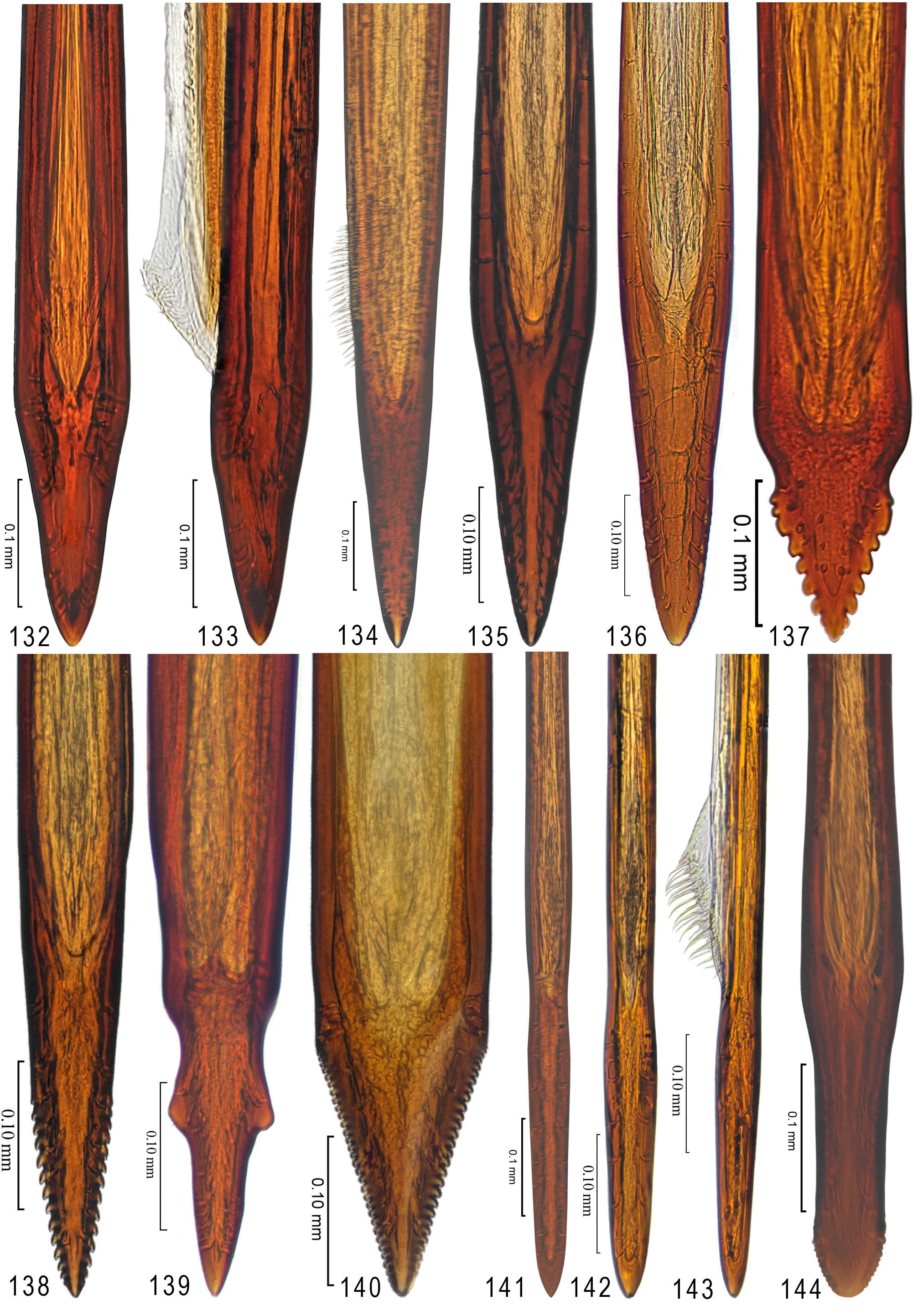
Figs. 11 View FIGURES 11–12 , 54 View FIGURES 43–54 , 109 View FIGURES 98–120 , 134 View FIGURES 132–144
urn:lsid:zoobank.org:act:E1986F6E-DE16-4F86-9D18-7E392CFAB135
Diagnosis. Anastrepha mitaraka can be distinguished from other species of Anastrepha by the following combination of characters: V-band complete, connected to S-band; subapical hyaline area in radial cells distal to crossvein r-m extending anteriorly into cell r 1; oviscape 0.79 times as long as mesonotum; aculeus 2.34 mm long, ventrally curved; tip 0.25 mm long, nonserrate. In the key of Zucchi et al. (2011) it runs to A. flavipennis Greene , from which it differs in having cell bm hyaline and the aculeus more than 2.0 mm long, or A. belenensis Zucchi , from which it differs in having the V-band complete and connected to the S-band, the subapical hyaline area in the radial cells extending into cell r 1, and lacking a constriction near midlength on the aculeus tip.
One sequence of A. mitaraka ( MT 763869 View Materials ), from the holotype from French Guiana, was included in the COI barcode analysis of Moore et al. (in prep.). The smallest interspecific K2P distance was 5.6% with A. flavipennis Greene .
Description. Mostly yellow to orange. Setae orange brown to red brown.
Head: Yellow to orange except brown ocellar tubercle. 3 frontal setae; 2 orbital setae, posterior seta well developed. Ocellar seta weak, approximately as long as ocellar tubercle. Facial carina, in profile, straight on dorsal twothirds. Antenna not extended to ventral facial margin. Palpus in lateral view dorsally curved, evenly setulose.
Thorax: Mostly yellow to orange, most of scutum orange; with following areas white to pale yellow (poorly differentiated in holotype): postpronotal lobe and lateral margin of scutum bordering it; medial scutal vitta, broadly quadrate posteriorly, extending laterally to dorsocentral seta; sublateral scutal vitta from transverse suture to posterior margin, including base of intra-alar seta; scutellum; dorsal margins of anepisternum and katepisternum; katepimeron; and most of anatergite and katatergite. Scuto-scutellar suture without brown spot medially. Subscutellum and mediotergite entirely orange. Mesonotum 3.32 mm long. Postpronotal lobe, notopleuron, scutum and scutellum entirely microtrichose; scutal setulae orange, evenly distributed sublaterally. Chaetotaxy typical for genus. Katepisternal seta absent.
Legs: Entirely yellow to orange.
Wing ( Fig. 54 View FIGURES 43–54 ): Length 7.09 mm, width 3.08 mm, ratio 2.30. Apex of vein R 1 at 0.55 wing length, proximal to level of anterior end of crossvein r-m. Cell c 1.19 times as long as pterostigma; pterostigma 3.41 times as long as wide. Vein R 2+3 slightly sinuous. Crossvein r-m at 0.64 distance from bm-m to dm-m on vein M 1. Vein M 1 strongly curved apically; cell r 4+5 at apex 0.90 times as wide as at level of dm-m, 0.69 times as wide as maximum subapical width. Cell cu a with distal lobe moderately long, length of cu a 1.66 times as long as anterior margin, lobe 0.87 times as long as vein CuA+CuP. Wing pattern mostly orange and moderate brown. C-band mostly orange, cells bc and c orange, pterostigma mostly brown, distal margin in cells r 1 and r 2+3 narrowly brown, with small ovoid dark brown spot on fork of vein Rs, and with elongate faint brown marking in cell br opposite cell bm and with posterior margin of band in cell br narrowly brown. C-band and S-band broadly connected along vein R 4+5; hyaline marginal spot in cell r 1 subtriangular, with apex aligned proximal to anterior end of crossvein r-m. Basal hyaline area in cell dm relatively small. Basal half of S-band relatively broad, mostly orange, anterobasal margin mostly narrowly brown in cells r 1, r 2+3 and br, posterodistal margin mostly narrowly brown, but broadly posteriorly in cell m 4 extending to apex of lobe of cell cu a, with weak incision in cell m 4 (this area with teneral streak in holotype, perhaps normally without incision); distal section mostly orange, with margins in cells r 1 and r 2+3 and all of part in cell r 4+5 moderate brown; medium width, at apex of vein R 2+3 0.56 times width of cell r 2+3, slightly broader along vein R 4+5, barely extended to apex of vein M 1, without marginal hyaline areas; hyaline area proximal to apex of band extended into cell r 1. V-band with proximal arm moderately broad, mostly brown except for most of part in cell r 4+5, except proximal margin, and bordering most of crossvein dm-m; broadly connected to S-band along vein R 4+5; on posterior margin extended approximately three-fourths distance to vein CuA+CuP; distal arm slender, brown except anteriorly, connected to proximal arm; hyaline area between arms of V-band and vein M 1 approximately one-third width of cell cell r 4+5.
Abdomen: Mostly orange, without brown markings.
Female terminalia: Oviscape 2.61 mm long, 0.79 times as long as mesonotum; entirely orange; spiracle at basal 0.33. Eversible membrane not everted, partially visible through oviscape, with approximately 20 dorsobasal denticles in subtriangular pattern. Aculeus ( Fig. 109 View FIGURES 98–120 ) slightly ventrally curved in lateral view, 2.34 mm long, 0.90 times oviscape length; in ventral view base distinctly expanded, triangular, 0.23 mm wide, shaft 0.12 mm wide at midlength; tip ( Fig. 134 View FIGURES 132–144 ) 0.25 mm long, 0.11 times aculeus length, 0.08 mm wide at base and preapically, 3.13 times as long as wide; in ventral view gradually tapered, nonserrate; 0.07 mm wide in lateral view, 0.88 times ventral width. Spermathecae not examined.
Distribution. Anastrepha mitaraka is known only from French Guiana.
Biology. The host plants and other aspects of the biology of this species are unknown.
Type data. Holotype ♀ ( MNHNP USNMENT00875225 ), FRENCH GUIANA: [Saint-Laurent du Maroni: Maripasoula,] Mitaraka , site MIT-DZ, 2°14’1.8”N 54°27’1”W, 306 m, 1 Mar 2015, SLAM, J. Touroult & E. Poirier / MITARAKA/218 La Planète Revisitée —MNHN/PNI Guyane 2015. GoogleMaps
Etymology. The name of this species is a noun in apposition, the name of the region where the holotype was collected.
Comments. This species has not been placed in a species group. In the COI analysis of Moore et al. (in prep.) it was placed as the sister group of A. flavipennis Greene.


| MT |
Mus. Tinro, Vladyvostok |
| MNHNP |
Museo Nacional de Historia Natural del Paraguay |
No known copyright restrictions apply. See Agosti, D., Egloff, W., 2009. Taxonomic information exchange and copyright: the Plazi approach. BMC Research Notes 2009, 2:53 for further explanation.
|
Kingdom |
|
|
Phylum |
|
|
Class |
|
|
Order |
|
|
Family |
|
|
Genus |