
Sicyonella antennata Hansen, 1919
|
publication ID |
https://doi.org/ 10.5281/zenodo.170671 |
|
DOI |
https://doi.org/10.5281/zenodo.6269039 |
|
persistent identifier |
https://treatment.plazi.org/id/C61387E9-1C52-FFF5-8C50-EB951B5E0E3C |
|
treatment provided by |
Plazi |
|
scientific name |
Sicyonella antennata Hansen, 1919 |
| status |
|
Sicyonella antennata Hansen, 1919 ( Figs. 1–5 View FIGURE 1 View FIGURE 2 View FIGURE 3 View FIGURE 4 View FIGURE 5 , 17 View FIGURE 17 A)
Sicyonella antennata Hansen, 1919: 30 , 31, pl. 2, fig. 5, pl. 3, fig. 1; Hayashi, 1992: 225 –227, figs. 120–122.
Type locality. Kai Islands, eastern part of the Banda Sea, Indonesia, 22 m.
Type specimens. Hansen did not designate type series.
Material examined. [ USNM Collection] USNM1026369: 1 male (cl 5.9 mm) and 2 females (cl 6.9 and 7.9 mm), Sitanki, Sibutu Island, Sulu Archipelago, Philippines, surface, electric light, 25 Feb. 1908, separated from USNM 260757. USNM173562: 1 male (cl 7.6 mm) and 1 female (cl 7.7 mm), Singapore, date unknown.
[ NSMT Collection] NSMTCr 16021: 1 male (cl 5.4 mm), Nagura Bay, Ishigaki Island, Okinawa, Japan, seagrass bed, 2–3 m, light trap, 27 Dec. 1998, coll. M. Tamaki. NSMTCr 14116: 5 males (cl 3.4–5.3 mm), pier of Ishigaki Tropical Station of Seikai National Fisheries Research Institute (ITS), Urasoko Bay, Ishigaki Island, Okinawa, Japan, sand bottom, 2–3 m, light trap, 1 Sept. 2000, coll. K. Fukuoka. NSMTCr 16022: 1 male (cl 5.6 mm), dissected, pier of ITS, Urasoko Bay, Ishigaki Island, Okinawa, Japan, sand bottom, 2–3 m, light trap, 18 Oct. 2000, coll. K. Fukuoka. NSMTCr 14117: 5 males (cl 4.3–4.9 mm), pier of ITS, Urasoko Bay, Ishigaki Island, Okinawa, Japan, sand bottom, 2–3 m, light trap, 5 Dec. 2000, coll. K. Fukuoka. NSMTCr 16023: 1 female (cl 5.7 mm), dissected, pier of ITS, Urasoko Bay, Ishigaki Island, Okinawa, Japan, sand bottom, 2–3 m, light trap, 26 Mar. 2001, coll. K. Fukuoka. NSMTCr 16024: 1 female (cl 5.8 mm), pier of ITS, Urasoko Bay, Ishigaki Island, Okinawa, Japan, sand bottom, 2–3 m, light trap, 26 Mar. 2001, coll. K. Fukuoka. NSMTCr 16025: 1 female (cl 6.9 mm), pier of ITS, Urasoko Bay, Ishigaki Island, Okinawa, Japan, sand bottom, 2–3 m, light trap, 23 Oct. 2001, coll. K. Fukuoka. NSMTCr 16026: 3 females (cl 4.0– 5.3 mm), Ibaruma Bay, Ishigaki Island, Okinawa, Japan, fishing port, surface by handnet under an electric light at night, 5 Nov. 2002, coll. K. Fukuoka. NSMTCr 16027: 1 female (cl 3.4 mm), pier of ITS, Urasoko Bay, Ishigaki Island, Okinawa, Japan, sand bottom, 2–3 m, light trap, 7 Nov. 2002, coll. K. Fukuoka.
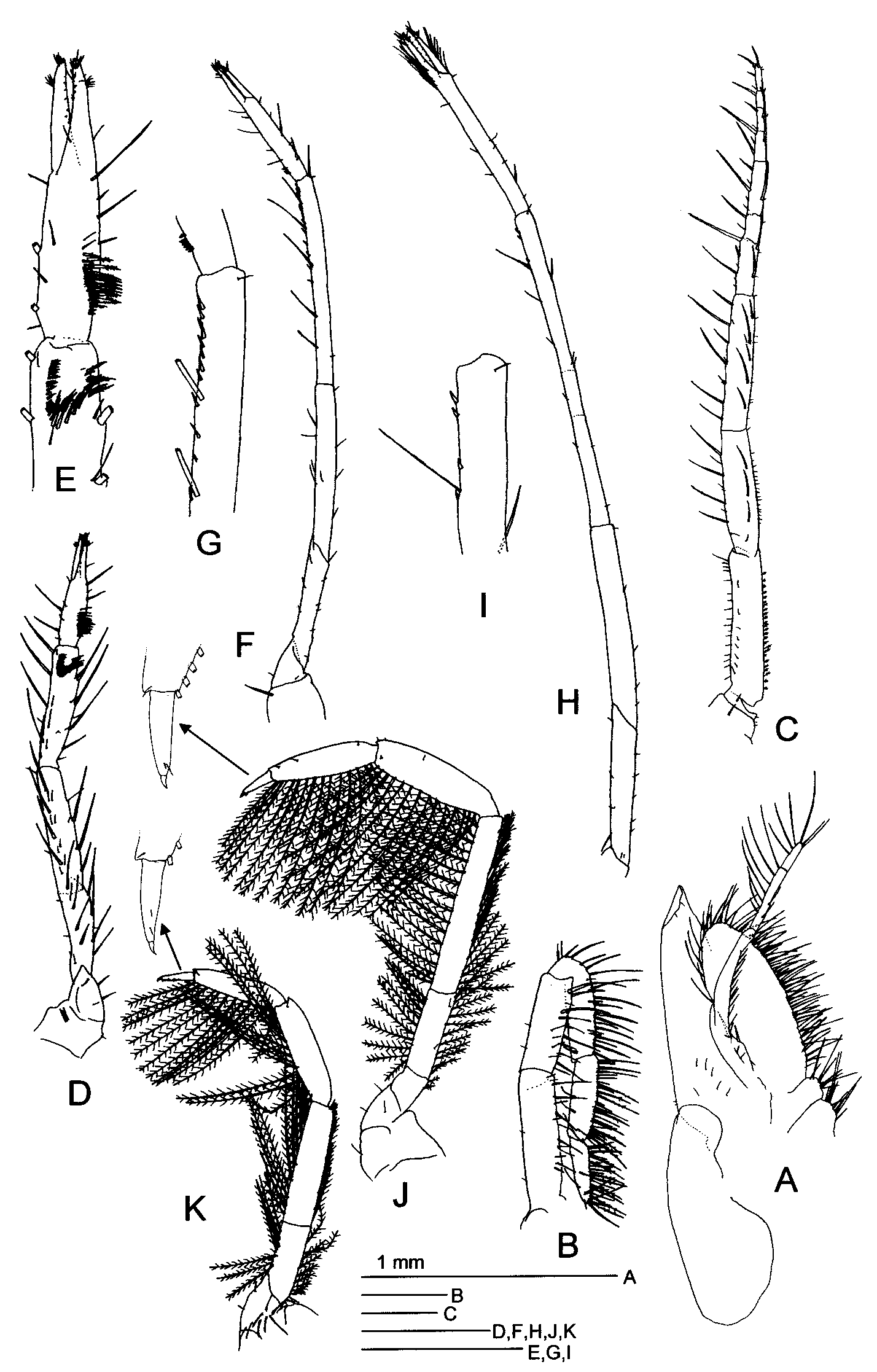
Description. Carapace with postorbital and hepatic spines; pterygostomian spine small. Rostrum short, extending to or slightly beyond base of antennular peduncle, with 2 teeth on dorsal margin ( Fig. 1 View FIGURE 1 A–C).
Abdomen smooth; sixth somite 1.8–1.9 times as long as fifth one, with indistinct dorsomedian carina except for posterior 0.3; pleuron of first to fifth somites expanded posteriorly, armed with setae on ventral margin. Telson tapering posteriorly to acute tip, 0.8 length of last abdominal somite, armed with 4 spines on distal 0.6 of ventral lateral margin ( Fig. 3 View FIGURE 3 K).
Eye elongated, 1.9–2.3 times as long as wide of cornea in male, and 2–2.3 times as long as wide of cornea in female ( Fig. 1 View FIGURE 1 A, B). Cornea large, 2.2–2.9 times as wide as base of stalk in male and 2–2.4 times as wide in female ( Fig. 1 View FIGURE 1 A, B). Eyestalk gradually wider distally, without orbital spines ( Fig. 1 View FIGURE 1 A–C).
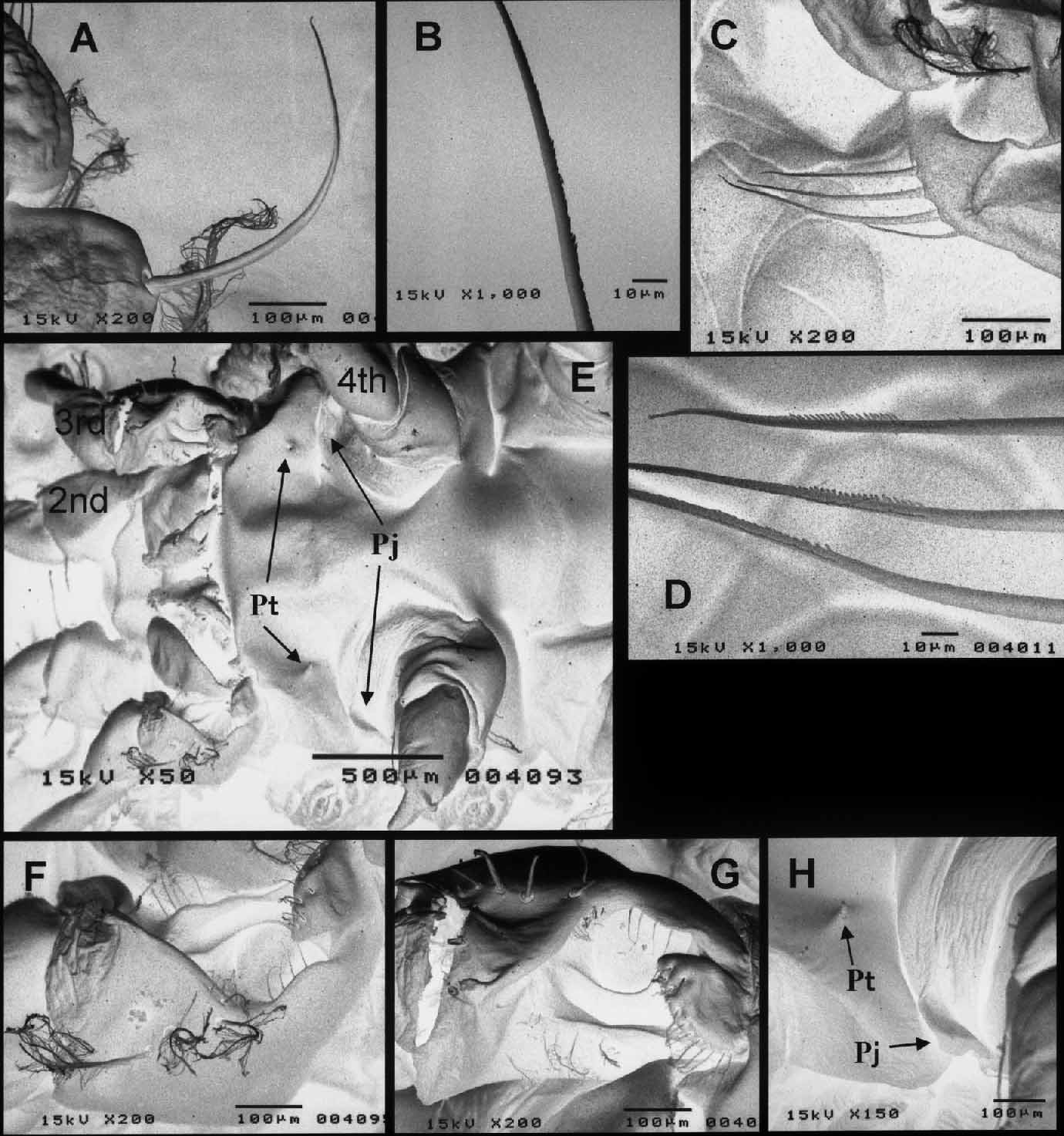
Antennular peduncle elongate ( Fig. 1 View FIGURE 1 A, B). Stylocerite short, not extending to middle of first segment of peduncle ( Fig. 1 View FIGURE 1 A). First segment of antennular peduncle reaching distal 0.3 of scaphocerite, with anterolateral spiniform process ( Fig. 1 View FIGURE 1 A, B). Second segment of antennular peduncle shows sexual dimorphic; in male long, overreaching apex of scaphocerite, 0.5–0.8 length of first segment, 2.6–3.5 times as long as wide ( Fig. 1 View FIGURE 1 A); in female robust, not reaching apex of scaphocerite, 0.4–0.5 length of first, 1.7–2.4 times as long as wide ( Fig. 1 View FIGURE 1 B). Third segment of antennular peduncle shows sexual dimorphic, elongated and 1–1.4 times longer than second segment in male, 0.9–1.1 times as long as second in female ( Fig. 1 View FIGURE 1 A, B). Distal two segments of antennular peduncle 1–1.7 times as long as proximal segment in male, and 0.8–1.1 times as long as proximal segment in female ( Fig. 1 View FIGURE 1 A, B). Mesial antennular flagellum of male with modified proximal part ( Fig. 4 View FIGURE 4 A); first segment armed on upper margin with 3 long, rather stout setae ( Fig. 4 View FIGURE 4 B); second segment short, armed on upper margin with 2 stout setae branched in terminal end ( Fig. 4 View FIGURE 4 B, C); third segment elongate, upper margin concave ( Fig. 4 View FIGURE 4 A), with 1 stout seta on predepression ( Fig. 4 View FIGURE 4 A, B), with 1 stout and 1 slender setae in middle ( Fig. 4 View FIGURE 4 A, D), and with 3 robust and 1 stout setae on postdepression, robust setae on postdepression scaly on posterior surface of distal part ( Fig. 4 View FIGURE 4 A, E).
Scaphocerite 3.6–3.7 times as long as wide, apical lobe triangular with rounded apex ( Fig. 1 View FIGURE 1 D). Antennal peduncle robust, extending to proximal 0.4 of scaphocerite ( Fig. 1 View FIGURE 1 D). Mandible with flattened, 3segmented palp; second segment of palp expanded in middle; third segment of palp 0.4 of second segment in length, 2–2.3 times as long as wide ( Fig. 1 View FIGURE 1 E). Incisor process developed, with acute process on distomesial corner, mesial margin smooth, divided into two portions ( Fig. 1 View FIGURE 1 E).
Maxillule with proximal endite spatulate with 3 long and several rather long, robust, spiniform setae on distal margin; distal endite broadened mesially, armed with numerous strong, spiniform setae on distal and mesial margins, and with long and short spiniform setae on distal half of lateral margin; endopod elongate, armed with 1 long, robust, naked, spiniform seta near apex of mesial margin and with 5–7 plumose setae on distal 0.3 of lateral margin to apex ( Fig. 1 View FIGURE 1 F).
Maxilla with endopod tapering distally, armed with 6–9 short spines on distal part; endite rudimentary; scaphognathite large, with rounded anterior and posterior lobes, armed densely with plumose setae on margin ( Fig. 1 View FIGURE 1 G).
First maxilliped with endopod long, slender, 3segmented, armed with 2 or 3 long spines on proximal expanded part; exopod large; epipod large, not bilobed; distal endite large, broadened distally, armed densely with setae on margin; proximal endite represented by two similar lobes, small, armed with setae on mesial margin ( Fig. 2 View FIGURE 2 A).
Second maxilliped without exopod and epipod. Ischium of endopod 1.1–1.3 times as long as merus; merus as long as carpus; distal 3 segments gradually shorter in length distally ( Fig. 2 View FIGURE 2 B).
Third maxilliped long, robust, extending to distal 0.3 of distal segment of antennular peduncle in male, and extending beyond distal end of antennular peduncle in female; ischium armed with short setae on mesial and lateral margins; merus 0.7–0.8 of ischium in length; carpus 1.1–1.2 times as long as merus; propodus as long as carpus, 3subsegmented by indistinct articulations; dactylus 0.7–0.8 length of carpus, 4subsegmented by indistinct articulations, terminating to robust, spiniform seta; distal 4 segments armed with short and long, robust, spiniform setae on mesial and lateral margins ( Fig. 2 View FIGURE 2 C).
First pereopod reaching middle of merus of third maxilliped, chelate; merus indistinctly divided from ischium; carpus slightly shorter than merus, armed on distal 0.3 of mesial surface with brushing setae arranged in a Vshape; propodus slightly shorter than carpus, armed with short and long brushing setae on proximal 0.3–0.4 of lower side of mesial surface; dactylus 0.7 of palm length, as long as propodal finger; cutting edge sparsely armed with minute setae ( Fig. 2 View FIGURE 2 D, E).
Second pereopod 1.3 times as long as first pereopod, chelate; merus indistinctly divided from ischium; carpus 1.3 times as long as merus, armed with small spines on distal half of lower margin; propodus 0.7–0.8 length of carpus, armed with minute, spiniform setae on near proximal end of lower margin; dactylus 0.4 of palm length, similar to propodal finger; cutting edge sparsely armed with minute setae ( Fig. 2 View FIGURE 2 F, G).
Third pereopod 1.3–1.4 times longer than second pereopod, chelate; merus divided from ischium by oblique articulation; carpus 1.7 times as long as merus, sparsely armed with spines on distal 0.2 of lower margin; propodus 0.7 of carpus in length; dactylus 0.3– 0.4 of palm length, as wide as propodal finger; cutting edge sparsely armed with short setae ( Fig. 2 View FIGURE 2 H, I).
Fourth pereopod 7segmented, 0.7 length of third pereopod, no chelate; ischium and merus armed with long plumose setae on lower and upper margins, merus 1.6 times as long as ischium; carpus and propodus armed with long plumose setae on lower margin; dactylus terminating in small claw ( Fig. 2 View FIGURE 2 J).
Fifth pereopod 7segmented, 0.7–0.8 length of fourth pereopod, similar to fourth pereopod in shape and armature ( Fig. 2 View FIGURE 2 K).
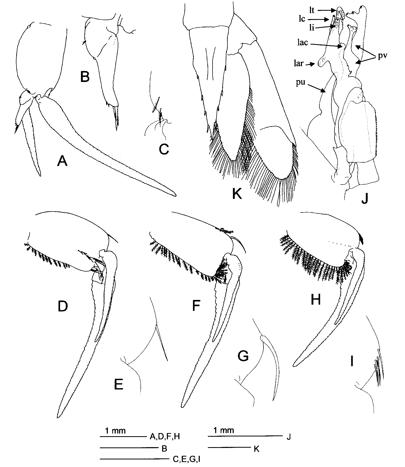
First pleopod lacking endopod; second to fifth pleopods biramous, exopod longer than endopod ( Fig. 3 View FIGURE 3 A, D, F, H). Sympod armed on distomesial angle with 2 or 3 short, plumose setae in second pleopod ( Fig. 3 View FIGURE 3 C), with long spiniform seta in third pleopod ( Fig. 3 View FIGURE 3 D, E), with extremely long, robust, posteriorly curved, spiniform seta in fourth pleopod ( Figs. 3 View FIGURE 3 F, G, 5A, B), and with 2–6 short and long, spiniform setae in fifth pleopod ( Figs. 3 View FIGURE 3 H, I, 5C, D), setae in fourth and fifth pleopods serrated in distal half ( Fig. 5 View FIGURE 5 B, D). Appendix interna present on proximal part of endopod of second male pleopod, robust, reaching proximal 0.4 of endopod, armed with 4 long and 4 short spines on distal margin ( Fig. 3 View FIGURE 3 A, B).
Uropodal endopod extending beyond apex of telson; uropodal exopod 1.2–1.3 times as long as endopod ( Fig. 3 View FIGURE 3 K).
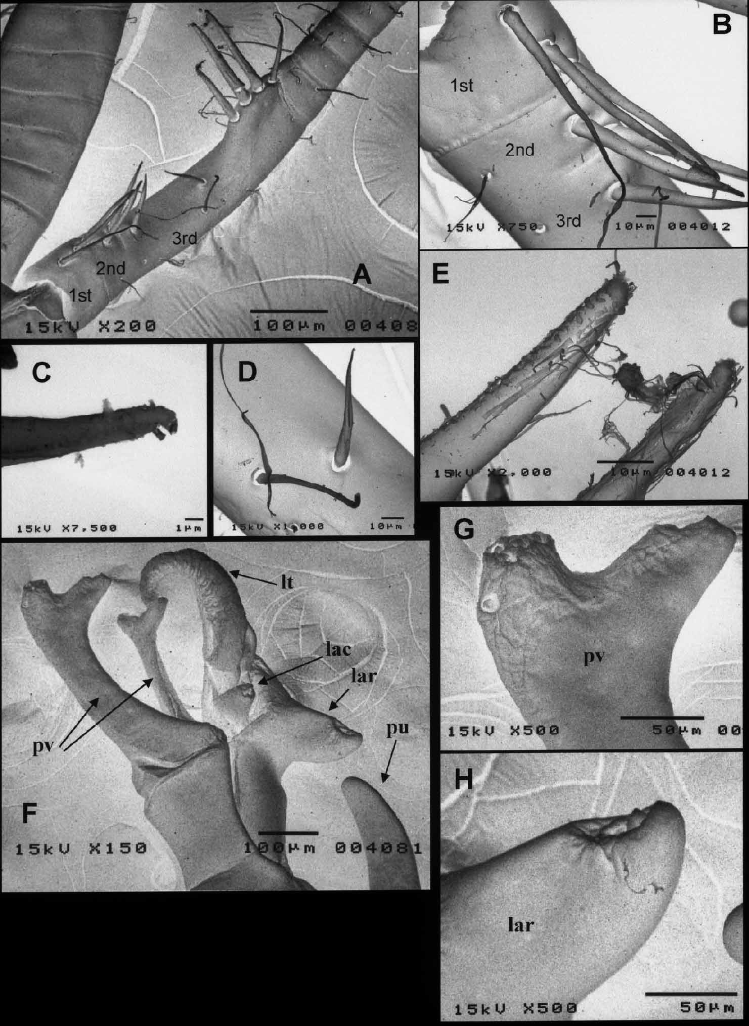
Petasma of male developed ( Figs. 3 View FIGURE 3 J, 4F–H). Processus unicifer (pu) 0.4 length of lamina externa, narrow, tapering to rounded apex ( Fig. 3 View FIGURE 3 J). Processus ventralis (pv) very long, divided into 2 branches ( Figs. 3 View FIGURE 3 J, 4F); posterior branch long, terminating into 2 secondary branches, mesial secondary branch short, armed with several hooks, lateral one directed laterally, longer than broad, armed with a few hooks on near end and distal end respectively ( Fig. 4 View FIGURE 4 F, G); anterior branch shorter than posterior one, narrow except expanded terminal part, divided into 2 secondary branches ( Fig. 3 View FIGURE 3 J, 4F). Lobus terminalis (lt) long, nearly spirally twisted, wrinkled ( Figs. 3 View FIGURE 3 J, 4F). Lobus connectens (lc) cylindrical, slender, with terminal hook ( Fig. 3 View FIGURE 3 J). Lobus inermis (li) almost as long as lobus connectens, robust, tapering distally, with terminal hook ( Fig. 3 View FIGURE 3 J). Lobus armatus (lar) and lobus accessorius (lac) with terminal hook ( Figs. 3 View FIGURE 3 J, 4F, H).
Thelycum of female ( Fig. 5 View FIGURE 5 E): sternite of sixth thoracic somite with pair of small, acute protuberances on posterior surface ( Fig. 5 View FIGURE 5 E, H); sternite of seventh thoracic somite projected laterally just in front of base of fourth pereopod ( Fig. 5 View FIGURE 5 E, H); coxa of third pereopod armed with several plumose setae on lateral surface, opened oviduct bearing several slender setae on lateral margin and a few short setae on lateral part of upper margin ( Fig. 5 View FIGURE 5 F, G).
Color. Body transparent with scattered red chromatophores. Antennal flagellum with single red band in stiff proximal portion ( Fig. 17 View FIGURE 17 A).
Distribution. Sicyonella antennata is recorded from East and Southeast Asia: Indonesia, the Gulf of Thailand ( Hansen, 1919), Japan ( Hayashi, 1992; this study), Singapore and the Philippines (USNM collection).
This species was collected from depths of 7–22 m ( Hansen, 1919) and from a seagrass bed ( Hayashi, 1992; this study).
Remarks. Sicyonella antennata was originally described from one male and one female specimen collected from off the Kai Islands, Indonesia, and several specimens collected from the Gulf of Thailand and deposited in the Copenhagen Museum ( Hansen, 1919). Subsequently, Hayashi (1992) recorded this species from a seagrass bed at Iriomote Island, Yaeyama Islands, Okinawa, southwestern Japan.
The present Japanese specimens agree with the original description and illustrations by Hansen (1919), except for the length of the antennular peduncle. In Hansen’s (1919) original description, the mesial margin of the combined length of the distal two segments is somewhat less than twice as long as that of the proximal segment in the male (bl 25 mm), and is distinctly longer than that of the proximal segment in the female (max. bl 31.5 mm). That of the Japanese specimens is shorter than Hansen’s description: 1.7 and 1.1 times as long as the proximal segment in the male and female, respectively.
In the Japanese specimens, the length of the distal two segments varies with body length. This character seems to vary with season, also. The combined length of the distal two segments was longer in individuals collected in September than in December ( Fig. 6 View FIGURE 6 ). The morphological development of the petasma also varied by seasons: it was perfectly development in the September specimen (cl 4.5 mm) and imperfect in the December specimen (cl 4.9 mm). These seasonal differences are probably caused by fluctuations in the growth rate with temperature.


No known copyright restrictions apply. See Agosti, D., Egloff, W., 2009. Taxonomic information exchange and copyright: the Plazi approach. BMC Research Notes 2009, 2:53 for further explanation.
|
Kingdom |
|
|
Phylum |
|
|
Class |
|
|
Order |
|
|
Family |
|
|
Genus |
Sicyonella antennata Hansen, 1919
| Fukuoka, Kouki, Tamaki, Motoya & Kikuchi, Tomohiko 2005 |
Sicyonella antennata
| Hayashi 1992: 225 |
| Hansen 1919: 30 |