
Potamotrygon adamastor, João Pedro Fontenelle & Marcelo R. De Carvalho, 2017
|
publication ID |
https://doi.org/ 10.11646/zootaxa.4310.1.1 |
|
publication LSID |
lsid:zoobank.org:pub:51059A1A-74EE-4DFC-90DD-B9130CE267CB |
|
DOI |
https://doi.org/10.5281/zenodo.5618458 |
|
persistent identifier |
https://treatment.plazi.org/id/C55887A8-1740-3B1C-CDB6-F95661F962E6 |
|
treatment provided by |
Plazi (2017-08-21 08:18:58, last updated 2024-11-27 11:35:53) |
|
scientific name |
Potamotrygon adamastor |
| status |
sp. nov. |
Potamotrygon adamastor View in CoL , sp. nov.
( Figs. 15–23 View FIGURE 15 View FIGURE 16 View FIGURE 17 View FIGURE 18 View FIGURE 19 View FIGURE 20 View FIGURE 21 View FIGURE 22 View FIGURE 23 , 42 View FIGURE 42 , 43 View FIGURE 43 ; Tables 3–4)
Holotype. MZUSP 104662 View Materials (adult female, 545 mm DW), rio Urariquera , rio Branco basin, municipal district of Boa Vista, upper Amazon Basin, state of Roraima, Brazil, 3°22’51.9594''N, 60°35’44.1594''W, Feb. 2007, coll. M.R. de Carvalho et al. ( Fig. 15 View FIGURE 15 ). GoogleMaps
Paratype. MZUSP 104664 View Materials (adult female, 450 mm DW), rio Branco basin, rio Urariquera , municipal district of Boa Vista, upper Amazon Basin, state of Roraima, Brazil, 3°22’51.9594''N, 60°35’44.1594''W, Feb. 2007, coll. M.R. de Carvalho et al. ( Fig. 16 View FIGURE 16 ). GoogleMaps
Diagnosis. Potamotrygon adamastor sp. nov. is distinguished from congeners, except P. limai , P. scobina , P. garmani sp. nov., and P. amazona sp. nov., by a combination of characters: dorsal disc dark gray covered with scarce small yellow to white ocelli with a slender dark contour; small irregular spots sometimes present on disc margin; rostral and caudal dermal denticles with a single pointed crown; a single irregular row of enlarged thorns on dorsal tail midline; tail base wide and robust; three angular cartilages of different sizes between jaw and hyomandibula; cartilaginous rod in tail relatively short (smaller than the distance between cloaca and caudal sting origin). Potamotrygon adamastor sp. nov. is distinguished from P. limai by having ocellated spots, not having a light polygonal pattern over the lower back, by having a wider and shorter tail, and fewer thorn rows over dorsal tail midline. From P. scobina and P. garmani sp. nov., by having a considerably thicker, more muscular disc, dermal denticles with fewer dichotomies and smaller basal plates, fewer spots, and a wider and shorter tail (mean tail width 19% DW vs. 13.4% DW in P. scobina and 14.1% DW in P. garmani sp. nov.; mean tail length 78.1% DW vs. 121.5% DW in P. scobina and 100.6% DW in P. garmani sp. nov.). Finally, P. adamastor sp. nov. is distinguished from P. amazona sp. nov. by having fewer dermal denticles on disc, a less elaborate dorsal color pattern (simple and usually with small ocellated spots with dark halos vs. numerous white spots forming clusters in P. amazona sp. nov.), and a shorter tail (mean tail length 78.1% DW vs. 86.1% DW in P. amazona sp. nov.) and internal cartilaginous rod.
Description. Disc oval, slightly longer than wide (DL 103.7–107.3% DW) ( Figs. 15 View FIGURE 15 , 16 View FIGURE 16 ). Anterior margin of disc broad, without a small protuberance on snout ( Fig. 17 View FIGURE 17 a). Disc robust, muscular and dorsoventrally thick. Eyes small and oval, around 2.5 times smaller than spiracles; spiracles obliquely positioned to eyes and trapezoidal to oval ( Fig.17 View FIGURE 17 a). Head region large and protruding, slightly more than 1/3 of disc length, with interorbital distance 14.1–17.6% DW, and interspiracular distance 16.5–19.7% DW. Jaw musculature robust, evident dorsally anterior to eyes ( Fig. 17 View FIGURE 17 a). Nasal curtain partially covering mouth. Mouth small and lightly undulated (mouth width 8.0– 9.3% DW), about equal to internasal distance. Labial ridges absent ( Fig. 17 View FIGURE 17 b). Five buccal papillae present, two posterior alternating with three anterior. Branchial basket wider than long, with space between first branchial slits 26.9–30.2% DW, and distance between fifth branchial slits 18.4–22.1% DW.
Teeth small and numerous in each jaw, wider than long, set in quincunx, in a narrowly arched upper tooth plate and a wide and trapezoidal lower tooth plate ( Fig. 18 View FIGURE 18 ). Tooth rows varying from 50–56 in upper jaw and 45–60 in lower jaw. Adult males presenting a single central pointed cusp on central teeth of both jaws. Lateral teeth, and teeth of juvenile males and females simple, presenting a single but more rounded cusp.
Pelvic fins broad (their length 52.0–54.2% DW), subtriangular, with rounded apices and a slightly undulated posterior margin. Pelvic fins extend beyond posterior disc ( Figs. 17 View FIGURE 17 c, d). Length of anterior margins of pelvic fins ranging from 17.9–25.5% DW. Claspers robust, their posterior tips slightly curved and rotated on clasper axis ( Fig. 17 View FIGURE 17 e). Clasper groove long, somewhat sinuous; apopyle at level of posterior margin of pelvic fins; hypopyle just anterior to clasper flap. Dorsal pseudosiphon oval, medial to clasper groove.
Tail width ranging from 17.6–20.8% DW. Tail long, narrowing more intensely from origin of caudal stings ( Figs. 15 View FIGURE 15 , 16 View FIGURE 16 ). Cartilaginous rod short and robust. Tail with many lateral, pointed denticles. Dorsal and ventral caudal folds present on tail tip. Length of caudal stings vary from 13.9–21.6% DW.
Coloration. ( Fig. 19 View FIGURE 19 ). Dorsal disc dark grayish to brown, with white to yellow ocelli, with a darker contour; ocelli small, up to 2/3 eye length and surrounded by smaller, irregular light colored spots. Spots more numerous on disc margins. Ventral disc white to beige, with lateroposterior margins covered by dark blotches; dark color pattern not reaching anterior disc margin. Intensity of pigmentation age and size dependent; older specimens more pigmented. The only male observed (MZUSP 104654) presents larger ocellated spots compared to females of equivalent size. This variation may be due to sexual dimorphism, however since only a single adult male specimen was examined, further investigation is necessary to verify this.
TABLE ³. Morphometric đata for P. adamastor sp. nov. SD: stanđarđ đeviation.
Pelvic fin dorsal surface with same color pattern as disc margin, with light colored posterior margin. Ventrally, pelvic fins whitish with a posterior darker margin. Claspers also lightly colored and covered posteriorly with darker blotches. Tail with dark dorsal background, similar to disc, but without ocelli. Numerous small irregular spots randomly present on tail. Tail with small groupings formed by tiny light colored irregular spots at sides, over dark background, without ocelli. Ventrally, tail lighter, covered by dark blotches on almost entire ventral surface; blotches increase in size from lateral to central tail. Sometimes with light colored blotches inside darker blotches.
Dermal denticles. ( Fig. 20 View FIGURE 20 ). Disc covered with dermal denticles in three different regions. Rostral region presenting simple denticles, with a single pointed crown. Basal plate (Bp) indistinct, with slight basal ridges (Br) when present. Head and mid-region of disc with denticles with a long coronal plate (Cp), posteriorly oriented, with a single anterior dichotomy composed of two well-developed coronal ridges (Cr). Caudal region with simple denticles, with a single pointed crown plate, without dichotomies. Basal plate circular, simple and indistinct, with small basal ridges sometimes present converging to denticle center.
Ventral lateral line canals. ( Fig. 21 View FIGURE 21 ). Hyomandibular canal (HYC) extending from anterior to nostrils, contacting orbitonasal component of supraorbital canal (CON), and projects to anterior disc margin before curving posteriorly. Anterior subpleural tubules (AST) not observed. Hyomandibular canal extends posteriorly (as the subpleural component of the hyomandibular canal; SPC), curving medially at about level of first branchial slits. Subpleural loop (SPL) not broad, deflected cranially just anterior to level of pelvic fin insertions. A single posterior subpleural tubule (PST) present between subpleural loop and jugular component of hyomandibular canal (JCH). Jugular component extends anteriorly, deflecting medially anterior to branchial slits; jugular component slightly undulated anterior to first branchial slit, forming angular component of hyomandibular canal (ACH). Angular component contacts nasal (NAS) and supraorbital canals (SOC) through the infraorbital canal (IOC). Infraorbital canal projects posteriorly, forming a compact infraorbital loop (IOL), then curves medially. Infraorbital canal parallel to jugular canal (JUG), forming a concise posterior jugular loop (PJL). Suborbital component of infraorbital canal (SOI) long and slightly curved toward disc interior, presenting undulations, and connected to the prenasal component of nasal canal (PNC) on anterior disc. Posterior nasal loop (PNL) and nasal interior loop (NIL) small and set anterior to the nostril.
Skeletal morphology. Neurocranium. Nasal capsules (NC) elongated, ventrolaterally expanded ( Fig. 22 View FIGURE 22 ). Anterior margin of nasal capsules oval and convex, with an internal ventromedial sept separating both capsules. Precerebral fontanelle (PCF) expanded and subcircular, posteriorly delimited by the epiphysial bar (EBP). Frontoparietal fontanelle (FPF) cone shaped, progressively narrowing, ending just posterior to postorbital processes. Both fontanellae together keyhole-shaped. Postorbital process (POP) long and narrow, diagonally projected anteriorly to level of angular cartilages. Prespiracular cartilage (PSC) posteriorly curved at its external margin.
Jaws and hyomandibular arch. Hyomandibular arch (HYO) elongated and laterally projected ( Figs. 22 View FIGURE 22 b, c). Three angular cartilages present. Anterior angular cartilage (AAC) slightly concave, 1/3 length of hyomandibula, and laterally projected. Posterior angular cartilage (PAC) not concave, 2/3 length of anterior angular cartilage. Lateral angular cartilage (LAC) smaller, semicircular, positioned between the posterior angular cartilage and hyomandibula. Meckel's cartilage (MC) robust, with rectangular corners on internal surface, bearing a prominent and oval ventrolateral process (VTP) at its posterior margin, not contacting angular cartilages; lateroanterior process (LAP) robust. Palatoquadrate (PQ) also robust, with a small posterior concavity, limited at sides by a small triangular projection. Minute ligamental cartilage (LC) between both palatoquadrates, not fused to antimeres. Synarcual cartilage. ( Fig. 22 View FIGURE 22 a). Anterior synarcual articulates with neurocranium by a small but prominent odontoid process (OTP). Medial crest (MDC) extended over entire synarcual. Articular surfaces of scapular processes (ASP) laterally projected, separated by an accentuated concavity. Anterior articular surface more prominent then posterior surface; posterior surface more slender, its extremity acutely rounded.
Pectoral girdle. ( Fig. 22 View FIGURE 22 d). Anterior process of the pectoral girdle more prominent, posterior process with a small, pointed extremity delimiting an adjacent concavity. Scapular process presents a slender medial bar, and its lateral portions present a well-developed central concavity, resembling an elongated "X". A robust and elongated propterygium (PRO) anteriorly articulates to this process, as well as a small central mesopterygium (MES), and a posteriorly set and elongated metapterygium (MET).
Pelvic girdle. ( Fig. 22 View FIGURE 22 e). Prepelvic process (PPP) projecting anteriorly to anteriormost third of metapterigium. Pubosquiadic bar (PIB) slender, with well developed and expanded extremities, bearing triangular lateral prepelvic processes (LPP) anteriorly. Iliac process (IP) robust, posterior to lateral prepelvic processes, with two round terminal expansions. Isquial process (ISP) slender, positioned medially. Four well-developed obturator foramina (OF) present between both processes.
Clasper skeleton. ( Fig. 23 View FIGURE 23 ). Basal segment 1 (B1) wider anteriorly, articulating with pelvic fins rays. Basal segment 2 (B2) subcylindrical and with a ventral concavity. Both basal segments about equal in length, but segment 2 more slender. Axial cartilage (AX) elongated and cylindrical, about 3/4 clasper total length, and curved posteriorly. Ventral marginal cartilage (VM) diamond-shaped, ventrally curved. Beta cartilage (BE) slender and cylindrical, about 1/3 clasper total length, articulating with basal segment 1. Well developed and oval dorsal marginal cartilage (DM), about 2/5 clasper total length, slightly curved internally. Dorsal terminal 2 (DT2) oval, curved interiorly, and projected dorsally. Terminal accessory cartilage (TA) suboval, presenting a narrow anterior margin, and posteriorly convoluted. Ventral terminal cartilage (VT) well developed, about 1/2 clasper total length, being the posteriormost robust element, with a posterior margin straight and ventrally curved, and anterior portion oval and dorsally curved.
Geographic distribution. Potamotrygon adamastor is currently known only from rio Urariquera, a large right-bank tributary of rio Branco, Amazon Basin, state of Roraima, Brazil ( Fig. 43 View FIGURE 43 ).
Etymology. Named after Adamastor, one of the giants of Greek mythology who opposed Zeus and Thetis and was thereby sent to Earth, acting as a raging storm over the Cape of Storms. The epithet adamastor is an adaptation of the Greek “Adamastos”, meaning untamed. A noun in apposition.
Additional material. (5 specimens). MZUSP 104654 View Materials (adult male, 483 mm DW), rio Branco, rio Urariquera, municipal district of Boa Vista , upper Amazon Basin, state of Roraima, Brazil, 3°22’51.9594”N, 60°35’44.1594”W, February 2007, coll. M.R. de Carvalho et al. GoogleMaps ; MZUSP 104657 View Materials (adult female, 581 mm DW), same data as MZUSP 104654 View Materials GoogleMaps ; MZUSP 104663 View Materials (adult female, 615 mm DW), same data as MZUSP 104654 View Materials GoogleMaps ; MZUSP 115212 (2 neonates from MZUSP 104654).

FIGURE 15. Holotype of Potamotrygon adamastor, sp. nov. (MZUSP 104662, adult female, 545 mm DW, rio Urariquera, rio Branco basin, municipal district of Boa Vista, upper Amazon Basin, state of Roraima, Brazil, 3 ° 22 ’ 51.9594 l N, 60 ° 35 ’ 44.1594 l W). A. Dorsal view. B. Ventral view.
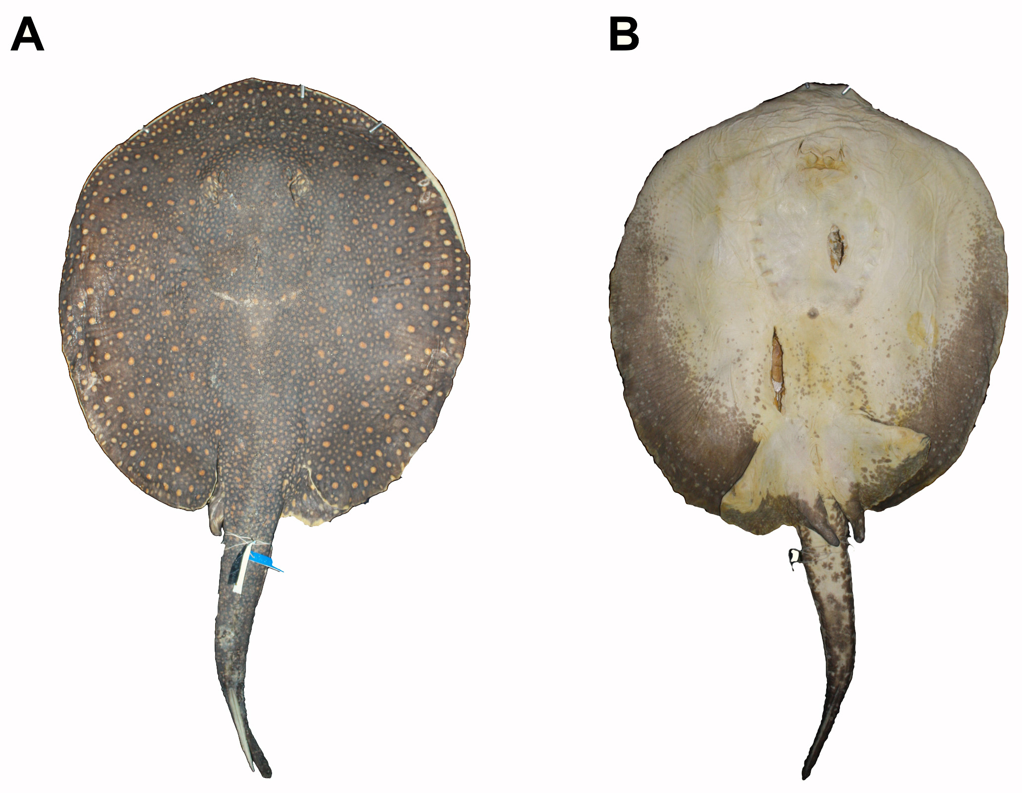
FIGURE 16. Paratype of Potamotrygon adamastor, sp. nov. (MZUSP 104664, adult male, 450 mm DW rio Urariquera, rio Branco basin, municipal district of Boa Vista, upper Amazon basin, state of Roraima, Brazil). A. Dorsal view. B. Ventral view.

FIGURE 17. Morphological details of Potamotrygon adamastor, sp. nov. (MZUSP 104662, adult female, 545 mm DW). A. Dorsal head and snout region. B. Mouth and nostrils. C. Dorsal view of tail origin, pelvic fins, and dorsal thorn rows. D. Ventral view of pelvic fins and cloaca. E. Dorsal view of clasper (MZUSP 104664, adult male, 450 mm DW).
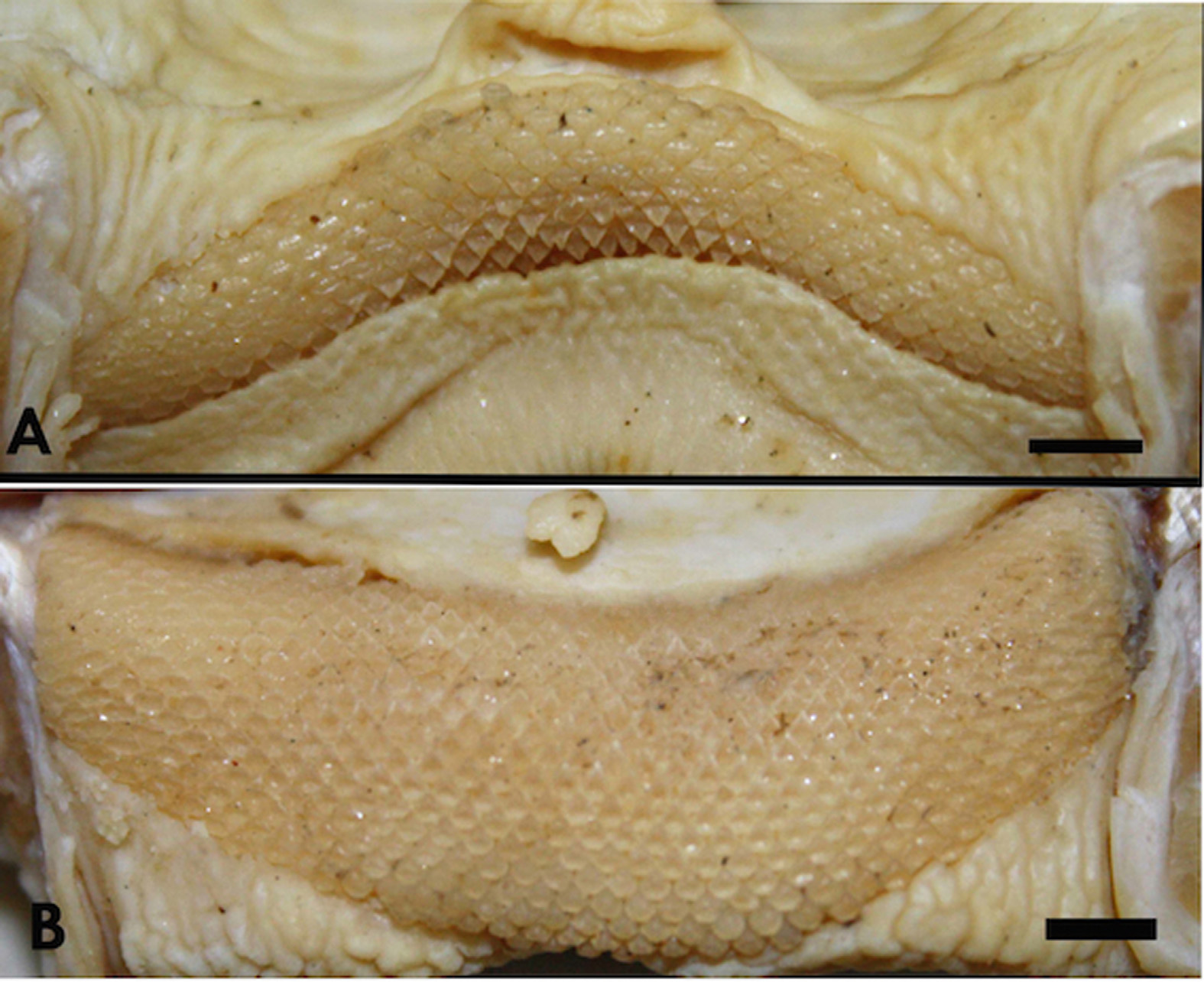
FIGURE 18. Teeth of Potamotrygon adamastor, sp. nov (MZUSP 104664, adult male, 450 mm DW). A. Upper jaw. B. Lower jaw. Scale bar = 2 mm.
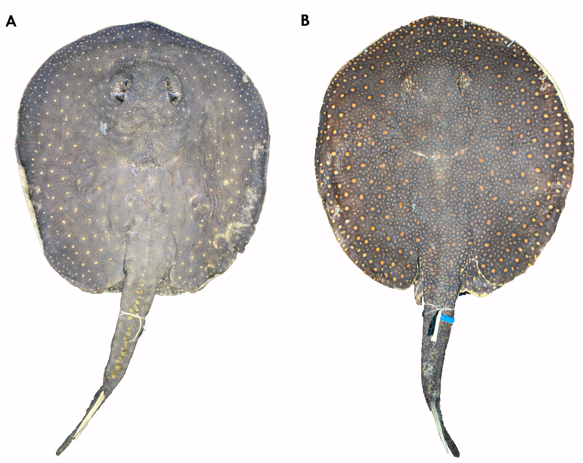
FIGURE 19. Color variation in Potamotrygon adamastor, sp. nov. A. MZUSP 104662 (adult female, 545 mm DW). B. MZUSP 104664 (adult male, 450 mm DW).
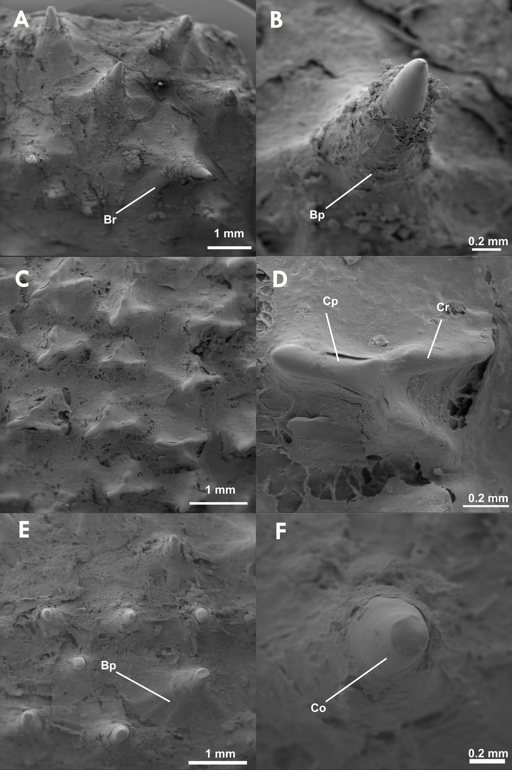
FIGURE 20. Scanning Electron Microscope (SEM) images of the dermal denticles of Potamotrygon adamastor, sp. nov. (MZUSP 104664). A. Rostral region. B. Rostral region in detail. C. Dorsal head region. D. Dorsal head region in detail. E. Dorsal caudal region. F. Dorsal caudal region in detail. Abbreviations: Bp, basal plate; Br, basal ridge; Cr, coronal ridge; Co, corona; Cd, coronal dichotomy; Cp, coronal plate.
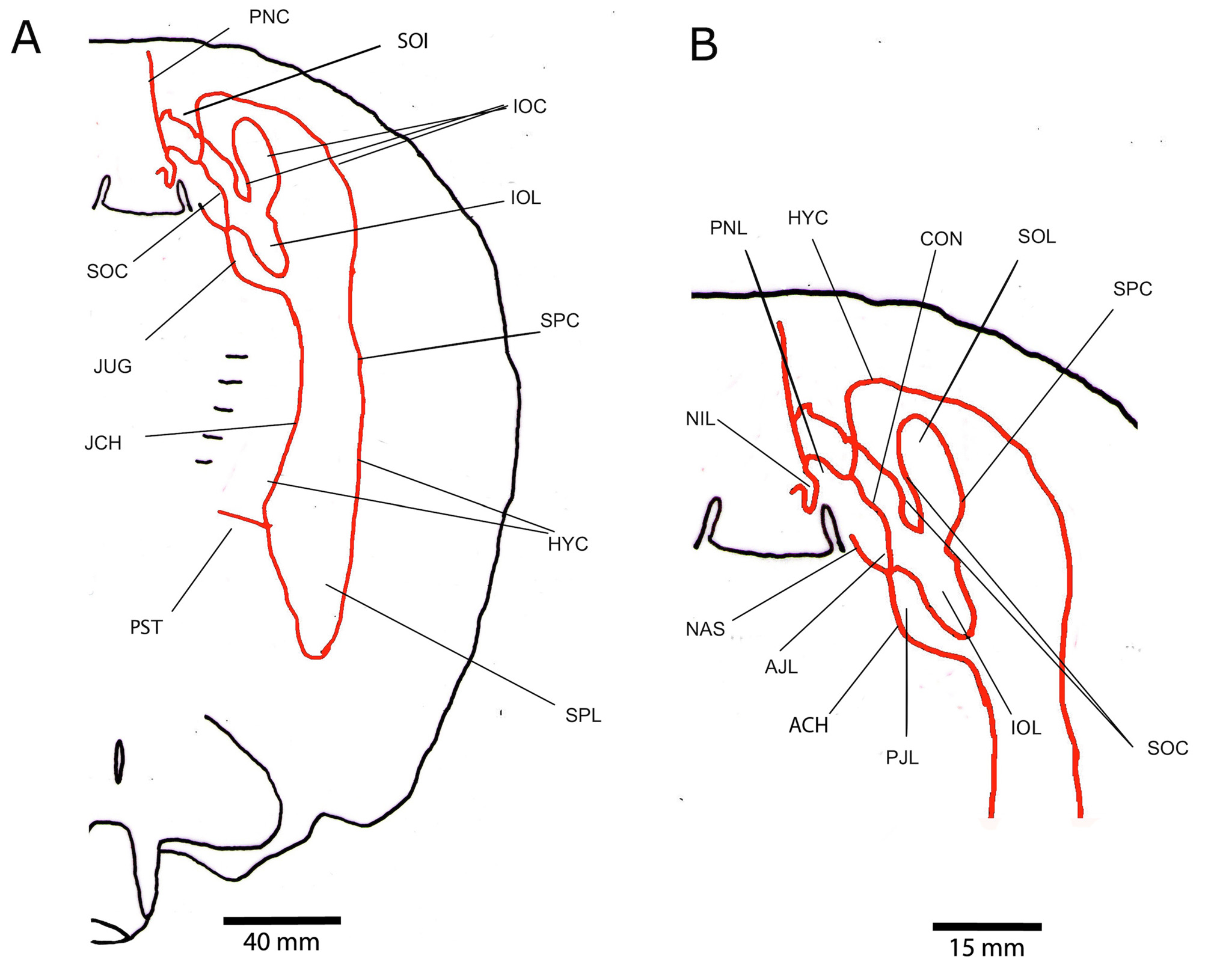
FIGURE 21. Ventral lateral-line canals of Potamotrygon adamastor, sp. nov. (based on MZUSP 104664). A. Overall arrangement. B. Anterior portion in detail. Abbreviations: ACH, angular component of hyomandibular canal; AJL, anterior jugular loop; AST, anterior subpleural tubules; CON, orbitonasal component of supraorbital canal; HYC, hyomandibular canal; IOC, infraorbital canal; IOL, infraorbital loop; JCH, jugular component of hyomandibular canal; JUG, jugular canal; NAS, nasal canal; NIL, nasointerior loop; PJL, posterior jugular loop; PNC, prenasal component of nasal canal; PNL, prenasal loop; PST, posterior subpleural tubules; SOC, supraorbital canal; SOL, suborbital loop; SOI, suborbital component of infraorbital canal; SPC, subpleural component of hyomandibular canal; SPL, subpleural loop; SRC, subrostral component of supraorbital canal.

FIGURE 22. Skeletal anatomy (from radiographs) of Potamotrygon adamastor, sp. nov. (MZUSP 104662, adult female, 545 mm DW). A. Ventral view of anterocentral disc. B. Ventral skull and jaws. C. Angular cartilages, right side. D. Ventral pectoral girdle. E. Ventral pelvic girdle and fins. Abbreviations: AAC, anterior angular cartilage; BCO, coracoid bar; BP, basipterigium; HYO, hyomandibula; LAC, lateral angular cartilage; MC, Meckel's cartilage; MES, mesopterygium; MET, metapterygium; MSC, mesocondyle; MTC, metacondyle; PAC, posterior angular cartilage; PIB, puboischiadic bar; PQ, palatoquadrate; PRC, procondyle; PRO, propterygium.
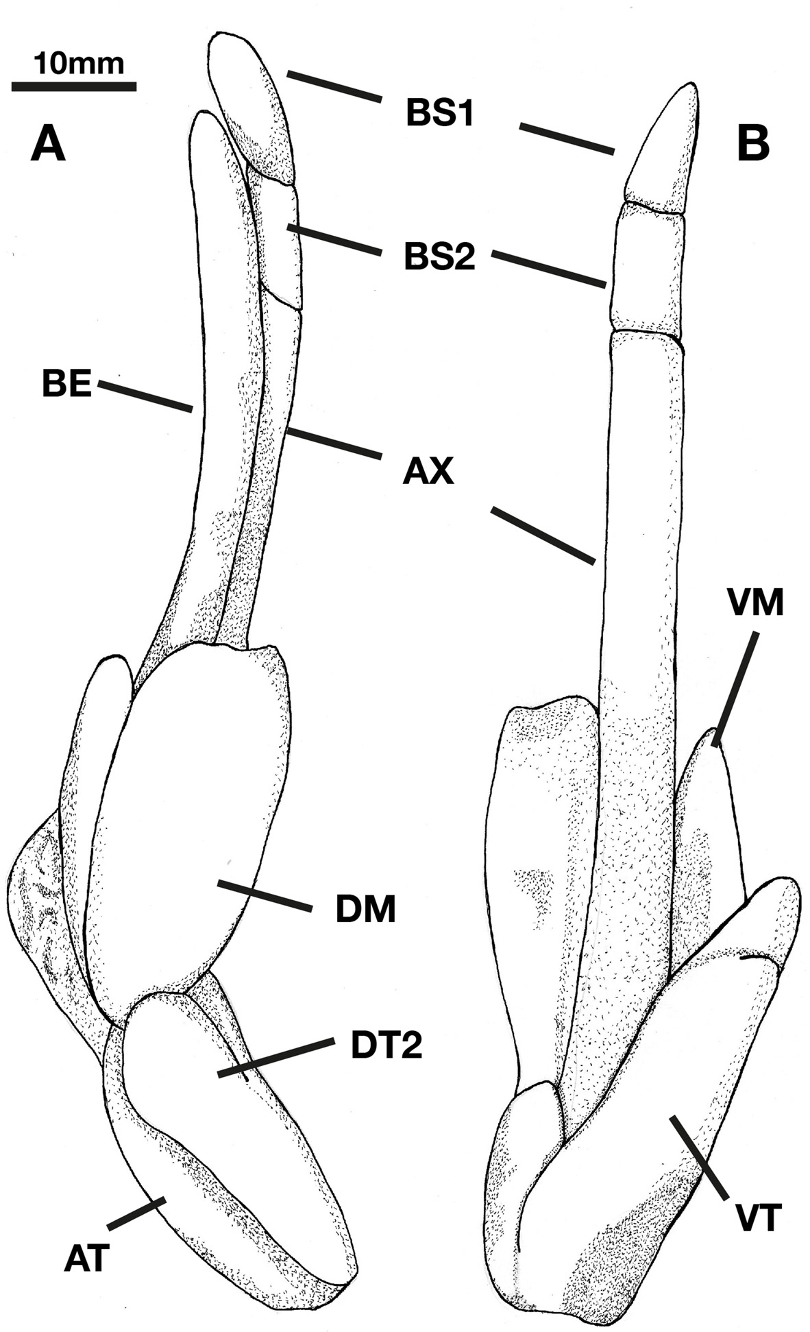
FIGURE 23. Illustration of the clasper skeleton of Potamotrygon adamastor, sp. nov. A. Dorsal view. B. Ventral view. Abbreviations: AT, acessory terminal; AX, axial cartilage; BE, beta cartilage; B 1, first basal segment; B 2, second basal segment; DM, dorsal marginal; DT 2, dorsal terminal 2; VM, ventral marginal; VT, ventral terminal.

FIGURE 42. Angular cartilages of species of the P. scobina complex. A. Holotype of Potamotrygon scobina (MCZ- 602 s, juvenile male, 238 mm DW). B. Potamotrygon scobina (MZUSP 104247, adult male, 503 mm DW). C. Potamotrygon adamastor, sp. nov. (MZUSP 104662, adult female, 545 mm DW). D. Potamotrygon limai (MZUSP 104068, juvenile male, 219 mm DW). E. Potamotrygon amazona, sp. nov. (MZUSP 117344, adult male, 532 mm DW). F. Potamotrygon garmani, sp. nov. (UNT 2174, adult? male, 341 mm DW). Abbreviations: AAC, anterior angular cartilage; LAC, lateral angular cartilage; PAC, posterior angular cartilage.
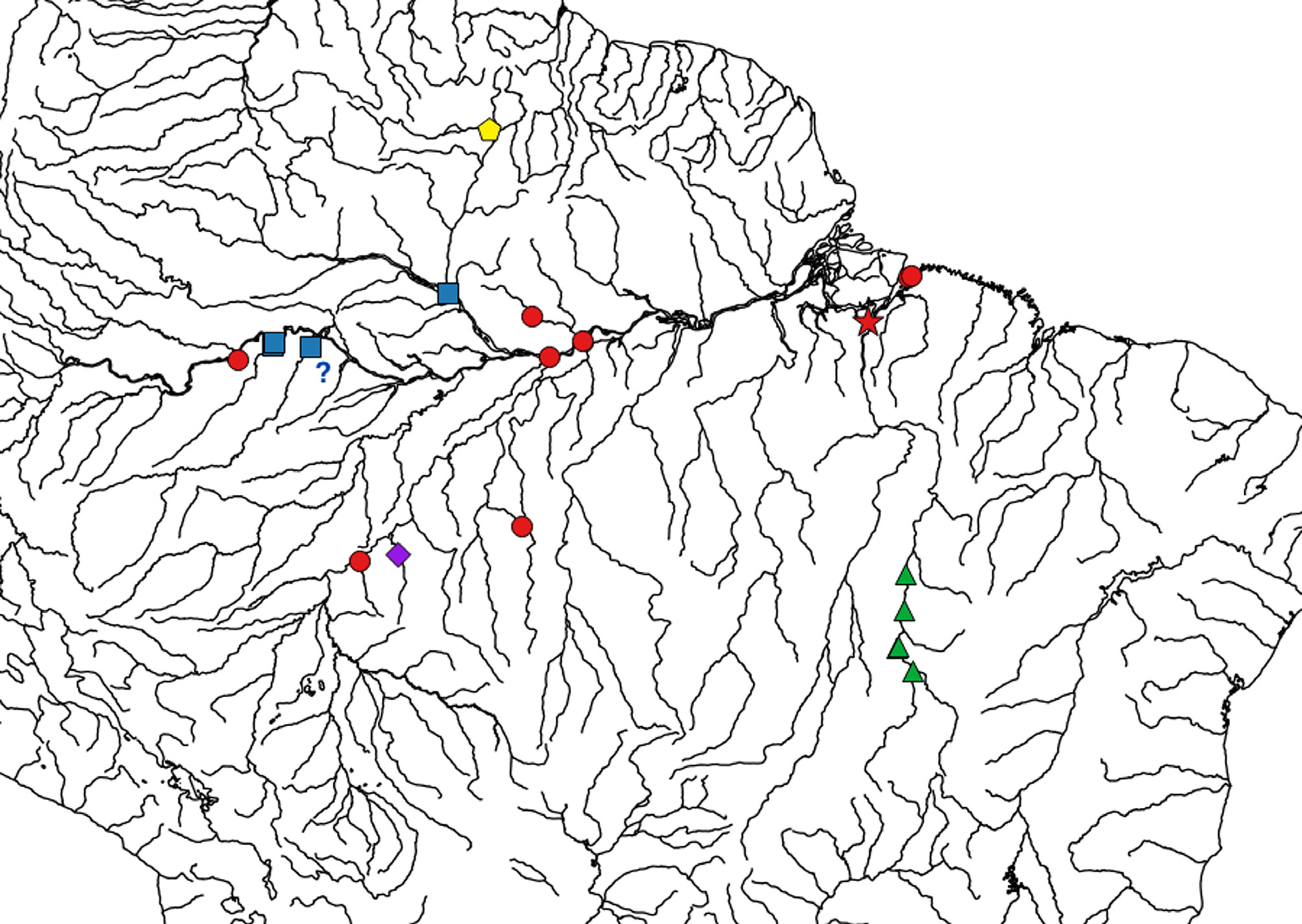
FIGURE 43. Distribution of species of the Potamotrygon scobina complex. Red circle: Potamotrygon scobina; red star: type locality of Potamotrygon scobina; purple diamond: Potamotrygon limai; yellow pentagon: Potamotrygon adamastor, sp. nov.; blue square: Potamotrygon amazona, sp. nov.; green triangle: Potamotrygon garmani, sp. nov.
No known copyright restrictions apply. See Agosti, D., Egloff, W., 2009. Taxonomic information exchange and copyright: the Plazi approach. BMC Research Notes 2009, 2:53 for further explanation.
|
Kingdom |
|
|
Phylum |
|
|
ParvPhylum |
Chondrichthyes |
|
Class |
|
|
Order |
|
|
Family |
|
|
Genus |
1 (by plazi, 2017-08-21 08:18:58)
2 (by ImsDioSync, 2017-09-15 16:31:58)
3 (by ExternalLinkService, 2019-09-26 03:09:51)
4 (by ExternalLinkService, 2021-10-29 01:42:54)
5 (by ExternalLinkService, 2021-10-29 04:13:19)
6 (by ExternalLinkService, 2021-10-29 15:56:10)
7 (by plazi, 2023-10-27 23:00:12)