
Sinocapnia kuankuoshui, Murányi, Dávid, Li, Weihai & Yang, Ding, 2015
|
publication ID |
https://doi.org/10.11646/zootaxa.4059.2.8 |
|
publication LSID |
lsid:zoobank.org:pub:550E33DB-F2EE-412E-8935-CF82065B4562 |
|
DOI |
https://doi.org/10.5281/zenodo.6102854 |
|
persistent identifier |
https://treatment.plazi.org/id/C37D87B5-6659-FF9A-FF01-F91AFB41FF24 |
|
treatment provided by |
Plazi |
|
scientific name |
Sinocapnia kuankuoshui |
| status |
sp. nov. |
Sinocapnia kuankuoshui View in CoL sp. n.
( Figs. 1–17 View FIGURES 1 – 2 View FIGURES 3 – 8 View FIGURES 9 – 13 View FIGURES 14 – 16 View FIGURES 17 – 18 )
Diagnosis. As for the genus.
Type material. CHINA, Guizhou Province, Zunyi City, Suiyang County, Kuankuoshui Nature Reserve, Maoya Town, Zhongping village, upstream section of Furong River, N 28°15’ E 107°10’, 700-890m, 26.0 3.2012, leg. Weihai Li: holotype male ( HIST), paratype: 1f ( HIST).
Description. Medium sized Capniidae , macropterous in both sexes. Measurements: male holotype forewing length 6.0 mm; female paratype forewing length 7.4 mm. Setation generally short and dense, longer setae occur on terminal segments; male epiproct bare, setation of tergal processes reduced. General colour brown, legs paler, wings hyaline with dark brown veins. Head with distinct, dark brown occipital rugosities, tentorial callosities and M-line; pale patch on the medial portion of occiput. Antenna uniformly brown, nearly as long as body with less than 30 segments, antennomeres cylindrical; palpi and mouthparts light brown, mentum darker. Pronotum with distinct C-shaped pattern of dark brown rugosities, median line, anterior and posterior sutures are also dark and distinct; nearly twice as wide as long, posteriorly dilated, corners rounded. Other sclerites of the thorax are with indistinct brown and dark brown pattern. Legs long, streched hind tibia overhangs abdomen. Width of femora less than fifth of their length; tibiae slightly longer than femora. Basitarsus and metatarsus are about of the same length. Claws symmetrical, smooth and gradually curved, arolium small.
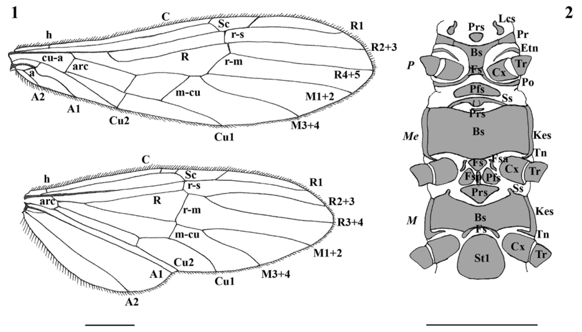
Ventral sclerites of thorax ( Fig. 2 View FIGURES 1 – 2 ): Prothorax: presternum small and rounded, not fused with the heart-shaped basisternum; precoxal bridge narrow, fused with basisternum; furcasternum quadrangular, fused with both basisternum and the stripe-like, curved postcoxal bridge; postfurcasternum large, elliptical, not fused with furcasternum. Mesothorax: spinasternum narrow, medially fused, not touching prothoracic postfurcasternum but fused laterally with the large, oblong basisternum; presternum very small, elliptical, not fused with basisternum; furcasternum triangular, weakly fused with basisternum, separated from the distinct furcasternal arms and from the furcasternal pit; postfurcasternum divided in two lateral, rounded parts by the furcasternal pit, the parts are not fused with other sclerites; katepisternum separated from basisternum but entirely fused with the ventrally elongated trochantin. Metathorax: presternum triangular, not fused with the large, oblong basisternum; spinasterum vestigial and fused with basisternum; furcasternum stripe-like and fused with basisternum, laterally projecting backwards but not fused with sternum I; katepisternum separated from basisterum but entirely fused with the ventrally elongated trochantin.
Wing venation ( Fig. 1 View FIGURES 1 – 2 ): Forewing: Costa simple, narrowing gradually from R4+5 towards Cu1; Subcosta parallel to Costa, there is one crossvein between them besides humeral crossvein, Subcosta curves abruptly beyond the last crossvein and join R1 before radiosubcostal crossvein; humeral crossvein is closer to arculus than to wing base; R1 is straight between its branching with Radial vein and radiosubcostal crossvein, with one leaning crossvein towards Costa well after the radiosubcostal crossvein; R2+3 and R4+5 branching just after radiosubcostal crossvein; Medial vein branches out to M1+2 and M3+4 before radiomedial crossvein, there is one crossvein between Medial vein and Cu1 besides arculus and mediocubital crossvein; Cu1 and Cu2 branching at arculus, Cu1 ends ½ of the wing length, Cu2 ends about ⅓ of wing length at the cubital crossvein, cubitoanal crossvein present between Cu2 and A1 just after anal crossvein; A1 ends around the cubital end of arculus, distinctly curved beyond anal crossvein, A2 reaches only as far as cubitoanal crossvein. Hindwing: Costa similar to the forewing; Subcosta has two crossveins towards Costa besides humeral crossvein, and joins R1 at radiocubital crossvein; R1 is straight, not branching with Radial vein but starts separately from wing base, has one crossvein towards Costa similar to the forewing; R2+3 and R4+5 branching far beyond radiomedial crossvein; Medial vein branches out from Radial vein after arculus, then branching to M1+2 and M3+4 well after radiomedial crossvein, there is no crossvein between Medial vein and Cu1 between arculus and mediocubital crossvein; Cu1 and Cu2 branching at arculus, Cu1 curved after cubital crossvein and ends ½ of the wing length, Cu2 parallel to A1 and ends at wing fold, cubitoanal crossvein absent; anal field small, the fold of the wing extending between the parallel Cu2 and A1; only A1 and A2 present, both are straight, A2 ends around the position of cubital crossvein.
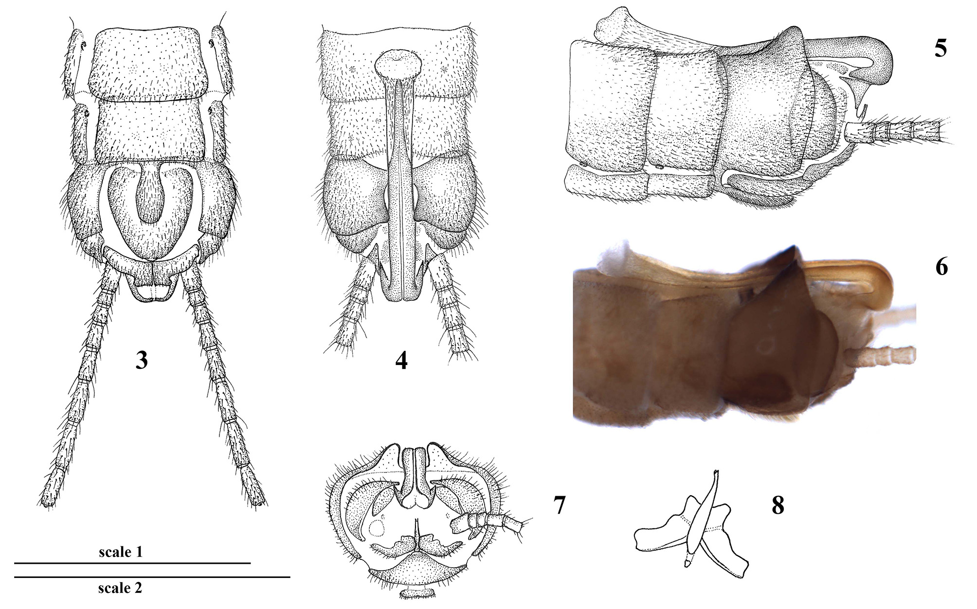
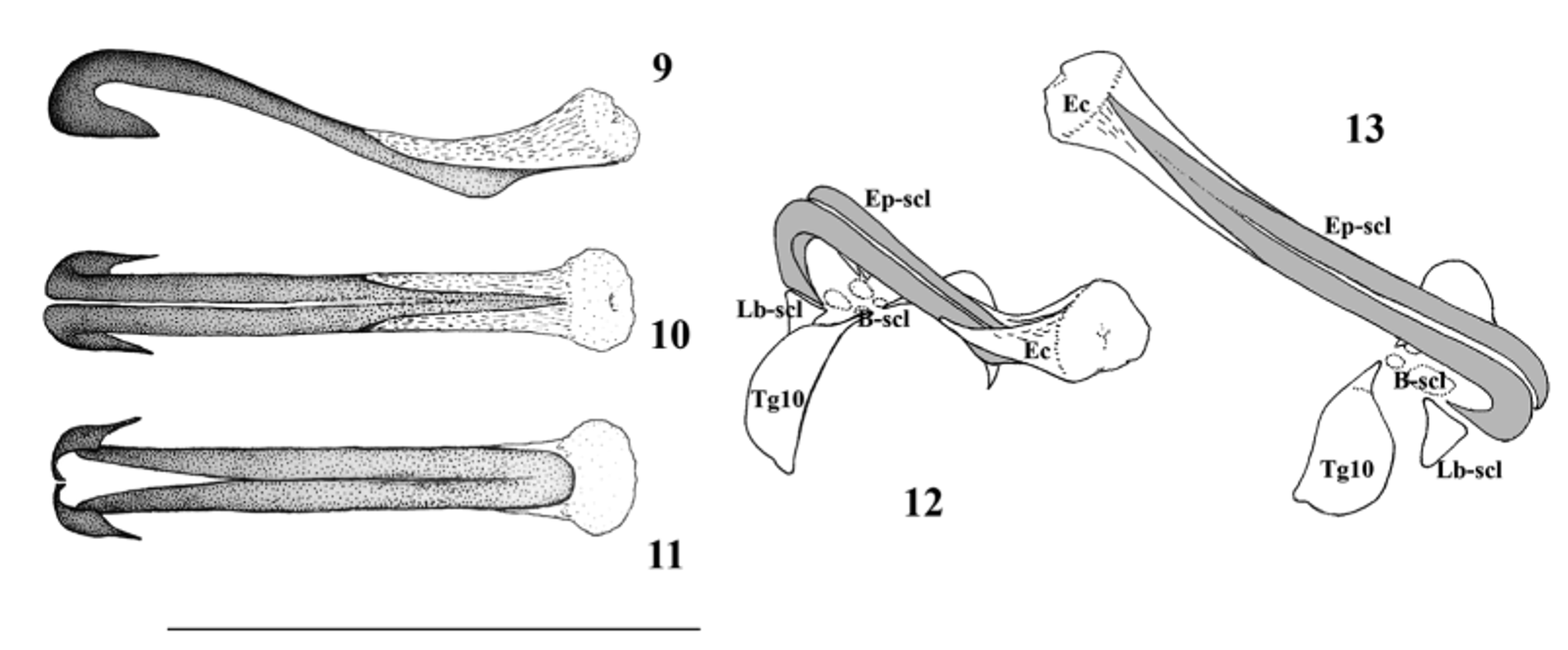
Male abdomen ( Figs. 3–13 View FIGURES 3 – 8 View FIGURES 9 – 13 ): Tergum I only laterally sclerotized, tergum II with medially interrupted antecosta and weak medial sclerotization. Terga III–VIII without defined antecosta and with wide medial membranous portion that is not clearly separated from sclerotized lateral portions; transverse row of four spots present on each of terga II–VIII, setation is increasingly longer towards the terminalia. Tergum IX is considerably darker than previous segments, with well sclerotized antecosta that is widely incised medially. Posterio-medially the segment is armed with a pair of processes that delimitate an oval medial membranous area; sclerotization of the processes is stronger at their apex and inner edge, their caudal face membranous with little setation. The processes as high as ⅓ of segment height, in dorsal view each process as wide as the medial membranous section of the tergum; processes gradually raised posteriorly, but apex of processes not overlapping edge of segment. Tergum X subdivided with the entire antecosta forming a ridge in the medial ⅓ of the segment’s width. Epiproct consists of a vestigial basal sclerite, a large but lightly sclerotized laterobasal sclerite and very long main epiproct sclerite that bears a huge and complex eversible crest but lacks an inner sclerite. The basal sclerite appears as two pairs of small sclerites separated from the basal fork of the main epiproct sclerite, leaving a large membranous area between the epiproct and tergum X. Lateral sclerite is triangular and ventrally directed, separated from the main epiproct sclerite. The main epiproct sclerite is strongly curved basally after a large basal fork, slightly curved ventrally at ½ of the length, then dorsally raised towards the apex; reaches back to anterior ½ of segment VII, bare and lacking both caudal setae and apical spines; base of the sclerite as wide as ⅓ of the width of segment IX, after the base it is evenly narrow with parallel edges in dorsal view, being as wide as 1/5 of the width of segment IX; dorsally divided in its basal ½, ventrally divided only in its base, while laterally entire, large ventral portion keeled in the apical ½, rounded tongue-shape in ventral view. Eversible crest is positioned dorsad on the apical ½ of the main epiproct sclerite, slightly raised towards the apex where it forms a hemispherical bulge; basal section is densely wrinkled, the apical bulge is bearing sensilla. Sternum I entire and unmodified but smaller than the additional sterna, with rounded corners. Sternum II consists of a large, rectangular posterior and two small anterior sclerites, sterna III– VIII lack anterior sclerites; spiracles present on sterna II–VIII. Sternum IX reduced to a well sclerotized arch connecting the ventro-basal part of tergum IX; in its medial part, the arch bears a large teardrop-shaped vesicle that nearly reaches paraproct base, while its width is 1/5 of the segment width. Subgenital plate separated from all other segments, rounded with triangular shape, apical part with small, rounded tip. Paraproct wide and long, with slightly separated apex that is blunt and subrectangular. Fusion plate is long and narrow, its apical tube is slightly curved upward with an acute tip. Retractoral plate small and rounded, divided from fusion plate. Cercus short, bearing only 8 segments. Cercal segments cylindrical but the terminal segment clubbed; basal segments with apical whorl of erect setae, terminal segment with an apical wart.
Female abdomen ( Figs. 14–16 View FIGURES 14 – 16 ): Tergum I unmodified. Terga II–VIII divided into two large lateral sclerites, and membranous only in the medial ⅓; tergum II with additional paired, small anterior sclerites. Terga IX–X entire and unmodified; epiproct separated from tergite X, simple and rounded. Sternum I entire and unmodified but smaller than the additional sterna, with rounded corners. Sterna II–VII consist of a large, rectangular posterior and two small anterior sclerites that are larger on sternum II than the following segments. Posterior sclerite of sternum VII developed into a large and entire pregenital plate overlapping anterior ⅓ of sternum VIII. The pregenital plate posteriorly bulging in lateral view, overlapping posterior portion rounded and darker in ventral view, otherwise the plate evenly brown; posterior edge is fused with the subgenital plate, hidden beneath the bulging portion. Sternum VIII with large and entire subgenital plate that lacks longitudinal keel and with two small lateral sclerites. The subgenital plate slightly overlapping sternum IX, sides converging from its anterior ⅓, posterior edge rounded; uniformly brown, but posterio-medially with a small unsclerotized area that makes the plate appearing incised. Lateral sclerites fused with posterio-lateral edges of the subgenital plate. Vaginal complex with genital opening narrower than ½ of the subgenital plate’s width, membranous genital cavity reaching back to anterior ½ of segment VII where it is branching into the oviducts; inner sclerite is lacking. Sternum IX fully sclerotized; paraproct with short, rounded tip. Cercus short, bearing only 7 segments. Cercal segments cylindrical but the terminal segment clubbed; basal segments with apical whorl of erect setae, terminal segment with an apical wart.
Larva: unknown.
Affinities. As for the genus.
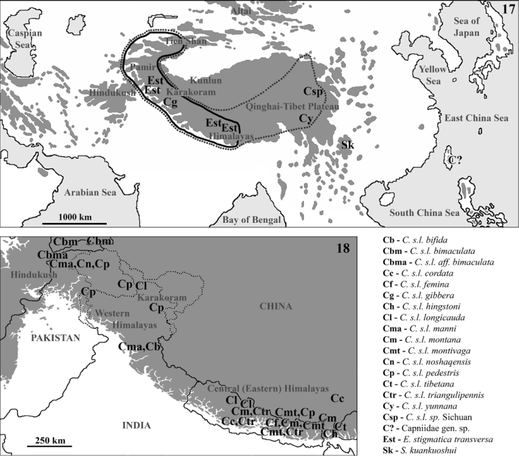
Distribution and ecology. The species was found at a single stream in Kuankuoshui Nature Reserve, Guizhou Province of southwest China ( Fig. 17 View FIGURES 17 – 18 ). This nature reserve is a medium high range on the transition of the Yunnan- Guizhou Plateau towards the Sichuan Basin and the hilly ranges of Hunan Province. Kuankuoshui is famous for its karst formations and natural forests, and many described species are only known from this area, e.g. the Kuankuoshui salamander: Pseudohynobius kuankuoshuiensis Xu & Zeng, 2007 (in: Xu et al. 2007). The type locality is a 4-5 ms wide upstream section by a bridge, about 3 walking hours distance from the sources.
Etymology. The species name refers to the type locality area, Kuankuoshui Nature Reserve. Used as a noun, gender neutral.
Commented checklist of the Capniidae known from the Oriental Realm ( FigS. 17–18 View FIGURES 17 – 18 )

No known copyright restrictions apply. See Agosti, D., Egloff, W., 2009. Taxonomic information exchange and copyright: the Plazi approach. BMC Research Notes 2009, 2:53 for further explanation.
|
Kingdom |
|
|
Phylum |
|
|
Class |
|
|
Order |
|
|
Family |
|
|
Genus |