
Zorotypus weiweii Wang, Li & Cai
|
publication ID |
https://doi.org/ 10.11646/zootaxa.4162.3.8 |
|
publication LSID |
lsid:zoobank.org:pub:173121D8-E1C3-481B-BE4A-421E59347483 |
|
DOI |
https://doi.org/10.5281/zenodo.6063038 |
|
persistent identifier |
https://treatment.plazi.org/id/C34887B1-B333-CD67-1BC1-F0B2FDC9FE42 |
|
treatment provided by |
Plazi |
|
scientific name |
Zorotypus weiweii Wang, Li & Cai |
| status |
sp. nov. |
Zorotypus weiweii Wang, Li & Cai View in CoL , sp. nov.
Figs. 1–23 View FIGURE 1 View FIGURES 2 – 10 View FIGURES 11 – 15 View FIGURES 16 – 23 , 28–30
Type material. Holotype apterous male, Malaysia: Sabah, Keningau, Borneo Jungle Girl Camp , 5˚26ʼ55.7ˮN, 116˚27ʼ08.6ˮE, rainforest, under bark of decaying log, 1115 m, 16. X. 2015, coll. Wang Jianyun & Zhang Weiwei . Paratypes, 4 dealate males, 2 dealate females, same location data as holotype; 2 apterous males, same rainforest, 5˚27ʼ03.9ˮN, 116˚26ʼ52.3ˮE, under bark of decaying log, 962 m, 9. X. 2015, coll. Liang Feiyang & Zhang Weiwei .
Diagnosis. The new species is similar to Z. impolitus , but it can be easily separated from the latter by the following characters: curly setae denser on vertex of male, forming a “V” shape (vs. male vertex setae relatively sparse); number of stout bristles on ventral surface of metafemur ten (vs. eight to nine); male ninth abdominal tergum (T9) sclerotized (vs. T9 scarcely sclerotized); male T11 with larger sclerotized area, extended to lateral margin (vs. T11 laterally scarcely sclerotized); and the male genitalia relatively stronger, ventral sclerite broader ( Figs. 2, 6, 9 View FIGURES 2 – 10 , 16, 20–23 View FIGURES 16 – 23 ).
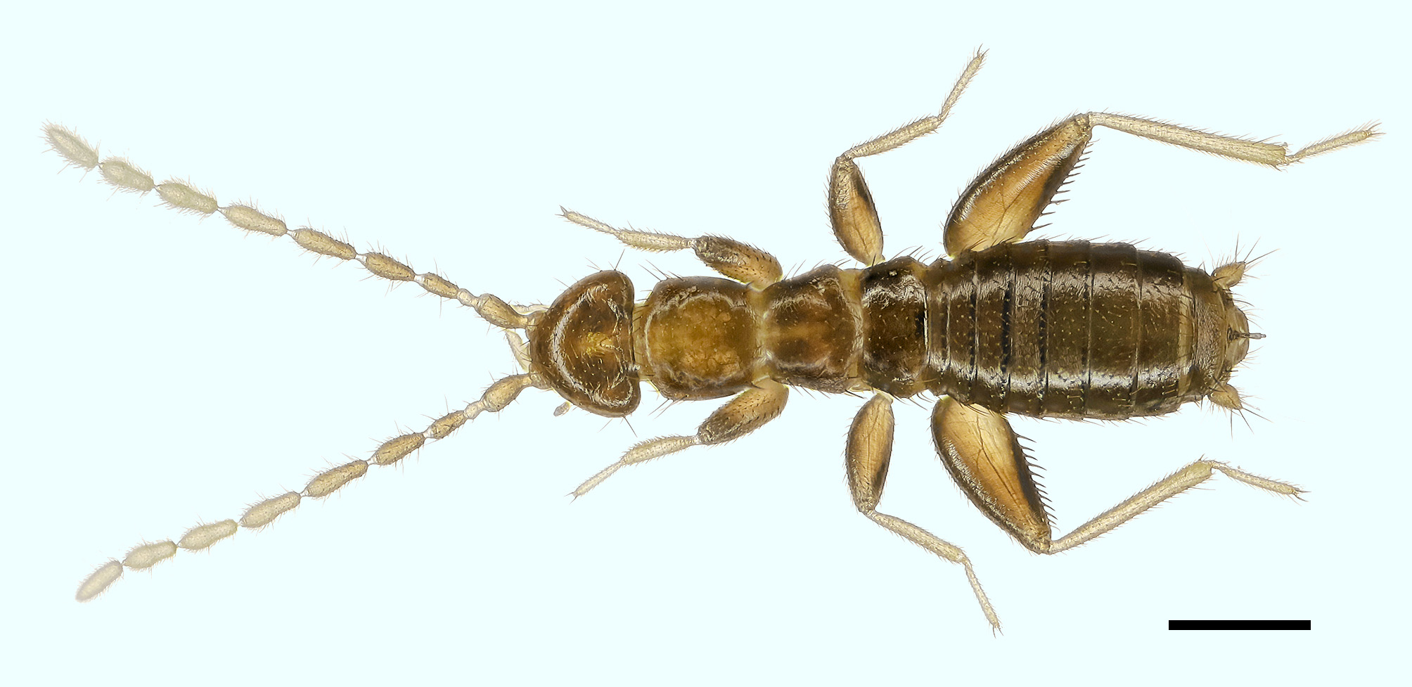
Description. Apterous male ( Figs. 1 View FIGURE 1 , 2, 4, 6, 7, 9 View FIGURES 2 – 10 , 11–23 View FIGURES 11 – 15 View FIGURES 16 – 23 , 28). Body length 2.3 mm, generally glossy brown except membranous regions, distal sections of antennae, tibiae, tarsi and cerci yellowish white to pale white. Head sub-triangular, wider than pronotum, vertex slightly concave, with curly setae grouped beside the midline, bases of those setae forming a “V” shape ( Fig. 2 View FIGURES 2 – 10 ); eyes and ocelli absent; antennae about as long as head and thorax combined, nine-segmented; antennomere I slightly curved outward at basal half, with a faint constriction at the basal half, antennomere II shortest, about half length of antennomere III, antennomeres III–IX longer than wide, antennomere V longest ( Fig. 11 View FIGURES 11 – 15 ); left mandible with five teeth, prostheca inserted on the ventral surface of subapical tooth, molar region without dentes; right mandible with five teeth, the largest tooth (third tooth) with three tiny granulated dentes, molar region with fine dentes ( Figs. 12, 13 View FIGURES 11 – 15 ); maxillae symmetrical, distal divided, apex of inner part bifurcate, apex of outer part with dense hairs; maxillary palpus five-segmented ( Fig. 14 View FIGURES 11 – 15 ); labial palpus three-segmented ( Fig. 15 View FIGURES 11 – 15 ).
Pronotum ( Figs. 1 View FIGURE 1 , 4 View FIGURES 2 – 10 ) sub-rectangular, posterior slightly narrowed; mesonotum trapezoidal, slightly shorter than pronotum, posterior broader than anterior; metanotum trapezoidal, shorter but wider than mesonotum, distinctly wider than long; thorax setose ( Fig. 4 View FIGURES 2 – 10 ). Legs with setae of moderate length, setae sparse on anterior surface of profemora and posterior surface of meso- and metafemora sparse, distal tarsomeres of all legs with short setae; posterior surface and dorsal surface of profemur covered with moderate-length setae, anterior surface and ventral surface with short setae; protibia with moderate-length setae, ventral surface of distal half with about 15 bristles, apically with two spurs; distal tarsomere about 2.5 times length of basal tarsomere. Mesofemur less strong than profemur, setae of mesofemur much like those of profemur; mesotibia with moderate-length setae and two small apical spurs. Metafemur ( Fig. 16 View FIGURES 16 – 23 ) swollen, much stronger than profemur or mesofemur, narrower apically, anterior and dorsal surface with moderate-length setae, apical half of ventral margin with three long, fine bristles on anterior surface, posterior surface nearly bare, with sparse, minute setae near the dorsal and ventral margin area, ventral surface with ten stout bristles, proximal first and third bristles longer than others; metatibia with moderatelength setae, dorsal surface subapically with one long bristle, ventral surface with two spurs apically; basal tarsomere with two spurs ventrally.
First abdominal tergum (T1) with single transverse row of short setae along hind margin and a few small setae laterally; T2–7 with regular vestiture of short and moderate-length setae and a pair of longer setae along hind margin; T8 with short and moderate-length setae and two pairs of erect long setae along hind margin, inner side setae longer and stronger ( Figs. 6 View FIGURES 2 – 10 , 17, 18 View FIGURES 16 – 23 ). T9 short, weakly sclerotized; T10 separated into anterior and posterior parts, anterior half sclerotized, with short and moderate-length setae laterally, posterior half mostly membranous, central region with medial spatula-like, upcurved projection; basal projection with several fine, short setae, apically with two setae ( Figs. 9 View FIGURES 2 – 10 , 17, 18 View FIGURES 16 – 23 ); T11 with long, strongly upcurved medial projection centrally, bent in the middle, apically with a tiny hook (= male mating hook) ( Figs. 17, 18 View FIGURES 16 – 23 ), projection of T11 narrower and longer than that of T10 ( Figs. 6, 9 View FIGURES 2 – 10 ); base of T11 projection broadly sclerotized, narrower laterally, posterior region of broadly sclerotized margin bearing four moderate-length setae on each side ( Fig. 6 View FIGURES 2 – 10 ); epiproct and paraproct unsclerotized; cercus ( Fig. 19 View FIGURES 16 – 23 ) 1-segmented, conical, with one long apical seta, three subapical moderate-length setae, several short setae, and very long, fine setae; surface covered with numerous minute spicules ( Fig. 19 View FIGURES 16 – 23 ) except at base and apex.
First abdominal sternum (S1) scarcely sclerotized; S2 weakly sclerotized centrally, with two or three short setae on each side; S3 with two transverse rows of short setae, basal row sparse; S4 with three transverse rows of short setae; S5–7 with short setae evenly scattered, S5 with pair of scarcely sclerotized circular areas; S8 and S9 fused, with shallow furrow, S8+9 subtriangular, with evenly scattered, moderate-length setae, posterior margin of S8 with a pair of longer setae; S10 invisible externally, beneath S8+9; S11 with two lateral sclerites, each with four short setae and two moderately long setae; on inner side setae longer, with a pair of tiny sensory setae on each side of inner membranous area ( Figs. 6, 9 View FIGURES 2 – 10 , 17–18 View FIGURES 16 – 23 ). Genitalia asymmetrical ( Figs. 20–23 View FIGURES 16 – 23 ), without elongate coiled flagellum or well-defined basal plate; dorsal sclerite twisted and elongated, slightly bent in middle; middle sclerite short, ventral sclerite broadly extended.
Apterous female. Generally as in male except as follows: head without curly setae on vertex. Abdominal T10 uniformly sclerotized, posterior margin with a pair of moderate length setae on each side, and with several short setae ( Fig. 10 View FIGURES 2 – 10 ); T11 without projection, mostly sclerotized, with several setae ( Fig. 10 View FIGURES 2 – 10 ); abdominal sterna with more setae than male; S8 not fused with S9, S9 short and trapezoidal, with several short setae and a pair of moderate length setae ( Figs. 8, 10 View FIGURES 2 – 10 ).
Alate. Body color generally darker than apteron (Fig. 29), color blackish brown. Compound eyes and three black ocelli present ( Fig. 3 View FIGURES 2 – 10 ). Pronotum wider than long, mesonotum and metanotum indistinctly divided into several areas: prescutum, scutum and scutellum ( Fig. 5 View FIGURES 2 – 10 ). Wings clothed and fringed with numerous microsetae.
Biology. Apterons, alates and dealates, as well as juveniles, were found living together under rotting bark of rain forest (Figs. 26, 27). Zorapterans mainly feed on fungal spores, organic matter or perhaps small arthropods. Alates and dealates were easily distinguished by nimble movements and compound eyes, even in juvenile stages (Figs. 28–30). We did not find any ecdysing juvenile individuals but zorapterans spent much of their time grooming almost every part of their bodies; thus, we speculate that they may be grooming to clear off fragments of exuviae. The antennae of Z. weiweii are nine-segmented, the last segment being developed at the apex of the second segment before the body color turns brown.
Etymology. The specific epithet refers to the Chinese entomologist Zhang Weiwei, one of the collectors of this new species, in honor of his great contributions to entomology.
Distribution. Sabah, East Malaysia.


No known copyright restrictions apply. See Agosti, D., Egloff, W., 2009. Taxonomic information exchange and copyright: the Plazi approach. BMC Research Notes 2009, 2:53 for further explanation.
|
Kingdom |
|
|
Phylum |
|
|
Class |
|
|
Order |
|
|
Family |
|
|
Genus |