
Martensopoda minuscula ( Reimoser, 1934 )
|
publication ID |
https://doi.org/10.11646/zootaxa.3937.3.9 |
|
publication LSID |
lsid:zoobank.org:pub:6B2517C5-C334-4FCB-B010-2BBF9ACEC66D |
|
DOI |
https://doi.org/10.5281/zenodo.3510346 |
|
persistent identifier |
https://treatment.plazi.org/id/C2631E54-B91D-FFCF-94F9-F3733E2A472C |
|
treatment provided by |
Plazi |
|
scientific name |
Martensopoda minuscula ( Reimoser, 1934 ) |
| status |
|
Martensopoda minuscula ( Reimoser, 1934) View in CoL
( Figs 4A–J View FIGURES 4 A – J , 5A–E View FIGURES 5 A – E , 6A–B View FIGURES 6 A – B )
Heteropoda minuscula Reimoser, 1934: 485 , fig. 9 (description and illustration of female). Spariolenus minusculus (Reimoser) . Jäger 2002: 58, figs 134–137 (designation of lectotype and paralectotype, transfer of female from Heteropoda View in CoL to Spariolenus View in CoL ).
Martensopoda minuscula (Reimoser) View in CoL . Jäger 2006: 341 View Cited Treatment , figs 13–25, 29–30 (description and illustration of female, transfer of female from Spariolenus View in CoL to Martensopoda View in CoL ).
Material examined. INDIA: Kerala: 1 male (with left legs III, IV and right leg IV missing), Pathanamthitta, Gavi, 9o26'09.07''N, 77o09'56.78''E, 1201 m, 21 December 2013, M.S. Pradeep leg. by hand ( ADSH 920134B); 1 female, same data as for male specimen ( ADSH 920234B); 1 female, Idukki, Pampadum Shola National Park, 10o08'54.66''N, 77o15'33.62''E, 1832 m, 0 2 January 2014, M.S. Pradeep leg. by hand ( ADSH 920334B).
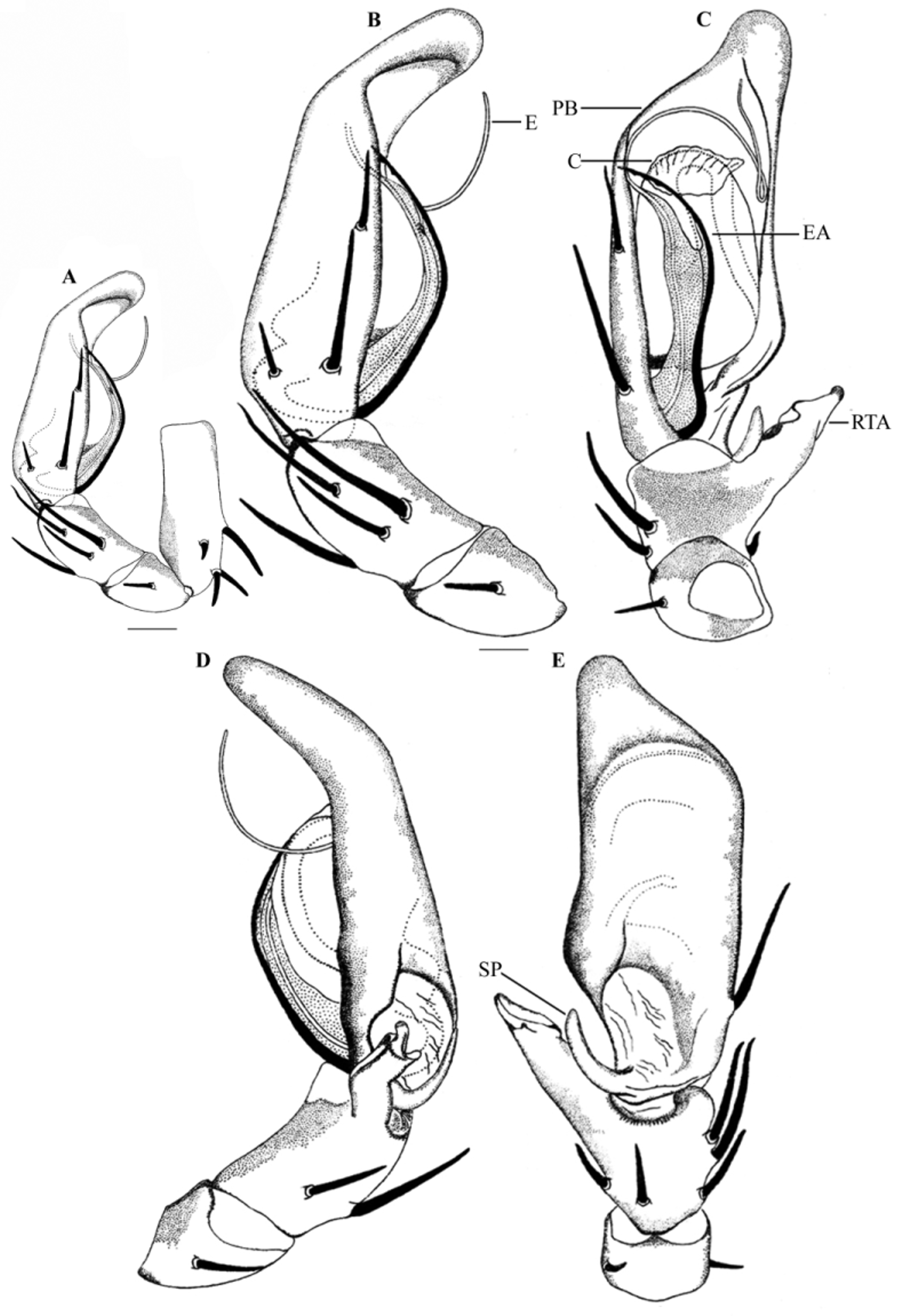
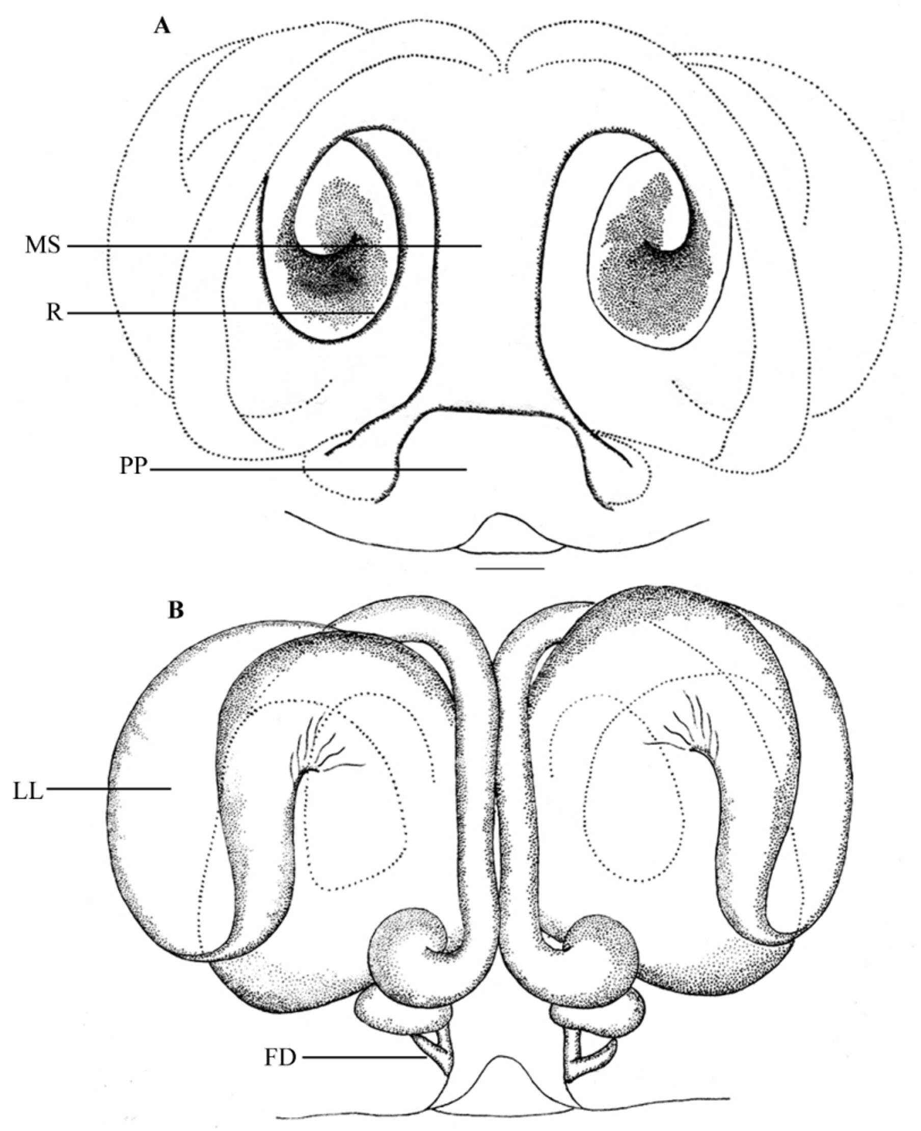
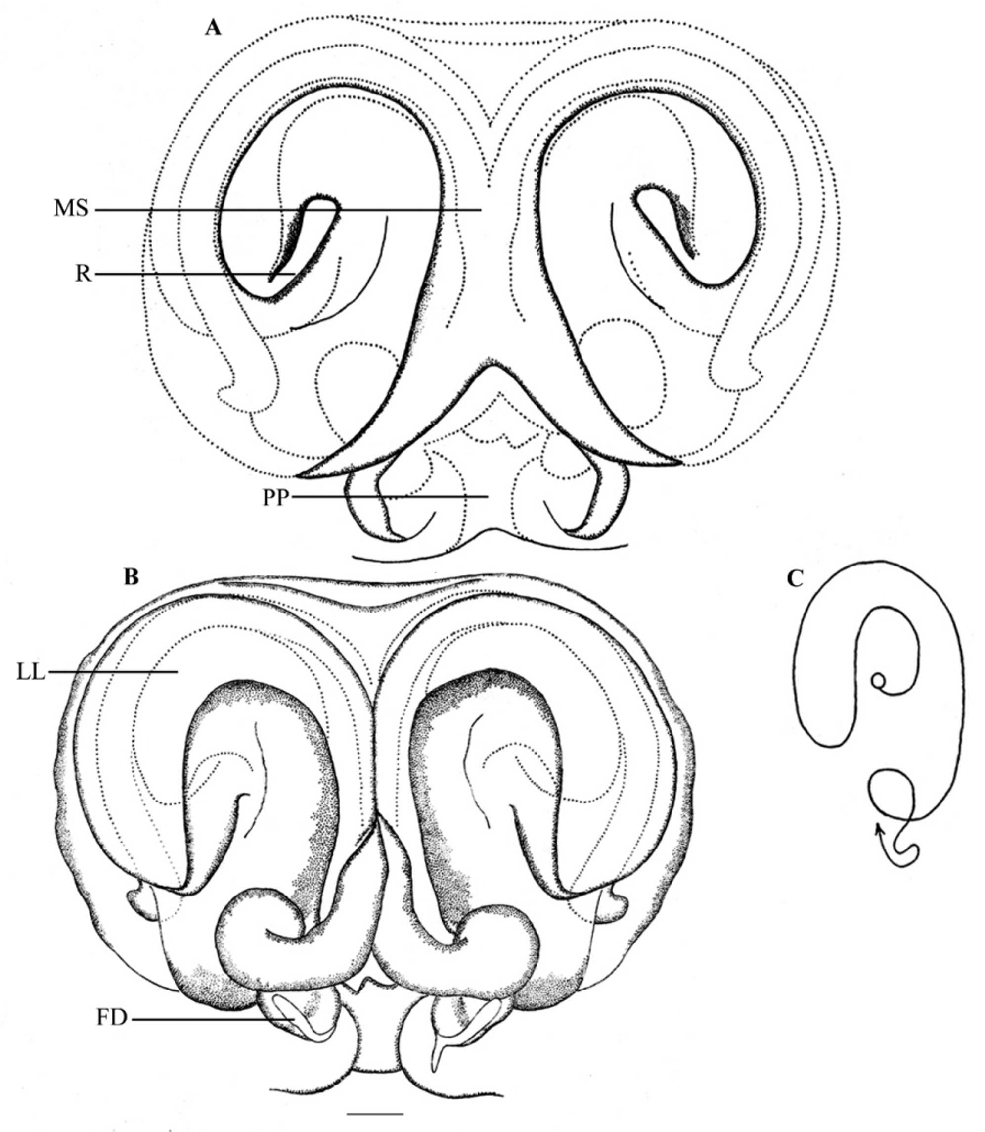
Diagnosis. Male of M. minuscula is closely related to M. transversa , but can be separated by the following combination of characters: less prominent prolateral bulge of cymbium ( Figs 4D View FIGURES 4 A – J , 5C View FIGURES 5 A – E ), in M. transversa , it is prominent ( Jäger 2006: fig. 5); embolus with a distal, oblique twist ( Figs 4D View FIGURES 4 A – J , 5C View FIGURES 5 A – E ), which is absent in M. transversa ( Jäger 2006: fig. 5); wider conductor ( Fig 5C View FIGURES 5 A – E ), in M. transversa , the conductor is reduced ( Jäger 2006: fig. 5); RTA short and directed at 1-o’clock position ( Figs 4D View FIGURES 4 A – J , 5C View FIGURES 5 A – E ), in M. transversa , RTA is long and directed at 11-o’clock position ( Jäger 2006: fig. 3). Females can be separated from all other described congeners by the median part of the internal duct system, which appears as paired, longitudinal tubes and lying along the entire length of the median line ( Figs 4J View FIGURES 4 A – J , 6B View FIGURES 6 A – B ), in M. transversa , it appears to be a median, flattened, longitudinal plate ( Jäger 2006: fig. 10) and in M. sanctor sp. nov., it is short and restricted to the posterior part of the median line ( Figs 1J View FIGURES 1 A – J , 3B View FIGURES 3 A – C ).
Description. Male: Prosoma uniformly yellowish-brown with narrow reddish- brown striae radiating from sides of fovea and with dark, faint markings ( Figs 4A–B View FIGURES 4 A – J ). Eye field yellowish-brown, hirsute; eyes with distinct dark patches. Clypeus, chelicerae, maxillae, labium, sternum yellowish-brown. Cheliceral promargin with 3 teeth, retromargin with 4 teeth; promargin provided with a row of long, curved hairs. Fang furrows provided proximally with a few denticles. Maxillae with reduced scopulae. Sternum without markings. Opisthosoma elongated oval, yellowish-brown, hirsute; dorsally and laterally with black shades. Spinnerets yellowish-brown. Leg segments yellowish-brown with inconspicuous dark brown patches; trochaters I–III notched; metatarsi I–III and tarsi I–III scopulated. Body length 6.43. Prosoma length 3.60, width 3.33. Opisthosoma length 2.83, width 1.85. Eye diameters: AME 0.22. ALE 0.39. PME 0.26. PLE 0.41. Eye interdistances: AME–AME 0.18. AME–ALE 0.05. AME–PME 0.20. ALE–PLE 0.28. PME–PME 0.26. PME–PLE 0.31. Clypeus height at AME 0.26, at ALE 0.21. Chelicera length 1.30. Measurements of palp and legs: Palp 5.25 [1.70, 0.58, 0.71, 2.26], I 12.86 [3.68, 1.57, 3.37, 3.04, 1.20], II 14.67 [4.34, 1.69, 3.80, 3.58, 1.26], III (right leg) 11.9 [3.50, 1.45, 3.06, 2.99, 0.90], IV missing. Leg formula 213... Spination. Palp: femur 131, patella 101, tibia 2111, tarsus 3000; legs: femora I–III 323; patellae I–III 001; tibiae I–II 2026, III 2126; metatarsi I–II 0 0 0 4, III 2024. Pedipalp ( Figs 4C–F View FIGURES 4 A – J , 5A–E View FIGURES 5 A – E ): Palpal segments yellowish-brown. Embolus forming a distal narrow loop ( Figs 4D View FIGURES 4 A – J , 5C View FIGURES 5 A – E ); tip of embolus directed at 1-o’clock position ( Figs 4D View FIGURES 4 A – J , 5C View FIGURES 5 A – E ); embolic apophysis separates distally from the embolus ( Figs 4D View FIGURES 4 A – J , 5C View FIGURES 5 A – E ); tip of embolic apophysis serrated ( Figs 5B–C View FIGURES 5 A – E ). Conductor wide, membranous, situated distally on the tegulum ( Fig 5C View FIGURES 5 A – E ). RTA massive with a narrow tip, directed at 2-o’clock position, with a short, disto-retrolateral branch ( Figs 4E View FIGURES 4 A – J , 5D View FIGURES 5 A – E ). Cymbial spur slender, curved ( Figs 4F View FIGURES 4 A – J , 5E View FIGURES 5 A – E ).
Redescription. Female: In all details like male except the following: Prosoma with dark, faint markings ( Figs 4G–H View FIGURES 4 A – J ). Eye field not hirsute. Sternum with faint dark markings. Opisthosoma oval, grayish; venter provided with scattered, inconspicuous dark brown patches. Spinnerets grayish. All trochanters notched; metatarsus IV less scopulated; all other metatarsi and all tarsi with well developed scopulae. Body length 8.51. Prosoma length 3.52, width 3.26. Opisthosoma length 4.99, width 3.40. Eyes diameter: AME 0.14. ALE 0.18. PME 0.16. PLE 0.25. Eye interdistance: AME–AME 0.12. AME–ALE 0.07. AME–PME 0.29. ALE–PLE 0.31. PME–PME 0.22. PME–PLE 0.34. Clypeus height at AME 0.19, at ALE 0.17. Chelicera length 1.48. Measurements of palp and legs: Palp 4.51 [1.31, 0.72, 0.94, 1.54], I 10.42 [2.86, 1.47, 2.61, 2.53, 0.95], II 11.18 [3.31, 1.28, 2.67, 2.83, 1.09], III 9.59 [3.13, 1.40, 2.32, 2.00, 0.74], IV 10.97 [3.20, 1.52, 2.66, 2.61, 0.98]. Leg formula: 2413. Copulatory organ ( Figs 4I –J View FIGURES 4 A – J , 6A–B View FIGURES 6 A – B ): Epigynal field transversally oval ( Figs 4I View FIGURES 4 A – J , 6A View FIGURES 6 A – B ). Median septum broad over its entire length, with rectangular posterior pit ( Figs 4I View FIGURES 4 A – J , 6A View FIGURES 6 A – B ). Rims around copulatory openings forming a large circle ( Fig 6A View FIGURES 6 A – B ). Lateral loops of the internal duct system may or may not extend posteriorly to receptacula ( Figs 4J View FIGURES 4 A – J , 6B View FIGURES 6 A – B ; Jäger 2006: figs 14, 15).
Note. According to Jäger (2006), the lateral loops of internal duct system of the female genitalia of M. minuscula extend posteriorly to the receptacula ( Jäger 2006: figs 14, 15). But in one of our female specimens, lateral loops of internal duct system extend posteriorly not to receptacula ( Figs 4J View FIGURES 4 A – J , 6B View FIGURES 6 A – B ).
Natural history. Both male and females of M. minuscula were collected from leaf litter in a semi-deciduous and semi-evergreen forests floor.
Distribution. Known only from southern India (Kerala & Tamilnadu) ( Fig 9 View FIGURE 9 ).


| ADSH |
Arachnology Division, Sacred Heart College |
No known copyright restrictions apply. See Agosti, D., Egloff, W., 2009. Taxonomic information exchange and copyright: the Plazi approach. BMC Research Notes 2009, 2:53 for further explanation.
|
Kingdom |
|
|
Phylum |
|
|
Class |
|
|
Order |
|
|
Family |
|
|
Genus |
Martensopoda minuscula ( Reimoser, 1934 )
| Sankaran, Pradeep M., Malamel, Jobi J., Joseph, Mathew M. & Sebastian, Pothalil A. 2015 |
Heteropoda minuscula
| Jager 2002: 58 |
| Reimoser 1934: 485 |