
Sphaerodoropsis aurantica, Capa, María & Rouse, Greg W., 2015
|
publication ID |
https://doi.org/10.11646/zootaxa.4019.1.9 |
|
publication LSID |
lsid:zoobank.org:pub:D4ECFEE2-BF9D-4B5F-8128-EFAAC2B728C4 |
|
DOI |
https://doi.org/10.5281/zenodo.6096003 |
|
persistent identifier |
https://treatment.plazi.org/id/C22DFF62-FFF4-FF82-FF21-FEA9FCAE9B4B |
|
treatment provided by |
Plazi |
|
scientific name |
Sphaerodoropsis aurantica |
| status |
sp. nov. |
Sphaerodoropsis aurantica n. sp.
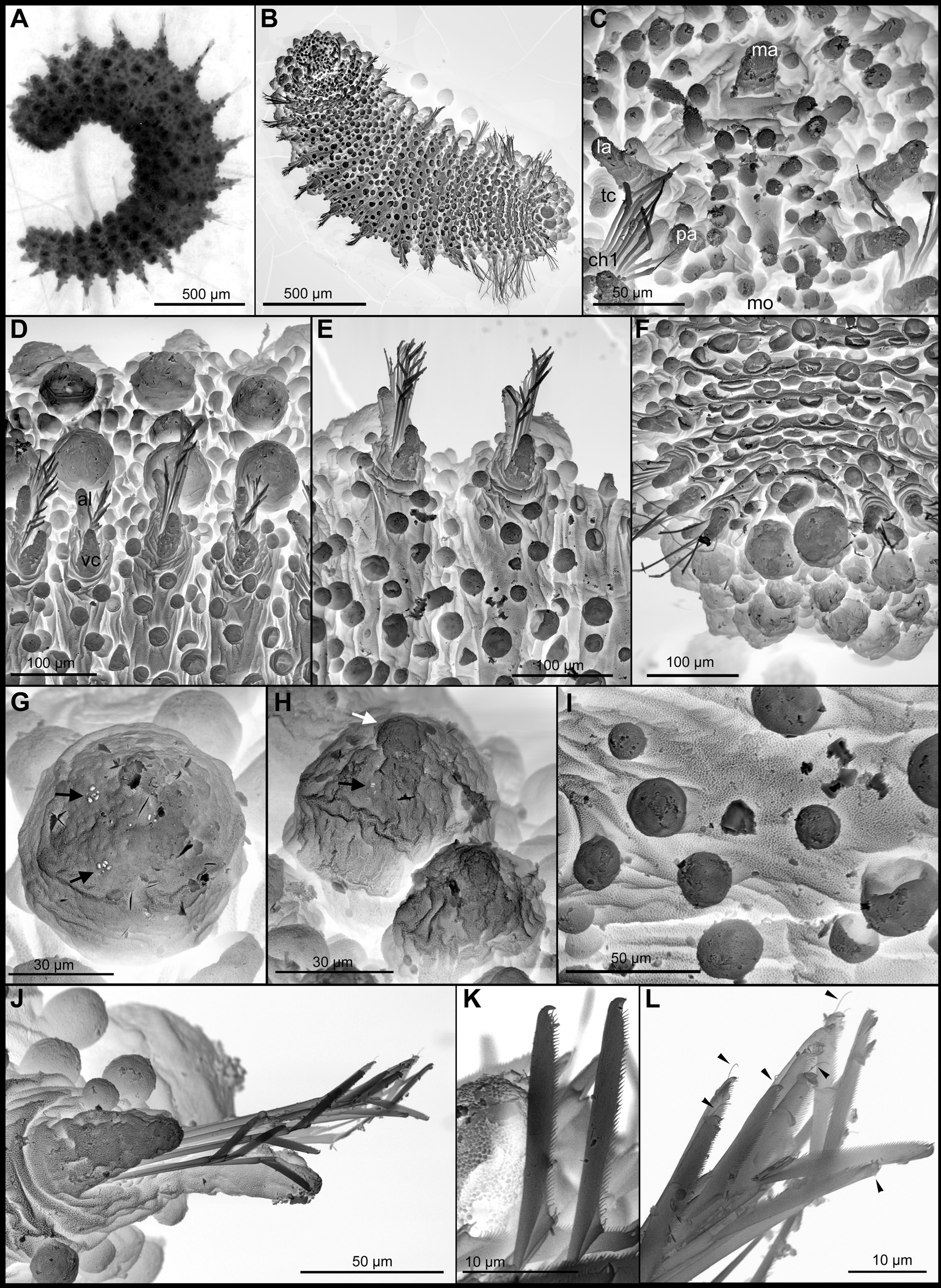
( Figs 2 View FIGURE 2 C, D, 3)
Sphaerodoropsis sp A.— Helm & Capa, 2015.
Type material. Holotype: AM W.44209, MI QLD 2380, on SEM stub. Paratype: AM W.44210, MI QLD 2380, posterior end used for DNA sequencing.
Other material examined. AM W.44218, MI QLD 2390, live photo, further used for confocal microscopy, Helm & Capa 2015; AM W.44220, MI QLD 2387, used for confocal microscopy, Helm & Capa 2015.
Diagnosis. Body ellipsoid, with strongly convex dorsum. Nine longitudinal rows of sessile and spherical dorsal macrotubercles, arranged in a single transverse row per segment row and four transverse rows (with up to 38) of spherical papillae per segment. Parapodia with digitiform acicular lobe, shorter ventral cirrus and around 10 spherical papillae. Over 10 compound chaetae per parapodium with thin shafts, long blades (6–8 times longer than its maximum width), with fine and short spinulation along superior edge and a distal recurved tip.
Description. Holotype 1.8 mm long after fixation, 0.6 mm maximum width; with 20 chaetigers. Body ellipsoid ( Fig. 3 View FIGURE 3 A–B); with convex dorsum and flattened ventrum. Tegument with transverse wrinkles, segmentation inconspicuous ( Fig. 3 View FIGURE 3 A–B). Head externally indistinct ( Fig. 3 View FIGURE 3 A–C). Anterior end bluntly rounded ( Fig. 3 View FIGURE 3 B–C). Prostomium with five appendages, including a pair of palps, in ventral most position, sub-conical and slightly wrinkled; a pair of lateral antennae, similar in shape and size to palps; and a median antenna, shorter (two thirds) and wider than lateral antennae and with a rounded distal end ( Fig. 3 View FIGURE 3 B–C). Antenniform papillae cannot be unequivocally recognised ( Fig. 3 View FIGURE 3 C). Around 30 digitiform small papillae confined by prostomial appendages and mouth in frontal view ( Fig. 3 View FIGURE 3 C). A pair of tentacular cirri, similar in shape and size to lateral antennae and palps, and several scattered papillae similar to prostomial. Macrotubercles, sessile, rounded or provided with an incipient rounded terminal papillae ( Fig. 3 View FIGURE 3 B, G–H), arranged in longitudinal rows along dorsum, and single transversal rows per segment. First and following chaetigers with nine macrotubercles, decreasing to eight in posterior chaetigers. Macrotubercles with two different sizes, following pattern in Fig. 2 View FIGURE 2 C; provided with pores arranged on groups ( Fig. 3 View FIGURE 3 G–H), some of them with incipient terminal papillae or at least pear-shaped ( Fig. 3 View FIGURE 3 H). Spherical papillae over dorsum, arranged in four transverse rows per segment; digitiform in anterior-most chaetigers and spherical in following ( Fig. 3 View FIGURE 3 C–D). Ventral surface with similar spherical papillae, arranged in about 5–6 irregular transverse rows, with a total of around 50 papillae per segment, in mid-body ( Fig. 3 View FIGURE 3 B). Parapodia sub-conical, increasing in size towards chaetiger 5 and around 1–2 times longer than wide, wrinkled ( Fig. 3 View FIGURE 3 C–D). Acicular lobe anterior to chaetal fascicle, projecting distally ( Fig. 3 View FIGURE 3 D–E, J). Ventral cirri sub-conical to pear-shaped, shorter than acicular lobe ( Fig. 3 View FIGURE 3 D–E, J). Mid-body parapodia with around 10 small spherical papillae, slightly different in size: one or two on dorsal surface, three on anterior surface, three on ventral surface and three on posterior surface ( Fig. 2 View FIGURE 2 D). Compound chaetae present in all chaetigers, arranged in a curved transverse fascicle around acicular lobe and numbering 10–22 per parapodium. Shaft with similar width all along, slightly widened distal end with delicate almost inconspicuous spinulation. Blades similar in length within fascicles (6–8 times longer than its maximum width), with fine and short spinulation along superior edge and a distal recurved tip ( Fig. 3 View FIGURE 3 J–L). Pygidium terminal, with mid-ventral digitiform anal cirrus and a pair of dorsal anal cirri, similar in shape to macrotubercles ( Fig. 3 View FIGURE 3 F). Mouth located ventrally near base of palps ( Fig. 3 View FIGURE 3 C). Gut visible by transparency with muscular pharynx occupying about four segments. Eyes and copulatory organs or gametes not seen.
Colour pattern. Live specimens white, with a bright orange transverse band on dorsum of chaetigers 12–13. Lateral most and dorsal most macrotubercles of all except for anterior and posterior two chaetigers also partially pigmented ( Fig. 3 View FIGURE 3 A). Colour lost in preserved material.
Variation. Paratype with 21 chaetigers. Number of epithelial tubercles and papillae matches those described in holotype. Parapodia of mid-body chaetigers with higher number of chaeate and papillae, within the range described in holotype. Pigmentation pattern varied slightly among described specimens and the orange band was more or less evident in some specimens, and within chaetigers 10–13 but the dorsolateral macrotubercles, were in all cases partially bright orange. Copulatory organs or gametes not observed in any specimen.
COI Barcode (Paratype, AM W.44210).
AAAATCAAAACAAATGTTGAAATAAAATAGGATCTCCTCCTCCTGCCGGATCAAAAAACCTAGTATTT AGATTACGATCAGTTAATAATATAGTAATTACACCTGCCAATACAGGCAAAGCTAATAATAATAAAATAG CTGTAATCAATACAGATCAAGTAAATAAAGGAATACGCTTAAACTTCATACCCACTACATGATCAAATA TAATAGTAACAATAAAATTAATAGCACCTAAAATAGAAGAAACACCAGCTATATGTAATGAAAAAATAG CTATATCAACAGAAGGCCCCGAATGAGTTATATTACTTGATAAAGGAGGATAAACTGTTCATCCTGTAC CAGCACCTTTCTCTACTAATGTAGATCCCAATAATAATATTAAAGAAGGAGGCAAAAATCAAAACCTTA TATTATTTAATCGAGGAAAAGCTATATCAATAGCACCTAACATTAAAGGAACTAATCAATTACCAAAAC CACCCATTATAACTGGTATTACAAGAAAAAAAATTATTAAAAAAGCATGACCAGTAACAATAGTATTAT ATAATTGATCTCTACCTAATAAACTACCTGGTTGACCTAATTCAGCACGAATCAACAAACTTATAGATGT ACCTAAAAATCCAGATCATATACCAAAAATAAAATACAAAGTACCAATATCTT
Remarks. Even though some macrotubercles were observed with distal papillae, this is not a constant attribute of all tubercles or present in all specimens of Sphaerodoropsis aurantica n. sp. examined and may be an artefact due to the collapse of some of these structures. Nevertheless, and as indicated previously (Capa et al. 2015; Capa & Bakken 2015), distinguishing Sphaerephesia and Sphaerodoropsis is confusing at present because there are species within each genus presenting pear-shaped macrotubercles that lack terminal papillae. This species would belong within the Group 2 proposed by Borowski (1994), together with other Sphaerodoropsis species with more than four longitudinal rows of macrotubercles arranged in a single transverse row per segment. There are nine other species in the group ( Borowski 1994; Aguirrezabalaga & Cebeiro 2005; Moreira & Parapar 2011), considering that S. minuta ( Webster & Benedict, 1887) and S. polypapillata Hartmann-Schröder & Rosenfeldt, 1988 , have recently been synonymised with other genera (Moreira & Parapar 2011; Capa et al. 2015). Species distinguished from S. aurantica n. sp. due to the lower (<9) number of longitudinal rows of macrotubercles are S. amoreuxi Aguirrezabalaga & Cebeiro, 2005 , S. benguellarum ( Day, 1963) , S. octopapillata ( Hartmann-Schröder, 1965) , S. sphaerulifer ( Moore, 1909) and S. uzintunensis Kudenov, 1987 . Of the remaining species, S. aestuarum Averincev, 1990 is distinguished from S. auranticus n. sp. because it has two transverse rows of additional papillae per segment in addition to the 8–10 macrotubercles and compound chaetae with blades around three times longer than wide; S. balticum ( Reimers, 1933) also has two transverse rows of papillae per segment in addition to the 7–9 rows of macrotubercles and short chaetal blades (less than twice as long as wide); S. gudmunduri Moreira & Parapar 2012 , lacks dorsal papillae among the nine macrotubercles and has short chaetal blades (around three times longer than wide); and S. katchemakensis Kudenov, 1987 bears two transverse rows of papillae in addition to the 8–9 macrotubercles and chaetae with blades ranging 3–4 times as long as wide ( Reimers 1933; Hartmann-Schröder 1996; Kudenov 1987a; Moreira & Parapar 2012).
Sphaerephesia gesae Moreira & Parapar, 2011 is the only species in that genus with more than four longitudinal rows of macrotubercles (Moreira & Parapar 2011; Alalykina 2015). It can be distinguished from S. aurantica n. sp. by the presence of two transverse rows of macrotubercles per segment, instead of only one found in the new species and the chaetal morphology with short blades (maximum twice as long and wide) in comparison to those described in S. aurantica n. sp.
Etymology. The species name refers to the bright orange pigmentation (orange in Latin= auranticus ) present in mid body tubercles.
Distribution. Species only known from type locality.
| DNA |
Department of Natural Resources, Environment, The Arts and Sport |
No known copyright restrictions apply. See Agosti, D., Egloff, W., 2009. Taxonomic information exchange and copyright: the Plazi approach. BMC Research Notes 2009, 2:53 for further explanation.
|
Kingdom |
|
|
Phylum |
|
|
Class |
|
|
Order |
|
|
Family |
|
|
Genus |