
Diplazontinae
|
publication ID |
https://doi.org/ 10.11646/zootaxa.3801.1.1 |
|
publication LSID |
lsid:zoobank.org:pub:E5F8C489-37F4-4A76-8E25-EFC65CDCA1D7 |
|
DOI |
https://doi.org/10.5281/zenodo.6135599 |
|
persistent identifier |
https://treatment.plazi.org/id/C1225000-FFAD-FFF0-B5BD-A535FA33F9F3 |
|
treatment provided by |
Plazi |
|
scientific name |
Diplazontinae |
| status |
|
Subfamily Diplazontinae
Classis Hexapoda Blainville
Ordo Hymenoptera Linnaeus
Subordo Apocrita Latreille
Superfamilia Ichneumonoidea Latreille
Familia Ichneumonidae Latreille
Subfamilia Diplazontinae Viereck
The subfamily Diplazontinae is one of the more easily recognized ichneumonid subfamilies. Its monophyly with respect to members of Cylloceriinae , Orthocentrinae , and several other pimpliform subfamilies has recently been demonstrated based on data from four molecular markers ( Klopfstein et al. 2011). Diagnostic features include the three-toothed mandibles, the rectangular, box-like first tergite, comparatively short propodeum, short ovipositor, and transverse head. The only diplazontine species that lacks the typical, three-toothed mandibles and has a comparatively long ovipositor, Episemura diodon , might be difficult to place in this subfamily. The other characters, however, clearly show its affiliation with this group. For identification at the subfamily level, the keys in “ Hymenoptera of the World” ( Goulet & Huber 1993; Wahl 1993) or Townes’ keys can be used ( Townes 1969). Seventeen of the 22 recent genera occur in the Western Palaearctic and are treated here. Fourteen of these have been included in recent molecular and morphological phylogenetic analyses of the subfamily ( Klopfstein et al. 2010a; Klopfstein et al. 2011). These studies recovered three strongly supported genus-groups, the Sussaba group consisting of Promethes and Sussaba , the Syrphoctonus group including Bioblapsis , Enizemum, Fossatyloides , Homotropus , Phthorima , and Syrphoctonus , and the Diplazon group encompassing Campocraspedon , Diplazon , Syrphophilus , Tymmophorus , and Xestopelta . Even though these groups were treated as informal, some morphological and behavioural characters were suggested to circumscribe these groups ( Klopfstein et al. 2010b; Klopfstein et al. 2011). I here use these informal groupings for reference, and add the three Western Palaearctic genera as follows: Episemura and Eurytyloides to the Sussaba genus group, based on a close association with Sussaba as suggested by, e.g., the lateral position of the spiracle of the second tergite, and Daschia to the Diplazon genus group based on the structure of the male terminal sclerites and several similarities to Campocraspedon (see genus diagnoses).
Assessment of morphological characters
For the identification of diplazontine genera, structural characters proved most useful, although colour patterns can also be very informative. Many genera, e.g. Campocraspedon Uchida and Phthorima Förster , can most easily be determined from the shape of the female metasoma, which is strongly modified, probably in order to reach their host in some substrate. These modifications are far less obvious in most male specimens, which also show a larger intra-specific variability than females in other characters. Males are thus often more difficult to place than females. An exception is the flavipes - pulchella species complex in the genus Sussaba . The shape and colour of the tyloids, i.e. male-specific structures involved in courtship behaviour, allow males to be distinguished very easily even in species where females are difficult to tell apart ( Klopfstein et al. 2010b; Steiner et al. 2010). The sexual dimorphism can be quite extensive in many species, especially where colouration or metasomal shape is concerned. In general, females have a mostly black face, often with some yellow markings, while the face of most males is entirely yellow. However, there are numerous exceptions to this rule, and the external genitalia should be taken into consideration when determining the sex of a specimen. In the following, I discuss the utility, the different states, intra-specific variability and visibility of different morphological characters or character systems in order to facilitate the use of the identification keys and species diagnoses. After discussing measurements, sculpture and colouration in general, I arrange the characters according to their appearance on the adult wasp, starting with the antennae and proceeding back from there.
Measurements: I refrained from the extensive use of morphometry for delimiting and identifying diplazontine species, as it requires a lot of work from the user of the keys, and furthermore, many morphometric characters are fairly variable even within species of Diplazontinae . In general, length and width were measured at the longest or widest part of a body part. The relative length and width of the first three tergites proved very useful in several genera, and the observed range of ratios is given for those. Tergite length was always measured in lateral view and along the side of the tergites, and width at the broadest position in the posterior quarter of the tergite.
Sculpture: The macro- and microsculpture of the face, mesosoma and of the tergites is important, especially for identification at the species level. Some closely related species in the genera Diplazon and Homotropus can only be identified by careful examination of the sculpture, and although especially males often show considerable intraspecific variation in sculptural characters, it is usually constant enough to allow reliable determination. However, the nomenclature for sculpture varies extensively between authors. Eady (1968) published an illustrated guide that allows some standardization of sculpture terminology. Not all his categories fully reflect the sculpture found in Diplazontinae . In general, I follow Eady’s nomenclature but additionally provide SEM micrographs showing the respective states as they are found in Diplazontinae ( Fig. 7 View FIGURE 7 ). Mostly, I use the terms “smooth” and “coriaceous” for microsculpture, and “rugose” and “punctate” for larger sculptural elements.
Colouration: Colouration of diplazontines is particularly variable and thus provides a large number of characters, some of which are highly informative at species and even at higher levels. I use the same expressions as previous authors to denote colours in diplazontines, with the exception of using “orange” for a colour that was usually referred to as “red” in the literature. This is first in order to more accurately report the situation in the observed specimens, and second to distinguish this prevalent color from a darker, truly red colouration found in some species. When using colour characters, one should bear in mind that older specimens often lose a lot of their colouration, especially if kept in alcohol, and a black metasoma can thus appear brown and a yellow spot almost white in older material. The expressions “dark”, “black” and “brown” can thus be interpreted as being largely interchangeable.
Antennal length: I here number the flagellomeres without counting the first two antennal segments (i.e. the scape and pedicel). The number of flagellomeres is a very important character in defining some of the species. Intra-specific variation was found to be relatively low, ranging between 0 to 3 flagellomeres. As expected, variation was lower in species with shorter and higher in species with longer antenna, and in general higher in males.
Ventral surface of antenna: Some females have the ventral surface of the middle to apical flagellomeres covered with conical sensilla of unknown function ( Fig. 5 View FIGURE 5 ). This character has been noted by Dasch (1964a) for Homotropus dimidiatus , but it is also present in many species of other genera and seems to be a rather homoplasious character. The conical sensilla are always distinctly shorter than the trichodeal sensilla and are often difficult to see in light microscopy, as they only appear as a matt, velvety pile. Their presence is more easily deduced from the absence of multiporous plate sensilla on the ventral surface, which can be assessed by comparing the ventral to the dorsal surface of the antenna.
Tyloids: In males, the presence or absence, location, shape and colouration of convex structures on the outer surface of the antenna, the tyloids, provide important characters for identification at the genus and species level. While the broad tyloids as found in the genera Sussaba and Enizemum Förster are very obvious, the narrow tyloids of Promethes Förster , Homotropus and Syrphoctonus Förster are more difficult to see, especially if the antenna is paler ventrally than dorsally, in which case the location of the colour transition often coincides with the tyloid location. Careful examination of the antenna from different angles is needed in such cases.
Face: The shape, microsculpture and colouration of the face provide some reliable characters. The shape is useful to delimit some species within the genera Sussaba and Woldstedtius , for which drawings are provided in the key. However, it has to be noted that some aspects of the shape cannot be captured by two-dimensional illustrations, as some distinct differences involve the third dimension as well. The face can be variously sculptured, ranging from being mostly smooth and shining to strongly sculptured and matt (cf. Fig. 7 View FIGURE 7 ). Moreover, there is a varying degree of punctation of the face. In most species of the genera Sussaba and Promethes , there are vertical impressions starting from the tentorial pits and pointing towards the antennal bases, thus separating the median, often elevated part from the remainder of the face. The impressions are most distinct towards the clypeus and become indistinct about at mid height of the face. The colouration of the face is also a good character, especially in females where the presence and absence of yellow markings along inner orbits provide a good means to distinguish most genera of the Diplazon genus group from the remainder. The yellow central face patch found in some species of each of the three genus groups is in contrast less reliable because of a larger intra-specific variability but can still provide a good character in some species.
Clypeus: The shape of the clypeus is often genus-specific, and figures are provided along with the key. Besides the extent to which the apical margin is emarginate and thus forms two lobes, the shape of the clypeus when viewed from the side is especially important. In most genera, the apical margin of the clypeus is thin, while it is conspicuously thickened in the genera Daschia Diller , Xestopelta Dasch and Campocraspedon . This condition might represent a synapomorphy of these three genera and thus point to them being closely related. However, recent phylogenetic analyses of the subfamily did not include Daschia , and are somewhat equivocal about the placement of Xestopelta ( Klopfstein et al. 2010a; Klopfstein et al. 2011). The clypeus is often either impressed right below a basal thickening, which gives it a concave appearance when viewed from the side, or it is impressed along the apical margin, which renders it convex basally. The former condition can be found in most genera of the Sussaba and Diplazon genus groups, the latter in Fossatyloides, Homotropus and Phthorima . However, there are a number of species which are intermediate to these two states, with the subbasal impression only present laterally and with an apical impression medially (e.g. in the genera Syrphoctonus , Enizemum and Woldstedtius ). The clypeus characters thus require some experience, especially in those genera, and are often less stable in males.
Mesoscutal colouration: The mesoscutum of most species is either black or brown, or it bears two yellow or whitish spots at the anterolateral corners, which I call “shoulder marks”. These can be very small spots to large, triangular shapes, and sometimes their inner corners extend back over most of the mesoscutum (cf. Fig. 13 View FIGURE 13. A D).
Notauli: While reaching over the entire length of the mesoscutum in some other ichneumonids, the notauli in Diplazontinae , if present, are quite short and often only present on the inclining part at the front of the mesoscutum ( Fig. 8 View FIGURE 8 ). They should thus be searched for not only from a dorsal but also from a lateral view. Usually, they represent reliable characters but vary in some species, for which I tried to control in the species keys (e.g. some male specimens of Syrphophilus tricinctorius (Thunberg)) .
Scutellum: The scutellum often offers quite reliable color characters, as it can bear a yellow, whitish or even orange apical spot which can cover up to the entire scutellum, or it has an apical spot and two lines along its sides. Furthermore, the scutellum is often partly enclosed laterally by carinae in the Sussaba genus group, while it has very short lateral carinae which only just cross the prescutellar groove in other genera.
Colouration of coxae: The coloration of the coxae, especially the hind coxa, is a useful character for some species and species groups. However, it can exhibit a lot of intra-specific variation, again especially but not exclusively in males. In some species with orange hind coxae, individuals with partly dark coxae can be found at higher altitude, and individuals with intermediate states occur regularly, i.e. with the orange hind coxa dark only basally. In other taxa, e.g. the former subspecies of Sussaba dorsalis , analysis supports the value of this character for species delimitation. The variability of coxa colouration is discussed in more detail in the species concerned.
Colouration of hind tibia: Many species have a yellow or orange hind tibia, often with the apex dark to varying degrees. In the genera Campocraspedon , Diplazon , Syrphoctonus and Homotropus however, there are species with whitish hind tibiae that can be variously brown or black banded. In Diplazon , either only the apex or more usually the apex and the base are dark. In Homotropus , many species have a dark apex and a subbasal dark spot. These colour patterns can be less distinct in males. It can also be difficult to distinguish between yellow and white on the tibia, especially in older museum specimens, but a comparison with the yellow or orange front and mid tibia can help in such cases. In the genera Woldstedtius and Enizemum , most species have black hind tibiae with a white base. The white colouration in these genera can extend further down on the inner side of the tibia, especially in males.
Wing venation: The venation both of fore and hind wings is very constant in diplazontines and ichneumonids in general when compared to other hymenopteran groups. In the fore wing, the presence or absence of the areolet (i.e., the presence or absence of vein 3rs-m) is useful for separating most specimens of the genera Enizemum , Homotropus and Phthorima from other genera. However, this character also shows considerable variation, with the outer vein (3rs-m) often not being pigmented at all. In some species (e.g. Syrphoctonus tarsatorius ), I have even found individuals with the areolet open in the right and closed in the left fore wing, or vice versa. To account for this variation, I allow some species to be keyed through both parts of the couplets. In any case, if there is some indication of vein 3rs-m that closes the areolet, no matter how incomplete, it should be regarded as present. Otherwise, wing venation is of minor value for species identification, as it is very constant. The few variable characters, e.g. the position of vein CU+cu-a opposite or distal of vein M or the length and position of vein 2/Cu in the hind wing, which have been used in the past, usually vary a lot within a species and thus proved to be of little use. The same applies to the ratio between the length of vein 2rs-m and its distance from vein 2m-cu. This ratio is comparatively small in most species of the Diplazon genus group but often does not provide reliable means for identification.
Basal hamuli of hind wing: The number of basal hamuli present distal to the costella in the hind wing is a good character to separate most species of the Sussaba genus group from the remaining diplazontines. These species have only one basal hamulus, with the exception of some specimens of Promethes nigriventris and P. melanaspis . No species of the other genus groups have only a single hamulus, exept for the very distinctive Bioblapsis polita , which can have 1–3.
Propodeum: The naming of the propodeal carinae and areas follows Townes (1969) ( Fig. 9 View FIGURE 9 ). The propodeum in Diplazontinae is shortened compared to other ichneumonids and the carination is reduced. A complete set of carinae in Diplazontinae encloses along the midline only the basal area and a large petiolar area, while the areola present in other ichneumonids is fused with the large petiolar area, as deduced from some specimens of Tymmophorus where it is still indicated ( Fig. 9 View FIGURE 9 c). The first and second lateral areas are only incompletely separated, and the third lateral and pleural areas are fused as well ( Fig. 9 View FIGURE 9 a). In many species, this carination is futher reduced ( Fig. 9 View FIGURE 9 d), up to an almost complete reduction of carinae in most Syrphoctonus and Woldstedtius species ( Fig. 9 View FIGURE 9 e). If the propodeum is rugose, it can sometimes be difficult to distinguish between the presence and absence of particular carinae. Especially in species which have their carination partly reduced (e.g. Homotropus signatus ), there is often considerable intra-specific variation. However, it is usually sufficient to assess whether the carination is reduced to traces of the lateral carinae, or if at least the basal and petiolar areas are fully enclosed by carinae.
Shape of metasoma: The metasoma of Diplazontinae is either dorsoventrally depressed or laterally compressed, or any state inbetween. In the genus Sussaba for instance, the metasoma of some species is laterally compressed only at the tip, gradually tapering from the third segment, while it is strongly compressed from the base of the third segment to the apex in others. The metasoma can be so strongly compressed that it is almost blade-like, visible only as a line when viewed from above. Lateral compression or strong depression is in some species combined with tergites that extend further back laterally than medially, rendering their hind margins concave when viewed from above. The functional significance of these modifications is not fully understood, but some species of Phthorima have been shown to be associated with syrphids that feed in aphid colonies which form galls or wax layers; they can only be reached with an especially thin or long metasoma. In Xestopelta gracillima , the degree of concavity of the tergites varies considerably between individuals, from indistinct to quite strong. For the other genera, this character is reliable at least in females. Concave hind margins of the tergites are also present in males of some species but are far less distinct, and males thus require special attention when it comes to this character.
Transverse impressions on tergites: In the genus Diplazon , tergites 1 to 3 or 4 bear transverse impressions that range in strength from deep impressions that bear transverse carinulae on the first tergite to a mere change in sculpture on the third and fourth tergites (cf. Fig. 7 View FIGURE 7 A and 7B). Although in general a reliable character, other genera of the Diplazon genus group can also have weak impressions on the first two tergites, which can make the distinction between these genera more difficult. For example, the impressions are rather weak in Diplazon pectoratorius (Thunberg) and Diplazon neoalpinus Zwakhals , where they are sometimes not stronger than in some specimens of Syrphophilus Dasch. I control for this in the keys.
Spiracles of the second and third tergites: The spiracles of both the second and third tergites vary in their location. In the genera Sussaba , Episemura Kasparyan & Manukyan , and Eurytyloides Nakanishi , they are both located on the laterotergites, below the lateral fold which separates the dorsal from the lateral part of the tergite; in Promethes , Campocraspedon and some Homotropus species, only the third spiracle is on the laterotergite; in all other diplazontines, they are both on the dorsal parts of the tergites, above the lateral folds. The laterotergite is usually inflexed in dried specimens; especially the spiracle of the second tergite might in such cases be difficult to see. Its location on the laterotergite can then be deduced from its absence from the dorsal part of the tergite, but care should be taken not to mistake the sometimes conspicuous, irregularly oval muscle insertion on the second tergite for a spiracle. For the third tergite, the situation is a bit more complicated. Especially in species with a laterally compressed metasoma, the fold separating the dorsal from the lateral part of the tergite is often indistinct at the level of the spiracle. The latter is then often located rather behind than below the lateral fold. However, if the spiracle is dorsal, it is often distinctly so, and in case of doubt, a positioning on the same level as the fold should be regarded as being below.
Shape of ovipositor sheaths: The shape of the ovipositor sheath, especially of the tip, can provide good characters for identification at genus level. In the genera Woldstedtius and Enizemum , the rather stout ovipositor sheaths are transversely truncate and open at the apex, not enclosing the ovipositor at the tip. In other genera, the ovipositor sheaths are closed at the tip and thus not exposing the ovipositor. In the genera Homotropus and Syrphoctonus however, there are a number of species that also have truncate ovipositor sheaths, but they are more tapered and thus narrower than in Woldstedtius and Enizemum . Futhermore, the truncation is more apical and thus only involving the already narrower part and not a broad truncation. Finally, the truncation is oblique rather than transverse in comparison to the axis of the ovipositor.
Terminal tergites and sternites of males: The use of the ninth and tenth tergites and the ninth sternite in males for genus or higher-level taxonomy has been demonstrated by various authors ( Beirne 1941; Dasch 1964a). The ninth and tenth tergites are fused to form a syntergum in the Sussaba and Diplazon genus groups, while they are present as separate tergites in the Syrphoctonus genus group ( Fig. 10 View FIGURE 10 ). The shape of the ninth sternite provides a good synapomorphy for the Sussaba genus group, in which it is convex apically, thus appearing unilobate. In the other two genus groups, it is emarginate, forming two lobes. Because these characters require dissection of the male metasoma, I have not included them in the keys but only refer to the respective states in the genus diagnoses. The value of these structures for species delimitation are not well understood. While considerable variation can be observed, it is unclear how much of it is inter- and how much intra-specific. Furthermore, preparation of the tergites can result in some artifacts because of convexity of the sclerites. I thus only used these character sources for higher-level taxonomy. For those species not already illustrated elsewhere ( Beirne 1941; Dasch 1964a), I provide figures of the male terminal sclerites.
Key to the Western Palaearctic genera of Diplazontinae
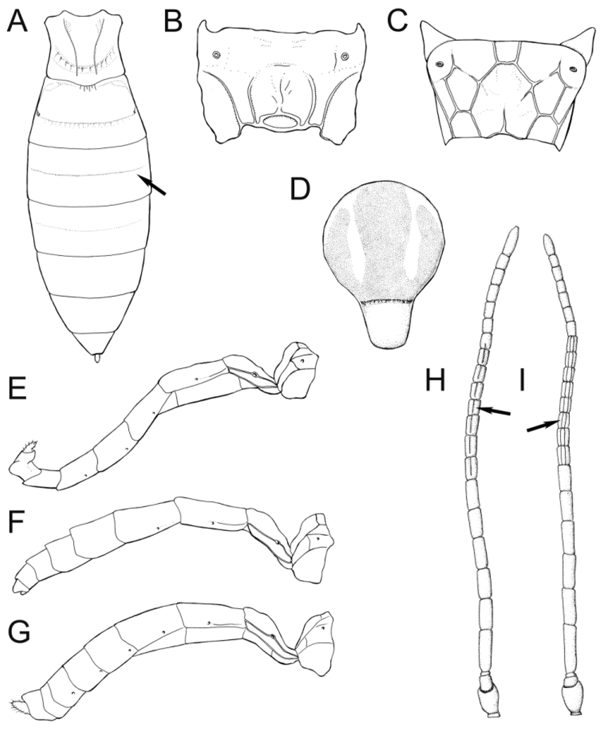
1. Metasomal tergite 2 with spiracle on laterotergite, well below lateral fold ( Fig. 11A View FIGURE 11. A ). Hind wing with one basal hamulus ( Fig. 11 View FIGURE 11. A C)................................................................................................ 2
- Metasomal tergite 2 with spiracle on dorsal part, above lateral fold ( Fig. 11 View FIGURE 11. A B). Hind wing with one or more basal hamuli ( Fig. 11 View FIGURE 11. A D)................................................................................................ 4
2. Female with metasomal tergites 4–6 with hind margins convex, extending at least as far back dorsally as laterally ( Fig. 11 View FIGURE 11. A E). Ovipositor sheaths at most 0.5 times as long as hind tibia. Scutellum with lateral carinae usually extending at least to middle ( Fig. 11 View FIGURE 11. A G). Face usually with large smooth areas; if entirely coriaceous ( S. punctiventris View in CoL and S. placita View in CoL ), then tyloids of male located around flagellomeres 6 to 8 and more than half as long as respective flagellomeres...................... Sussaba View in CoL
- Female with metasomal tergites 4–6 with hind margins concave, extending further back laterally than dorsally ( Fig. 11 View FIGURE 11. A F); if indistinct (sometimes in Episemura View in CoL ) then ovipositor sheaths at least 0.6 times as long as hind tibia. Scutellum with carinae distinct only on about basal third ( Fig. 11 View FIGURE 11. A H). Face largely to entirely coriaceous. Tyloids of male either restricted to first three flagellomeres ( Eurytyloides View in CoL ) or less than half as long as respective flagellomere ( Episemura View in CoL )......................... 3
3. Ovipositor sheath more than 0.6 times as long as hind tibia, basal half transversely striate ( Fig. 11 View FIGURE 11. A I). Mesoscutum, scutellum and mesopleuron strongly punctate, distance between punctures less than their diameter...................... Episemura View in CoL
- Ovipositor sheath less than 0.5 times as long as hind tibia, smooth and polished ( Fig. 11 View FIGURE 11. A J). Mesoscutum, scutellum and mesopleuron mainly smooth and shining, at most with some weak punctures that are more than their diameter apart..................................................................................... Eurytyloides umbrinus sp. nov.
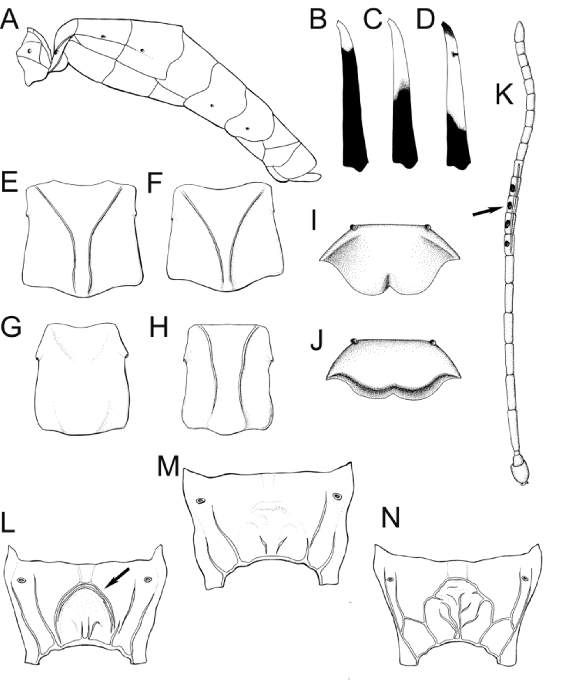
4. Mesoscutum with notauli distinctly impressed, although quite short ( Fig. 12A View FIGURE 12. A ). Fore wing areolet always open ( Fig. 12 View FIGURE 12. A C).. 5
- Mesoscutum with notauli absent ( Fig. 12 View FIGURE 12. A B). Fore wing areolet open or closed ( Fig. 12 View FIGURE 12. A D)............................ 10
5. Face smooth and shining, sometimes weakly punctate, with two vertical impressions arising from tentorial pits ( Fig. 12 View FIGURE 12. A E). Spiracle of third tergite on laterotergite ( Fig. 11 View FIGURE 11. A B). Hind wing often with only one ( Fig. 11 View FIGURE 11. A C) but sometimes with more basal hamuli...................................................................................... Promethes View in CoL
- Face not entirely smooth, at least distinctly punctate but usually coriaceous and matt, never with vertical impressions arising from tentorial pits ( Fig. 12 View FIGURE 12. A F). Spiracle of third tergite on dorsal or on lateral part. Hind wing usually with two or more basal hamuli ( Fig. 11 View FIGURE 11. A D)...................................................................................... 6
6. Clypeus with apical margin thin and often with a basal elevation, concave or flat in profile; apical margin often bilobed but sometimes truncate or evenly concave ( Figs 12 View FIGURE 12. A G, 12H). Females with face black with yellow along inner orbits and sometimes with a central yellow spot.............................................................................. 7
- Clypeus with apical margin thickened, convex and protruding in profile; apical margin convex, truncate or weakly concave, at most weakly bilobed ( Figs 12 View FIGURE 12. A I, 12J, 12K). Female with face usually entirely black but sometimes with yellow along inner orbits.............................................................................................. 8
7. Hind tibia white with a black apical band, or black-white-black banded ( Fig. 12 View FIGURE 12. A L), or black-white-black-orange banded. Tergites 1–3 and often 4 of metasoma with distinct preapical transverse impressions ( Fig. 13A View FIGURE 13. A ); if impressions not very distinct ( Diplazon neoalpinus View in CoL and Diplazon pectoratorius View in CoL ), then propodeal carinae reduced, not enclosing basal and petiolar areas ( Fig. 13 View FIGURE 13. A B)................................................................................... Diplazon View in CoL (most)
- Hind tibia mainly orange or yellow with apex dark. At most with indistinct transverse impressions on tergite one or one and two. Propodeum always with a full set of carinae defining basal, lateral and petiolar areas ( Fig. 13 View FIGURE 13. A C)........ Tymmophorus View in CoL
8. Scutellum mainly yellow or white. Female metasoma laterally compressed. Mesoscutum centrally impunctate and strongly shining, punctures restricted to front and sides. Yellow shoulder marks often with inner corners extending back over mesoscutum as two parallel lines ( Fig. 13 View FIGURE 13. A D).......................................................... Xestopelta View in CoL (part)
- Scutellum usually black, rarely with a small apical yellow spot. Metasoma dorsoventrally depressed in both sexes. Mesoscutum with obvious punctures over entire surface, shining between punctures or finely coriaceous and matt. Shoulder marks, if present, with inner corner never extending back............................................................. 9
9. Metasomal tergites 3–5 with hind margins concave even in males, extending conspicuously further back laterally than dorsally ( Figs 13 View FIGURE 13. A E, 13F). Spiracle of third tergite on laterotergite (cf. Fig. 11 View FIGURE 11. A B). Clypeus with apical margin truncate or weakly concave ( Fig. 12 View FIGURE 12. A J). Metasoma entirely black, or in males sometimes with yellow on posterior part of some tergites....................................................................................................... Campocraspedon View in CoL
- Tergites 3–5 with hind margins at most very weakly concave, extending about as far back dorsally as medially ( Fig. 13 View FIGURE 13. A G). Spiracle of third tergite on dorsal part. Clypeus with apical margin convex ( Fig. 12 View FIGURE 12. A I). Metasoma dark orange at least on apical parts of tergites 2 and 3......................................................... Daschia brevitarsis (Thomson) View in CoL
10. Fore wing areolet closed, although vein 3rs-m usually unpigmented ( Fig. 12 View FIGURE 12. A D). Male antenna always with tyloids, which can be narrow or broad ( Figs 13 View FIGURE 13. A H, 13I)....................................................................... 11
- Fore wing areolet open ( Fig. 12 View FIGURE 12. A C). Male antenna with or without tyloids......................................... 14
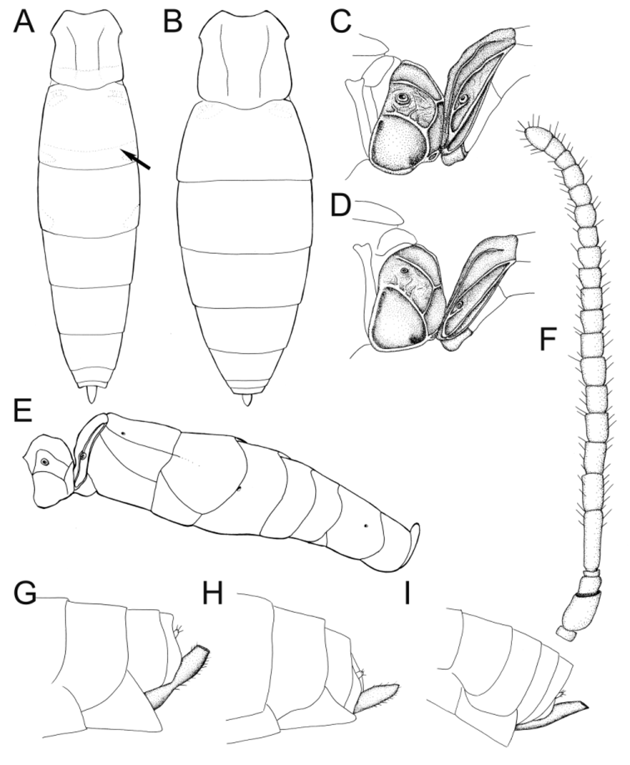
11. Female with metasomal tergites 3–5 with hind margins clearly concave, extending conspicuously further back laterally than dorsally ( Fig. 14A View FIGURE 14. A ) (usually indicated also in males). Face shining, finely coriaceous only along inner orbits, distinctly punctate......................................................................................... Phthorima View in CoL
- Metasomal tergites convex or truncate in both sexes. Face matt, punctures usually almost disappearing among the coriaceous sculpture.......................................................................................... 12
12. First metasomal tergite with median dorsal carinae strongly converging over basal half and very close to each other on apical half ( Figs 14 View FIGURE 14. A E, 14F). Male with tyloids usually bar-like and broad ( Fig. 13 View FIGURE 13. A I, except for E. schwarzi View in CoL ). Female with hind tibia black with a white base ( Fig. 14 View FIGURE 14. A B), male similar or with white part extending over half the tibia, especially on ventral side ( Fig. 14 View FIGURE 14. A C). Clypeus when viewed in profile concave laterally, rather flat centrally, and with apical margin somewhat elevated ( Fig. 14 View FIGURE 14. A I).................................................................................. Enizemum View in CoL (most)
- First tergite of metasoma with median dorsal carinae absent or strongly reduced ( Fig. 14 View FIGURE 14. A G), or if strong ( crassicornis group), then they are almost as far apart from each other as from the lateral margins of the tergite ( Fig. 14 View FIGURE 14. A H). Male with tyloids usually narrowly linear ( Fig. 13 View FIGURE 13. A H, except for the rare H. venustus and H. tauriscorum ). Hind tibia usually orange or white with subbasal and apical dark bands ( Fig. 14 View FIGURE 14. A D). Clypeus usually apically impressed, basal three-quarters convex ( Fig. 14 View FIGURE 14. A J)...... 13
13. Male with narrow tyloids each with an adjacent dark pit ( Fig. 14 View FIGURE 14. A K). Propodeum with two or more parallel, arcuate carinae around petiolar area ( Fig. 14 View FIGURE 14. A L). Female usually with a yellow line on lower mesopleuron.................................................................................................... Fossatyloides gracilentus (Holmgren)
- Male with narrow tyloids without adjacent pits ( Fig. 13 View FIGURE 13. A H). Propodeum various, often lacking transverse carinae ( Fig. 14 View FIGURE 14. A M), or with regular carinae around petiolar area ( Fig. 14 View FIGURE 14. A N). Female with mesopleuron usually entirely black.... Homotropus (part)
14. Propodeum with a full set of strong carinae delimiting at least basal and petiolar areas ( Fig. 13 View FIGURE 13. A C)...................... 15
- Propodeum with carinae reduced, not delimiting complete set of areas, at least basal or petiolar area not fully enclosed by carinae ( Fig. 14 View FIGURE 14. A M)...................................................................................... 17
15. Female with face black with yellow along inner orbits. Male antenna without tyloids. Tergites one and two often with weak subapical transverse impressions ( Fig. 15A View FIGURE 15. A ). Clypeus basally convex, remainder concave ( Figs 12 View FIGURE 12. A G, 12H)..... Syrphophilus View in CoL
- Female with face entirely black or with a yellow central spot. Male antenna with tyloids which are usually narrow and linear ( Fig. 13 View FIGURE 13. A H). Metasomal tergites without transverse impressions ( Fig. 15 View FIGURE 15. A B). Clypeus usually apically impressed, basal threequarters convex ( Fig. 14 View FIGURE 14. A J).............................................................................. 16
16. Propodeal spiracle and spiracle of first tergite with conspicuously enlarged and sometimes pale margins ( Fig. 15 View FIGURE 15. A C). Female with tergites 3–5 with hind margins concave, extending conspicuously further back laterally than dorsally ( Fig. 15 View FIGURE 15. A E). Antenna of female stout, apical flagellomeres wider than long, and with strong setae almost as long as the diameter of the segments ( Fig. 15 View FIGURE 15. A F).................................................................................... Bioblapsis View in CoL
- Propodeal spiracle not enlarged, with margin inconspicuous ( Fig. 15 View FIGURE 15. A D). Female with metasomal tergites convex or truncate. Antenna slenderer, with setae at most half as long as the diameter of a flagellomere................... Homotropus (part)
17. Ovipositor sheaths broadly and transversely truncate, thus apically open; last visible sternite in females large and triangular ( Fig. 15 View FIGURE 15. A G). Hind tibia usually black with a white base ( Figs 14 View FIGURE 14. A B, 14C) but sometimes entirely black or orange with a dark apex. Clypeus usually flat or with a subbasal transverse impression which is often only present on lateral parts, clypeus thus convex or flat centrally but concave in cross section laterally ( Fig. 16 View FIGURE 16. A D). Tyloids on male antenna, if present, usually broad ( Fig. 13 View FIGURE 13. A I)........................................................................................... 18
- Ovipositor sheaths either pointed or rounded and closed apically ( Fig. 15 View FIGURE 15. A H), or diagonally truncate ( Fig. 15 View FIGURE 15. A I); last visible sternite, if triangular, then less conspicuous. Hind tibia mainly white, yellow or orange, often with apex dark. Clypeus various but often (genus Homotropus ) with a preapical impression, rendering basal three-quarters convex ( Fig. 16 View FIGURE 16. A C). Tyloids on male antenna, if present, usually narrow ( Fig. 13 View FIGURE 13. A H).............................................................. 19
18. First tergite at most with short median dorsal carinae which are broadly separated. Male antenna without tyloids. Face including clypeus, mesosoma, legs and metasoma finely and evenly coriaceous and matt, punctures indistinct....... Woldstedtius View in CoL
- First metasomal tergite with median dorsal carinae reaching at least to middle and very close to each other on apical half ( Figs 14 View FIGURE 14. A E, 14F). Male antenna with tyloids which are most often bar-like ( Fig. 13 View FIGURE 13. A I). Sculpture not entirely fine, mesopleuron with both strong punctures and smooth parts........................................................ Enizemum View in CoL (part)
19. Female with yellow along inner orbits. Male antenna without tyloids........................................... 20
- Female with face black, often with a central yellow spot. Male antenna with narrow tyloids ( Fig. 13 View FIGURE 13. A H)................. 21
20. Clypeus with apical margin thin and often with a basal elevation, making it concave or flat in profile ( Figs 12 View FIGURE 12. A G). Epicnemial carina complete. Mesoscutum distinctly punctate on a smooth and polished background, yellow shoulder marks with inner corners not extending over most of mesoscutum.................................................... Diplazon View in CoL (part)
- Clypeus with apical margin thickened, convex and protruding in profile; apical margin convex ( Fig. 12 View FIGURE 12. A K). Epicnemial carina broadly interrupted behind fore coxa. Mesoscutum mostly smooth and shining, with punctures very weak or indistinct, yellow shoulder marks usually with inner corners extending over most of mesoscutum ( Fig. 13 View FIGURE 13. A D)............... Xestopelta View in CoL (part)
21. Spiracle of third tergite dorsal, above lateral fold. Epicnemial carina complete ventrally ( Fig. 16A View FIGURE 16. A ). Clypeus with a preapical impression, rendering basal three-quarters convex ( Fig. 16 View FIGURE 16. A C).................................... Homotropus (part)
- Spiracle of third tergite level with or below the fold separating the lateral from the dorsal part. Epicnemial carina usually broadly interrupted ventrally behind fore coxae ( Fig. 16 View FIGURE 16. A B). Clypeus usually flat or with a subbasal transverse impression which is often only present on lateral parts, clypeus thus convex or flat centrally but concave in cross section laterally ( Fig. 16 View FIGURE 16. A D)...................................................................................... Syrphoctonus View in CoL


No known copyright restrictions apply. See Agosti, D., Egloff, W., 2009. Taxonomic information exchange and copyright: the Plazi approach. BMC Research Notes 2009, 2:53 for further explanation.