
Pericharax crypta, Leocorny, Pedro, Alencar, Aline, Fromont, Jane & Klautau, Michelle, 2016
|
publication ID |
https://doi.org/ 10.11646/zootaxa.4175.4.2 |
|
publication LSID |
lsid:zoobank.org:pub:9052926E-1A4A-4CBC-B7FB-AA3B49577731 |
|
DOI |
https://doi.org/10.5281/zenodo.5661787 |
|
persistent identifier |
https://treatment.plazi.org/id/C06287E2-FFD1-FFA9-819C-856A127FD08C |
|
treatment provided by |
Plazi |
|
scientific name |
Pericharax crypta |
| status |
sp. nov. |
Pericharax crypta View in CoL sp. nov.
Etymology. From the Latin noun crypta (=grotto, covered passage/gallery), for the external shape of this sponge.
Diagnosis. Pericharax with a conspicuous striated surface and reduced atrial cavity. The skeleton is composed of two categories of triactines and three categories of tetractines. Spherulous cells are present.
Type Material. Holotype, WAM Z49226; from 33º30.72’S, 115º02.46’E to 33º30.71’S, 115º02.15’E in Geographe Bay , Western Australia GoogleMaps ; station: Trawl 2; depth: 36–37 meters; collector: Sampey, A.; date: 21/May/ 2007. Fragment from Holotype deposited under UFRJPOR 7129.
Type Locality. Geographe Bay , Western Australia, Australia.
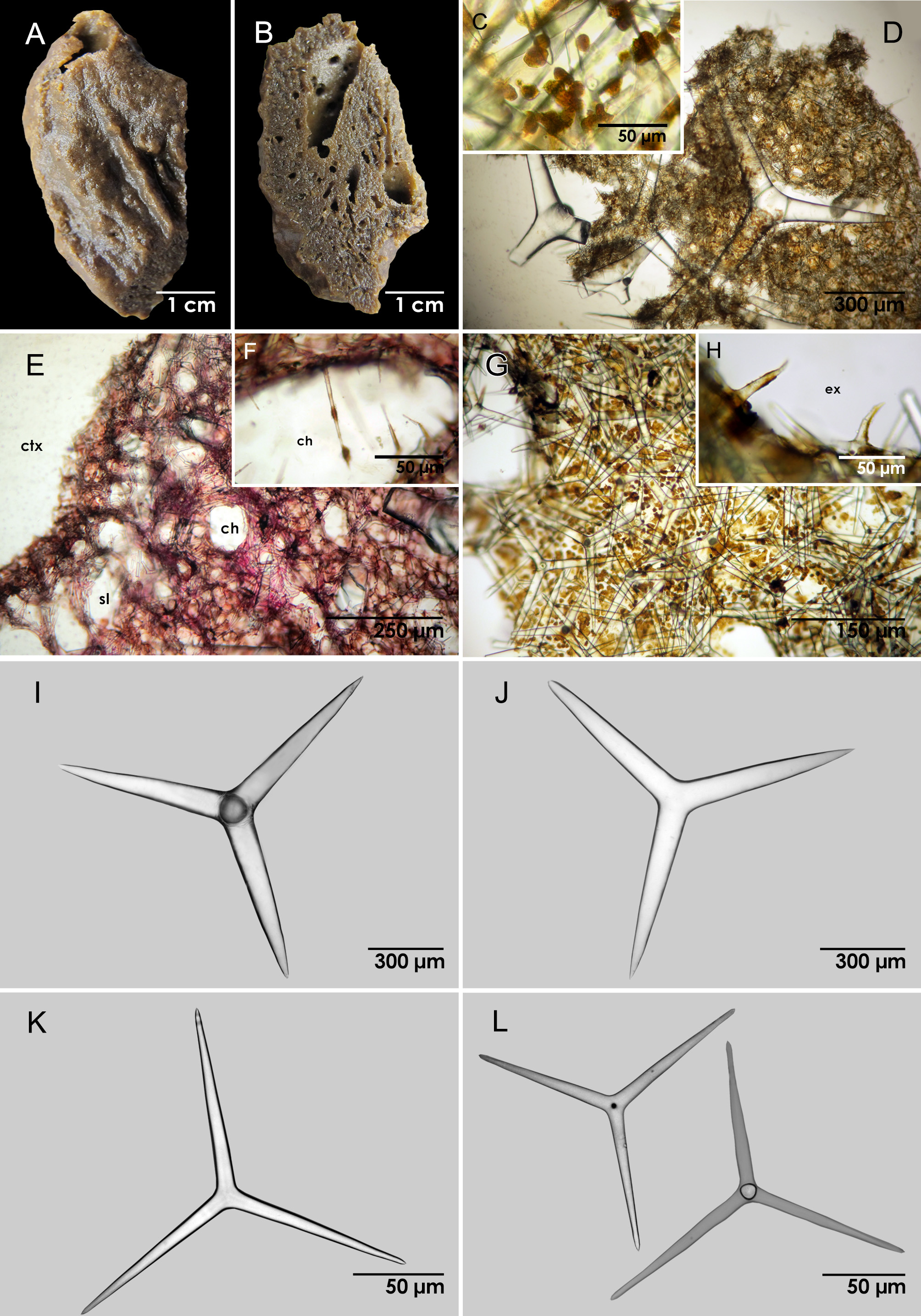
Description. Sponge dark brown in ethanol with a massive form (3.1 cm x 5.6 cm) and single apical osculum measuring 1.0 cm in diameter ( Figure 4 View FIGURE 4 A). The surface is ridged and the atrial cavity is reduced ( Figure 4 View FIGURE 4 B). Spherulous cells are present throughout the sponge body ( Figure 4 View FIGURE 4 C). The aquiferous system is leuconoid.
The skeleton is disorganized. The cortical skeleton is composed of tangentially arranged large and small triactines and large tetractines ( Figure 4 View FIGURE 4 D). Subcortical lacunae are present ( Figure 4 View FIGURE 4 E). The choanosomal skeleton is composed of few, sparsely distributed, large triactines, many small triactines and small tetractines I. The small tetractines I mainly surround the inhalant canals where their apical actines project inwards ( Figures 4 View FIGURE 4 E, 4F). The atrial skeleton is principally composed of large triactines and small tetractines II ( Figure 4 View FIGURE 4 G), and the exhalant canals are supported by small tetractines II ( Figure 4 View FIGURE 4 H).
Spicules. ( Table 4). Large tetractines ( Figure 4 View FIGURE 4 I): regular. The basal actines are conical with sharp tips (basal: 400–900–1175 µm / 75–198–288 µm). The apical actines are conical with sharp tips, always shorter than the basal ones. It was not possible to measure this actine.
Large triactines ( Figure 4 View FIGURE 4 J): regular. Actines are conical with sharp tips (750–956–1175 µm / 113–150–213 µm).
Small triactines ( Figure 4 View FIGURE 4 K): regular. Actines are conical with blunt tips (95–179–226 µm / 13–20–25 µm).
Small tetractines I ( Figure 4 View FIGURE 4 L, left): regular. The basal actines are conical with sharp tips. The apical actines are slightly conical and smooth with sharp tips. They are much longer and thinner than the apical actines of the small tetractine II (basal: 125–168–190 µm / 15–18–23 µm; apical: 90–173–508 µm / 4–6–8 µm).
Length (µm) Width (µm)
Spicule Actine Min Mean sd Max Min Mean sd Max n WAM Z49226
Large triactines 750.0 955.6 131.6 1175.0 112.5 150.0 32.5 212.5 10 Small triactines 95.0 178.8 25.9 222.5 12.5 20.2 2.8 25.0 20 Large tetractines Basal 400.0 900.0 293.4 1175.0 75.0 198.4 75.1 287.5 10 Small tetractines I Basal 125.0 167.9 15.6 190.0 15.0 17.6 2.0 22.5 20
Apical 90.0 173.0 95.4 507.5 3.7 5.9 1.2 7.5 20 Small tetractines II Basal 137.5 189.1 26.9 240.0 15.0 20.8 2.7 25.0 20
Apical 50.0 70.8 13.3 92.5 10.0 13.8 2.8 20.0 20 Small tetractines II ( Figure 4 View FIGURE 4 L, right): regular. The basal actines are slightly conical with sharp tips. The apical actines are conical and curved with sharp tips. They are shorter and thicker than the apical actines of small tetractine I (basal: 138–189–240 µm / 15–21–25 µm; apical: 50–71–93 µm / 10–14–20 µm).
Remarks. Pericharax crypta sp. nov. is distinguished by the presence of numerous large tetractines that occur throughout the various parts of the sponge skeleton. These spicules separate this new species from P. carteri Poléjaeff, 1883 and P. orientalis Van Soest & de Voogd, 2015 . Although P. carteri and P. orientalis have very similar external morphologies to P. crypta sp. nov., their spicule components are very different in shape and size ( Table 5).
Length (µm) Width (µm)
Spicule Actine Min Mean Max Min Mean Max
Large triactines 494.0 964.9 1852.5 24.7 58.6 132.5
Small triactines (regular) 51.0 88.8 121.5 4.9 7.2 7.3
Small triactines (sagittal) Unpaired 50.0 109.4 190.0 - 6.0 - Paired 55.0 110.8 155.0 - 6.0 -
Small tetractines Basal 80.0 152.8 190.0 7.5 9.9 10.0
Large triactines 360.0 834.2 1560.0 25.0 67.9 132.0
Small triactines (regular) 60.0 159.3 228.0 7.0 12.2 18.0
Small triactines (sagittal) Unpaired 61.0 95.1 132.0 6.0 8.6 13.0 Paired 60.0 83.2 138.0 5.0 8.1 12.0
Small tetractines Basal 126.0 177.8 228.0 7.0 9.9 14.0 Apical 57.0 87.4 111.0 6.0 7.4 9.0
| WAM |
Western Australian Museum |
No known copyright restrictions apply. See Agosti, D., Egloff, W., 2009. Taxonomic information exchange and copyright: the Plazi approach. BMC Research Notes 2009, 2:53 for further explanation.
|
Kingdom |
|
|
Phylum |
|
|
Class |
|
|
Order |
|
|
Family |
|
|
Genus |