
Allodrilus, Evangelista, Olivia, Flórez-V, Camilo & Sakakibara, Albino M., 2014
|
publication ID |
https://doi.org/ 10.11646/zootaxa.3847.4.2 |
|
publication LSID |
lsid:zoobank.org:pub:46E41A86-A877-4690-80D9-1C91149A4F8E |
|
DOI |
https://doi.org/10.5281/zenodo.6124150 |
|
persistent identifier |
https://treatment.plazi.org/id/C0248799-FFE1-687B-FF38-CE0A67ADF82C |
|
treatment provided by |
Plazi |
|
scientific name |
Allodrilus |
| status |
gen. nov. |
Key to species of Allodrilus gen. nov. (with emphasis on males):
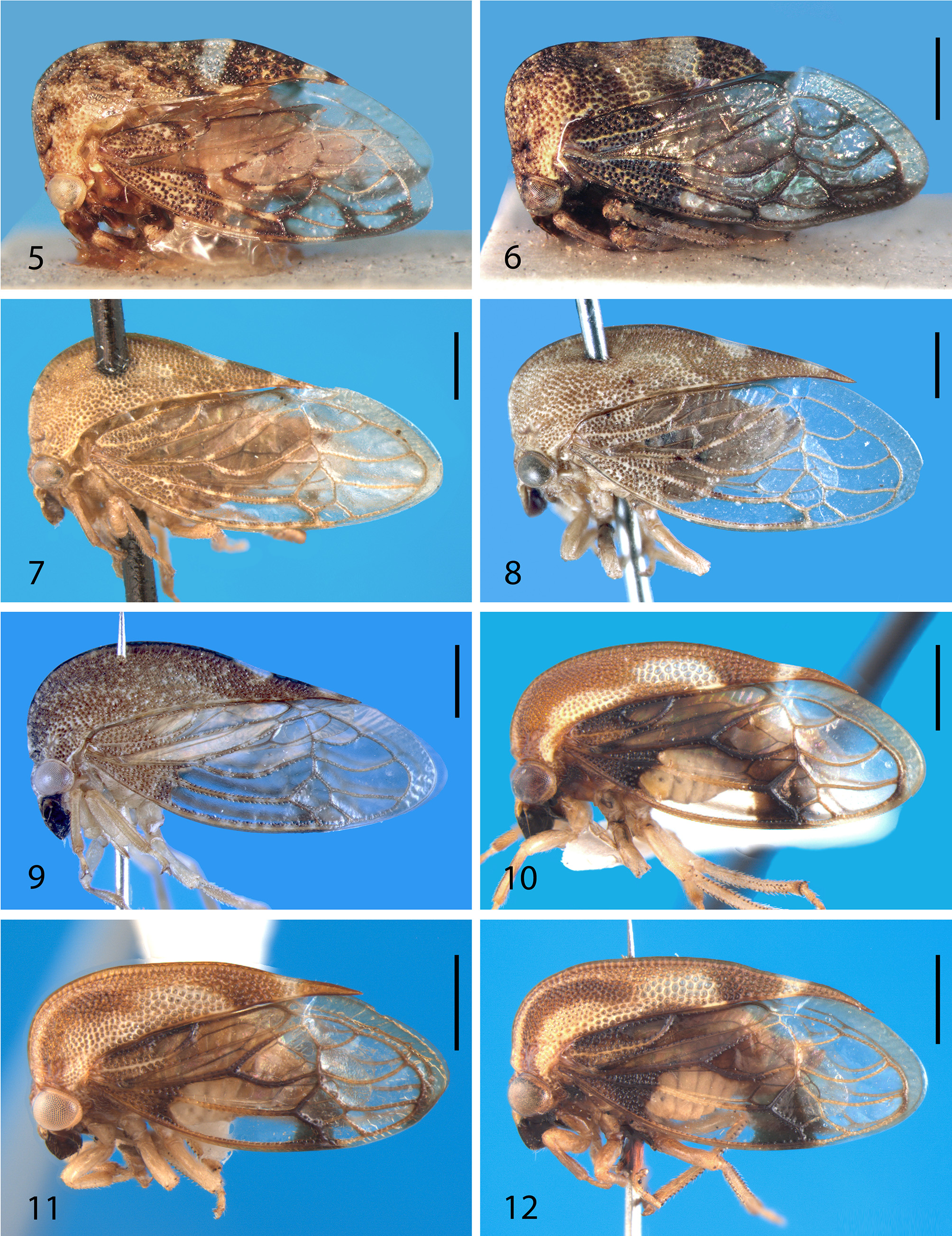
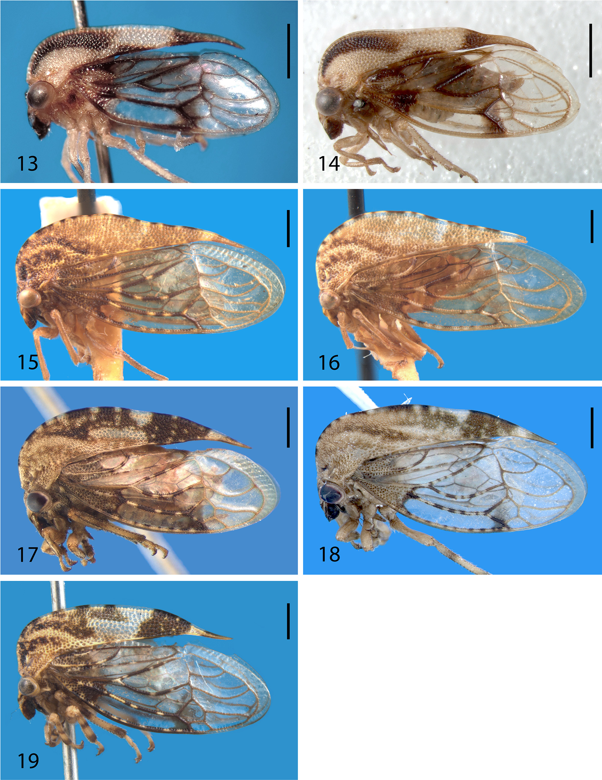
1 Pronotum mostly yellow or castaneus, often showing yellow spots or patches ( Figs. 7–14 View FIGURES 5 – 12 View FIGURES 13 – 19 , 22 View FIGURES 20 – 22 ).........................2
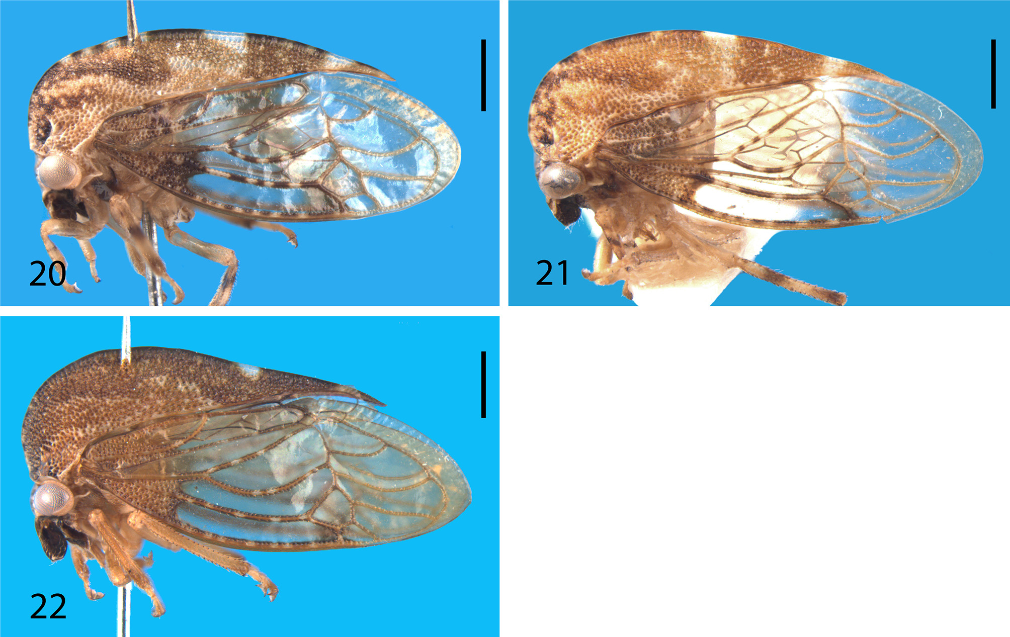
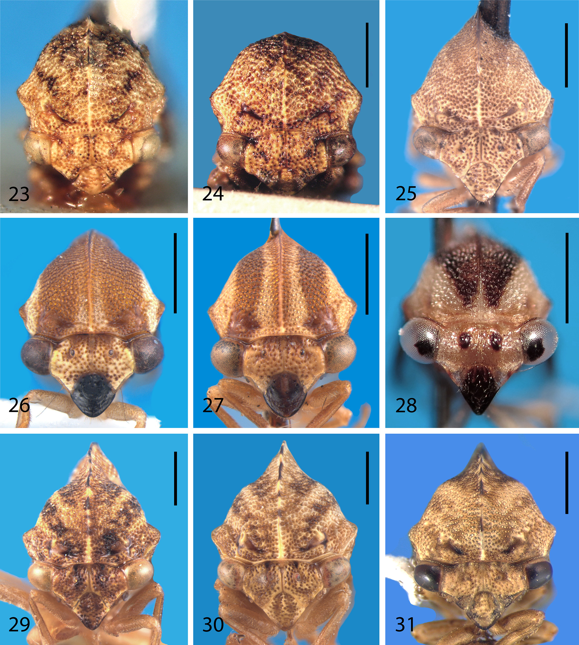
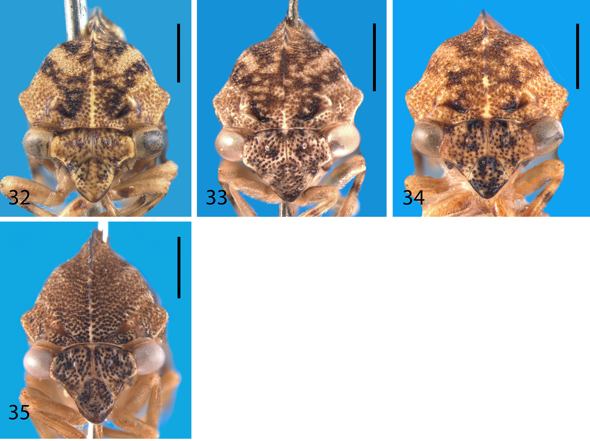
1’ Pronotum variegated yellow, brown, and testaceous, usually showing two pairs of irregular V-shaped dark brown bands on metopidium ( Figs. 15–21 View FIGURES 13 – 19 View FIGURES 20 – 22 ; 29–34)......................................................................... 5
2 Frontoclypeus entirely dark ( Figs. 26–28 View FIGURES 23 – 31 ); pronotum low, lateral margins broadly yellow ( Figs. 10–14 View FIGURES 5 – 12 View FIGURES 13 – 19 )................. 3
2’ Frontoclypeus concolorous with rest of vertex ( Figs. 25 View FIGURES 23 – 31 , 35 View FIGURES 32 – 35 ); pronotum moderately elevated and tectiform, mostly castaneous, testaceous or both ( Figs. 7–9 View FIGURES 5 – 12 , 22 View FIGURES 20 – 22 ).........................................................................4
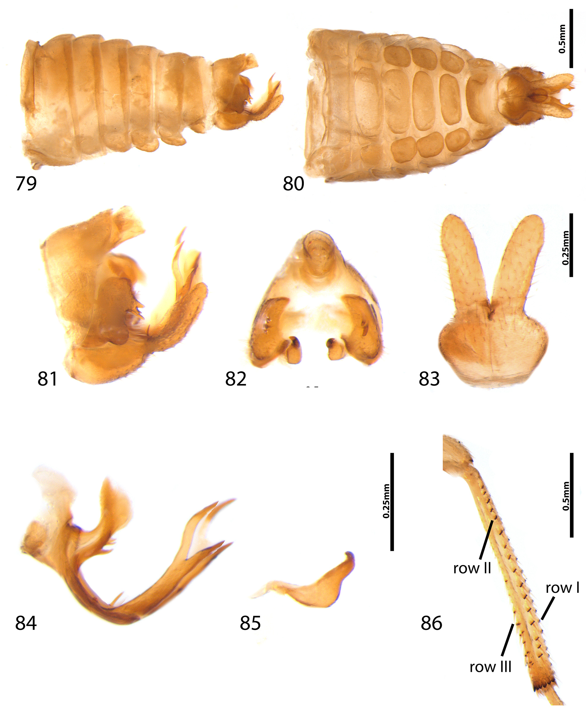
3 Pronotum testaceous with yellow patches, dorsum feebly arched ( Figs. 10–12 View FIGURES 5 – 12 ); apex of frontoclypeus rounded ( Figs. 26–27 View FIGURES 23 – 31 ); branches of aedeagal apophysis bifurcate pre-apically ( Fig. 84 View FIGURES 79 – 86 )............................ A. alboferrugineus sp. nov.
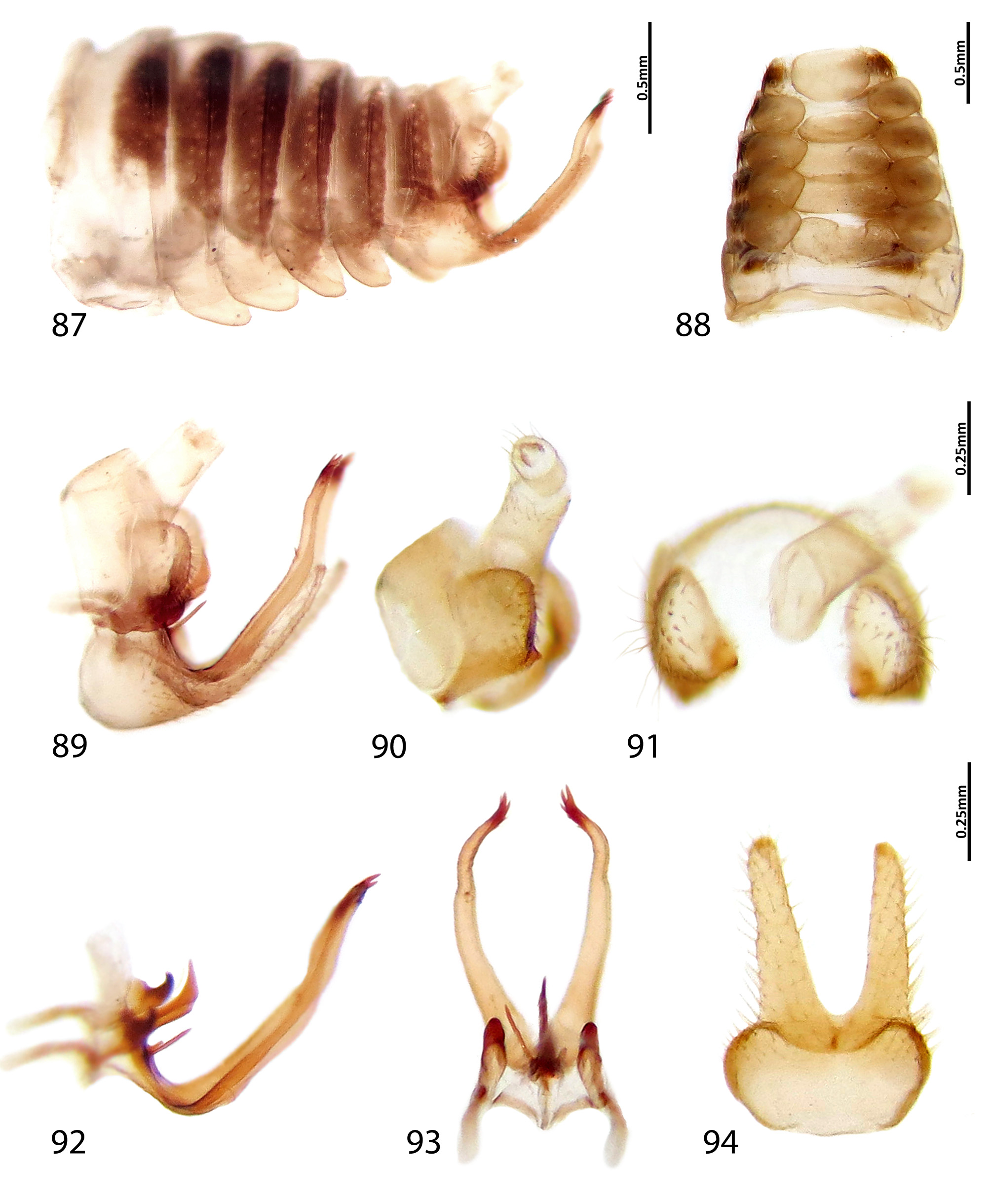
3’ Pronotum mostly yellow with dark brown patches, dorsum relatively straight ( Figs. 13–14 View FIGURES 13 – 19 ); apex of frontoclypeus acute ( Fig. 28 View FIGURES 23 – 31 ); branches of aedeagal apophysis not bifurcate pre-apically ( Figs. 92–93 View FIGURES 87 – 94 )................... A. colombiensis sp. nov.
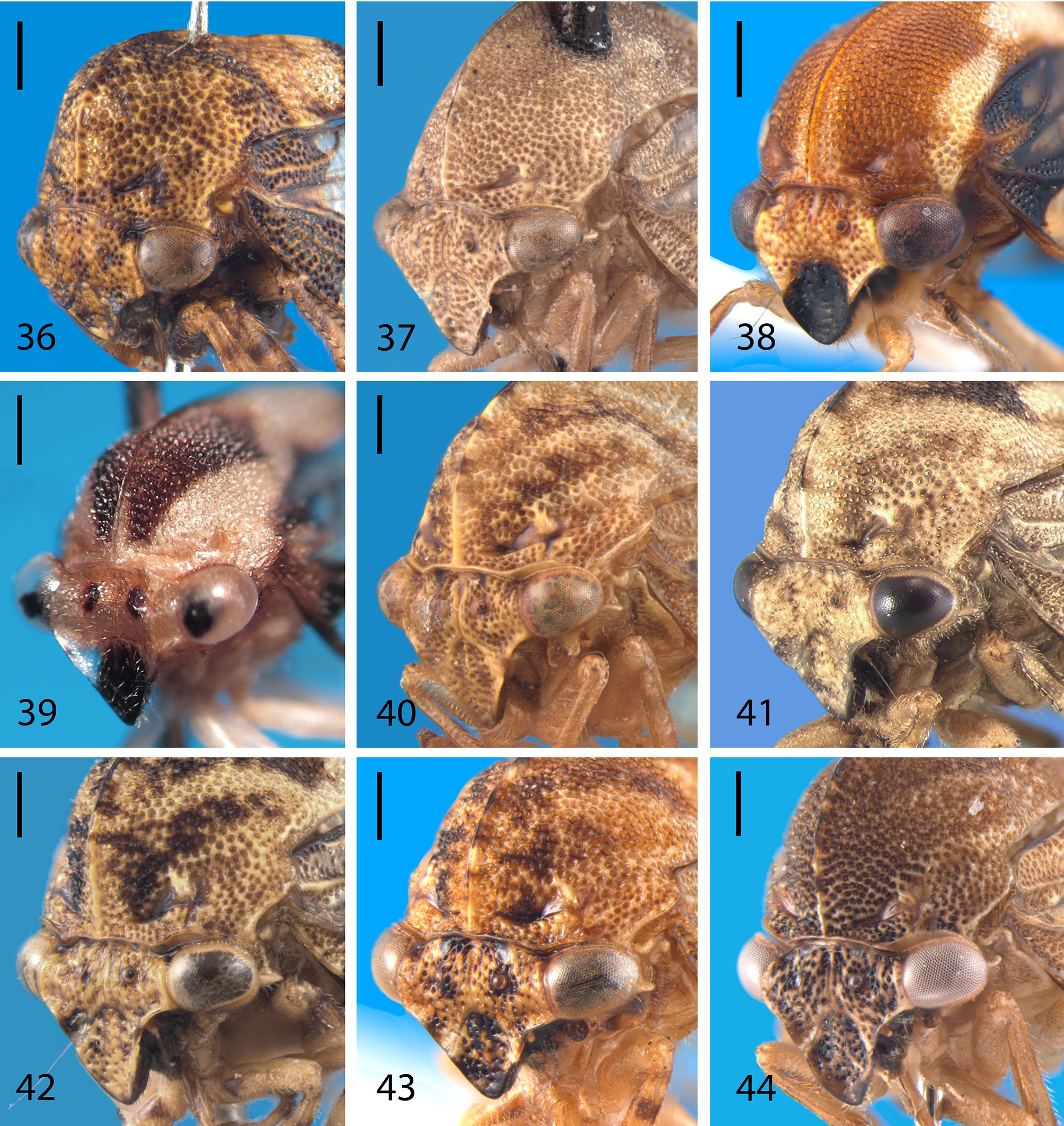
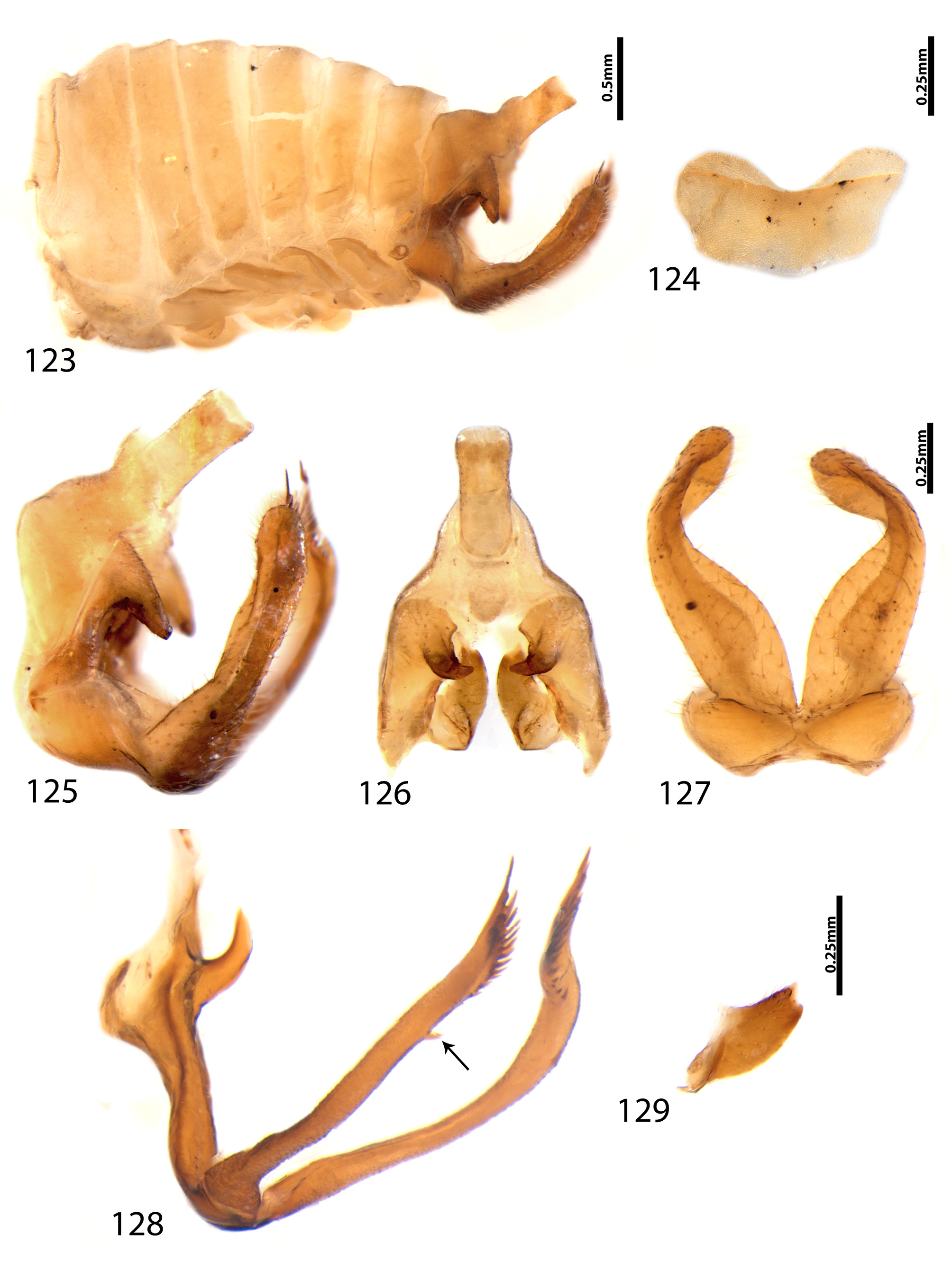
4 Vertex strongly concave below ocelli ( Fig. 44 View FIGURES 36 – 44 ); males: lateral plate strongly excavated medially, dorsal process triangular, strongly curved inwards, hook-shaped in caudal view ( Figs. 125–126 View FIGURES 123 – 129 ); branches of aedeagal apophysis with a ventral spine ( Fig. 128 View FIGURES 123 – 129 )............................................................................. A. similis sp. nov.
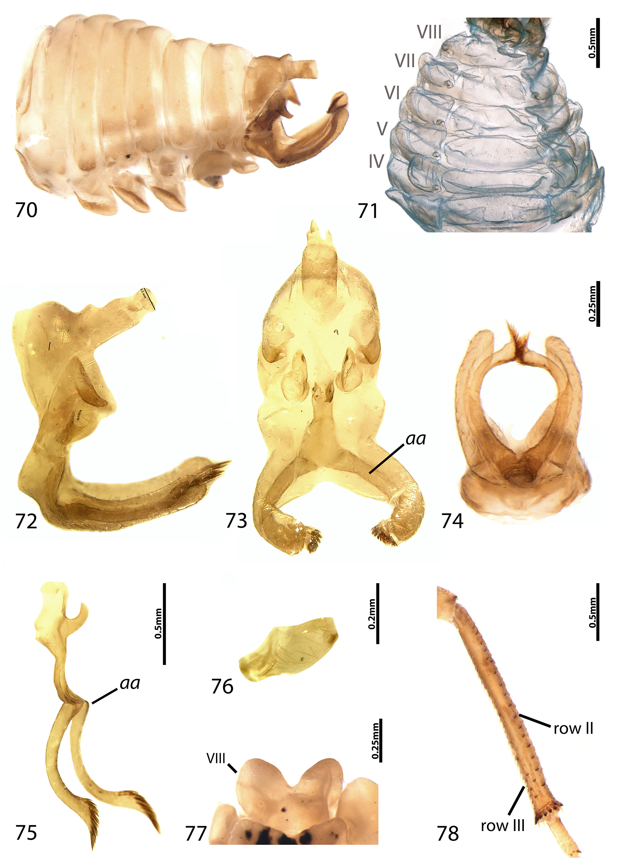
4’ Vertex usually not strongly concave below ocelli ( Fig. 37 View FIGURES 36 – 44 ); males: lateral plate weakly excatated medially, dorsal process finger-shaped, slightly curved inwards ( Figs. 72–73 View FIGURES 70 – 78 ); branches of aedeagal apophysis lacking spines ( Fig. 75 View FIGURES 70 – 78 )................................................................................... A. nitidipennis (Funkhouser) comb. nov.
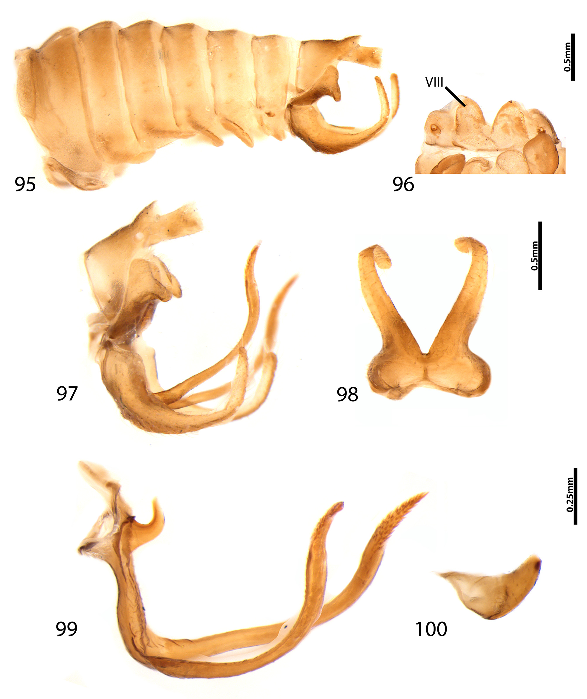
5 Lower margins of vertex straight or feebly sinuate in frontal view ( Figs. 29–31 View FIGURES 23 – 31 , 33–34 View FIGURES 32 – 35 ); dorsum uniformly arched ( Figs. 15–18 View FIGURES 13 – 19 , 20–21 View FIGURES 20 – 22 ); males: branches of aedeagal apophysis apically directed upwards and sideways ( Figs. 99 View FIGURES 95 – 100 , 106 View FIGURES 101 – 108 , 120 View FIGURES 115 – 122 ).......6
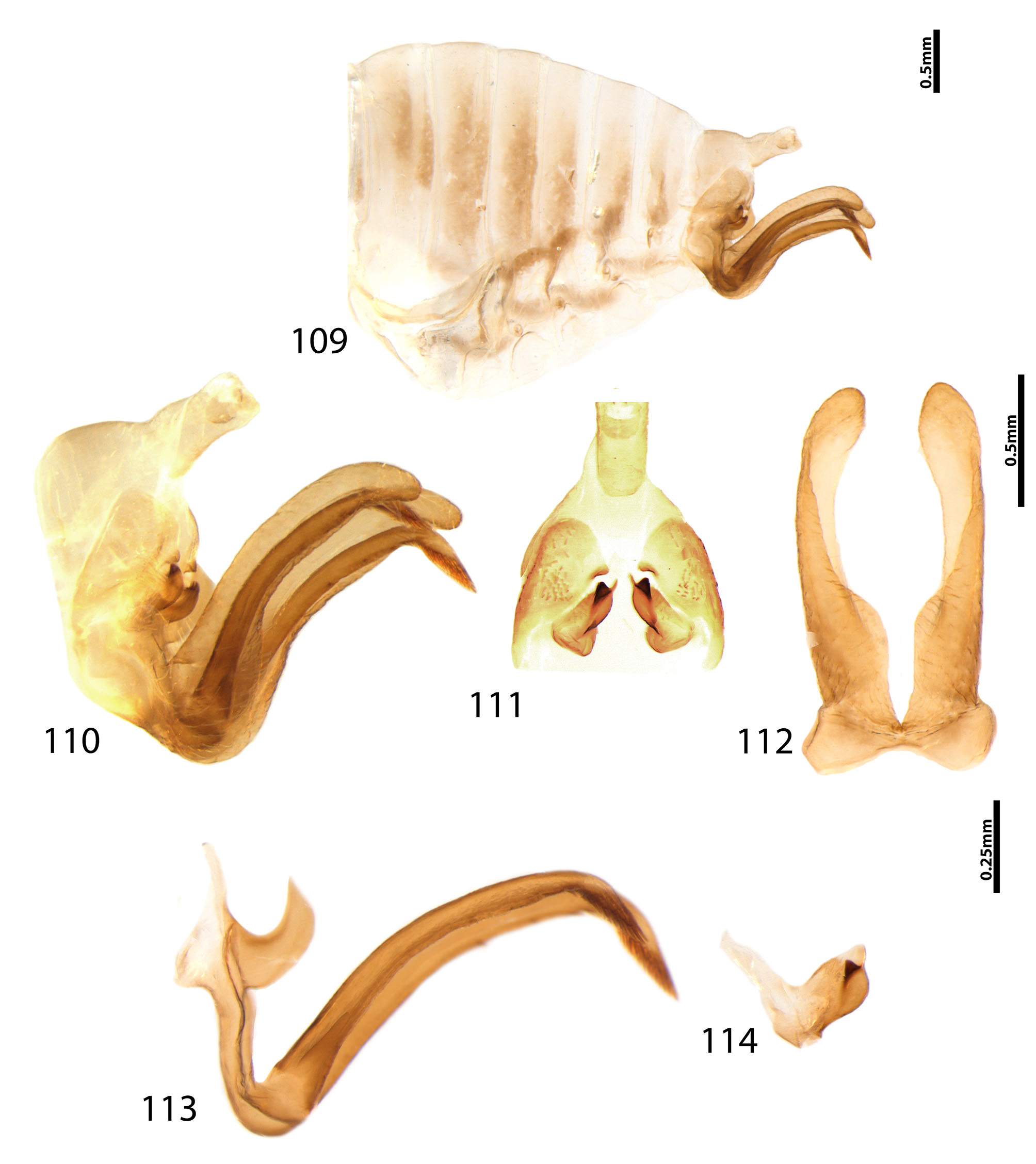
5’ Lower margins of vertex strongly sinuate in frontal view ( Fig. 32 View FIGURES 32 – 35 ); dorsum relatively straight, posterior process constricted pre-apically ( Fig. 19 View FIGURES 13 – 19 ); apex of aedeagal apophysis curved downwards ( Fig. 113 View FIGURES 109 – 114 ).................. A. horizontalis sp. nov.
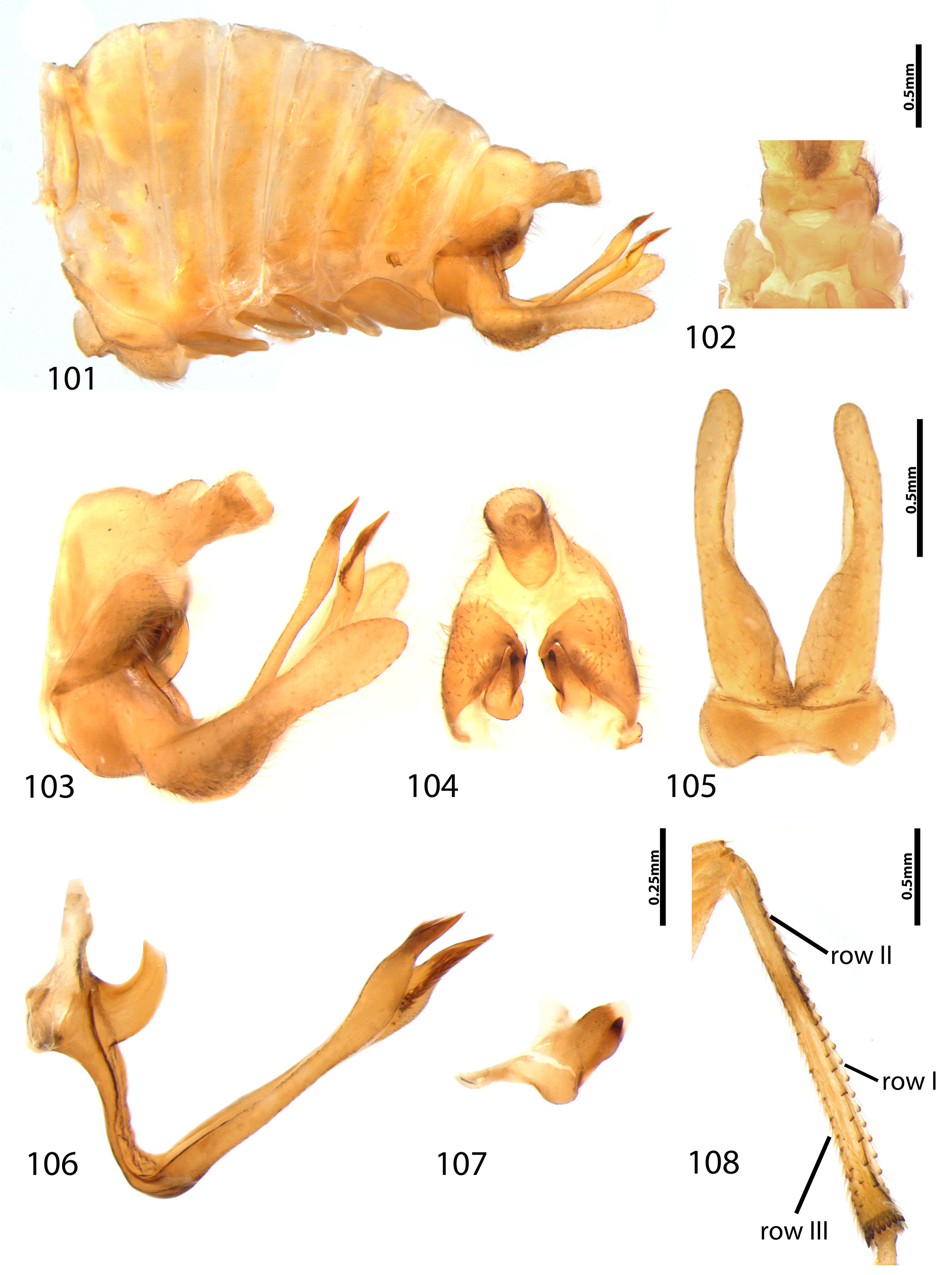
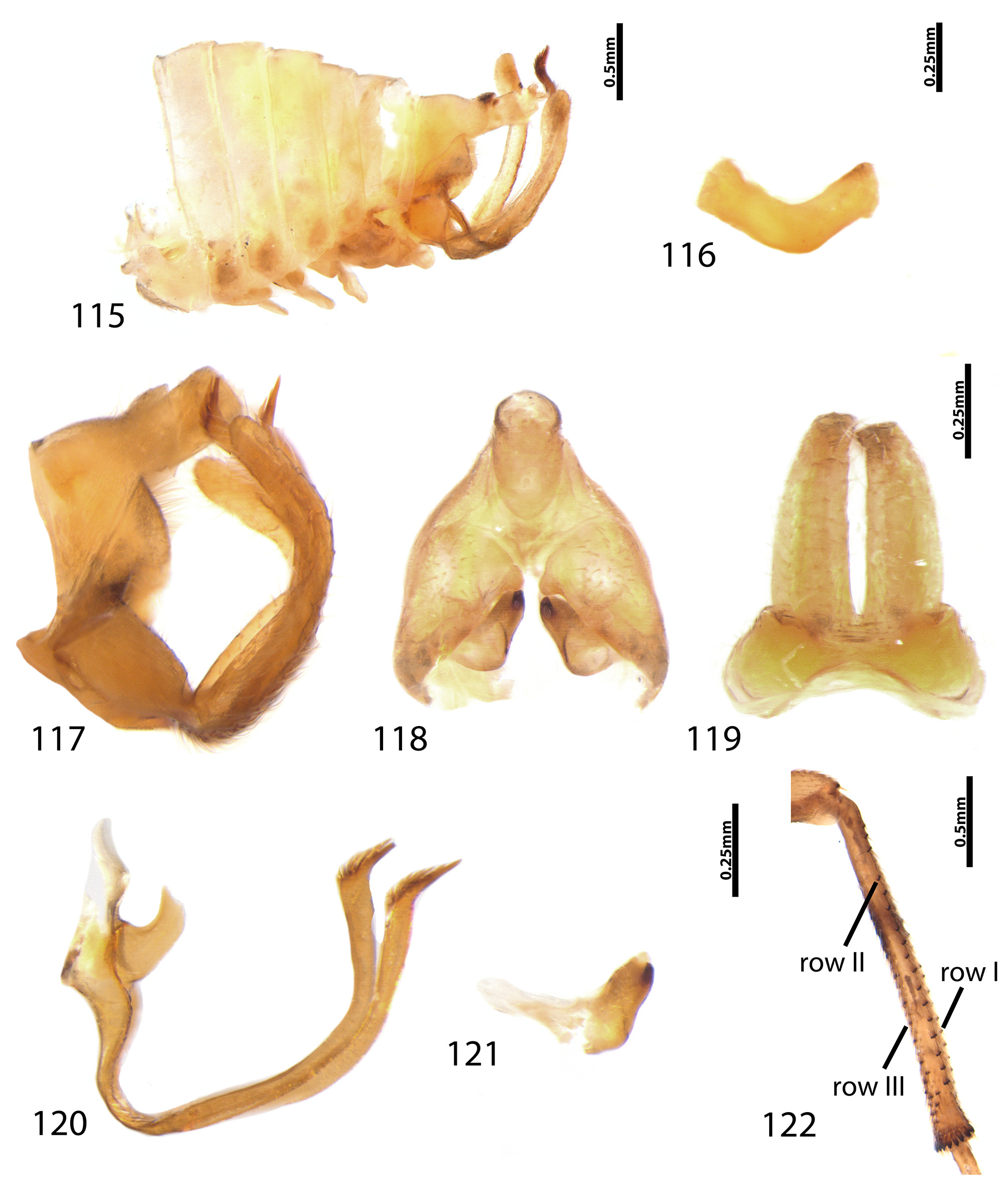
6 Males: Lateral plate dorsally arched, bearing small tubercle or tooth-like projection mostly visible in caudal view ( Figs. 103–104 View FIGURES 101 – 108 , 115, 117–118 View FIGURES 115 – 122 ); styles with small apical ventral lobe, curved sideways ( Figs. 107 View FIGURES 101 – 108 , 121 View FIGURES 115 – 122 )....................... 7
6’ Males: Lateral plate dorsally sinuose, bearing finger-like elongated rectangular process, truncate apically, visible in lateral view ( Figs. 95, 97 View FIGURES 95 – 100 ); styles with ventral lobe inconspicuous........................................ A. deitzi sp. nov.
7 Males: lateral plate not distinctly inflated, dorsal projection hook-shaped, directed inwards, visible in both lateral and in caudal view ( Figs. 103–104 View FIGURES 101 – 108 ); branches of aedeagal apophysis slightly larger pre-apically ( Fig. 106 View FIGURES 101 – 108 )......... A. granulatus sp. nov.
7’ Males: lateral plate inflated, forming round tubercle, as seen in lateral view ( Fig. 117 View FIGURES 115 – 122 ), with small round projection, visible only in caudal view ( Fig. 118 View FIGURES 115 – 122 ); branches of aedeagal apophysis not enlarged pre-apically ( Fig. 120 View FIGURES 115 – 122 ).. A. intermedius sp. nov.
No known copyright restrictions apply. See Agosti, D., Egloff, W., 2009. Taxonomic information exchange and copyright: the Plazi approach. BMC Research Notes 2009, 2:53 for further explanation.