
Hoplasoma mindanense Medvedev, 2002
|
publication ID |
https://doi.org/ 10.5281/zenodo.210044 |
|
DOI |
https://doi.org/10.5281/zenodo.6178218 |
|
persistent identifier |
https://treatment.plazi.org/id/BF36D22B-FF86-F218-6C85-FA3E4BE4A978 |
|
treatment provided by |
Plazi |
|
scientific name |
Hoplasoma mindanense Medvedev, 2002 |
| status |
|
Hoplasoma mindanense Medvedev, 2002
( Figs 6 View FIGURES 2 – 6 , 14 View FIGURES 10 – 17 , 22 View FIGURES 18 – 23 , 30 View FIGURES 26 – 32 , 37 View FIGURES 33 – 39 )
Holpasoma [sic!] mindanensis Medvedev, 2002: 62 (original description).
Type locality. “Mindanao, S. Cotabato Prov., Manobo Tamaday Forest Reserve, Mt. Temlofung”.
Type material. Holotype 3 ( LMRM), labelled: “ Philippines – Mindanao / S. Cotabato Prov., Manobo / Tasaday Forest Reserve, / Mt. Temlofung, alt. 1300 m. / 19-24.X.1994, Pascal Lays leg. [w, p] // HOLOTYPUS [p] / Hoplasoma / mindanensis m. [h] / L. Medvedev det. [p] 2000 [red label, w, h]”. Paratypes: Ƥ ( SMNS), labelled: “ Philippines – Mindanao / S. Cotabato Prov., Manobo / Tasaday Forest Reserve, / Mt. Temlofung, alt. 1300 m. / 19–24.X.1994, Pascal Lays leg. [w, p] // PARATYPUS [p] / Hoplasoma / mindanensis m. [h] / L. Medvedev det. [p] 2000 [red label, h]”; Ƥ ( MSNG), labelled: “ Philippines – Mindanao / South Cotabato Prov. / Manobo Tasaday Forest / Reserve - Mt. Tasaday [w, p] // 124°32´E.- 6°18´N. / alt. 1000-1100 m. / Pascal LAYS [w, p] // 3-31.v.1993 [h] / secondary ve- / getation [w, p] // PARATYPUS [p] / Hoplasoma / mindanensis m. [h] / L. Medvedev det. [red label, p] // Ex coll. Medvedev / Acquisto XI.2000 [w, p]”.
Additional material studied. 12 specimens — PHILIPPINES (MINDANAO IS.): AGUSAN DEL NORTE PROV.: 1 3 1 Ƥ, Butuan, without the date of collecting, Baker leg. ( USNM); LANAO DEL NORTE PROV.: 3 33 5 ƤƤ, Iligan, without the date of collecting, Baker leg. ( USNM); DAVAO PROV.: 1 Ƥ, Davao, Mt. Mayo, iv.1927, R. C. McGregor leg. ( USNM); 1 3, Davao, without the date of collecting, Baker leg. ( USNM).
Description. Completely orange, except apices of mandibles black.
Measurements. Male: 7.90-9.50 mm (holotype 9.10 mm); female: 8.50-10.40 mm.
Male (holotype, Fig. 6 View FIGURES 2 – 6 ). Labrum transverse, anterior margin almost straight, slightly oblique to left. Interocular space narrow, 1.10 times as wide as transverse diameter of eye. Antennae 0.72 times as long as body, length ratio of antennomeres equals 23-9-23 -33-32-30-29-27-27-23-28.
Pronotum 1.28 times as broad as long, widest at first quarter, lateral margins almost straight. Anterior margin slightly concave.
Elytra shiny, 2.50 times as long as wide and 0.76 times as long as body. Apex right angled.
Protarsomere 1 enlarged, subparallel, 1.85 times as long as wide, 1.30 times as wide as protarsomere 2. Length ratio of protarsomeres 1–4 equals 26-16-10-21. Metatarsomere 1 enlarged, parallel-sided, 2.80 times as long as wide, as wide as metatarsomere 2. Length ratio of metatarsomeres 1–4 equals 31-17-9-20.
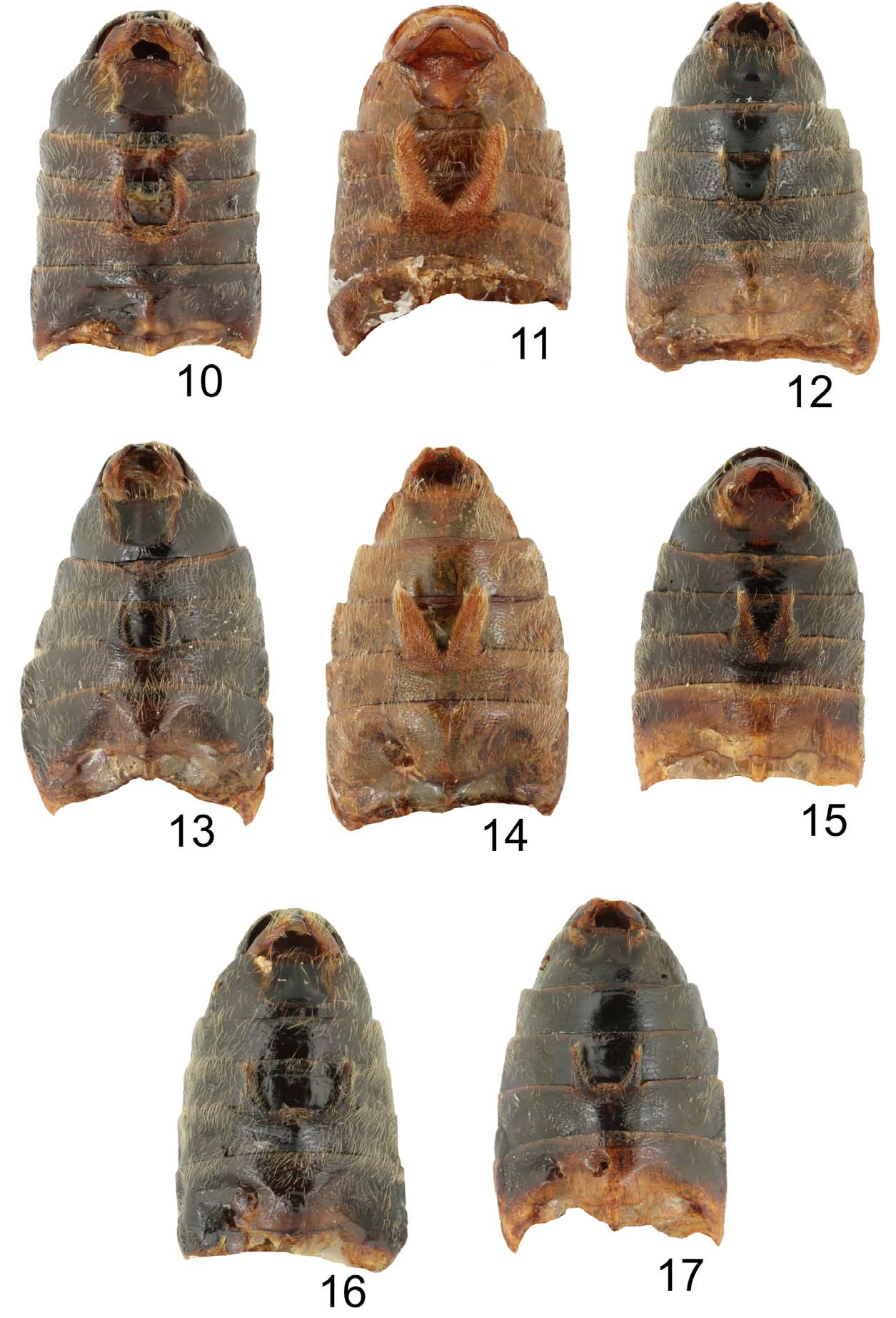
Abdominal processes broad, divergent, not separated basally, reaching slightly beyond base of ventrite 4, appendages subparallel, with lateral margins slightly converging distally, internal angle of divergence 45° ( Fig. 14 View FIGURES 10 – 17 ). Median plate of last ventrite without median ridge.
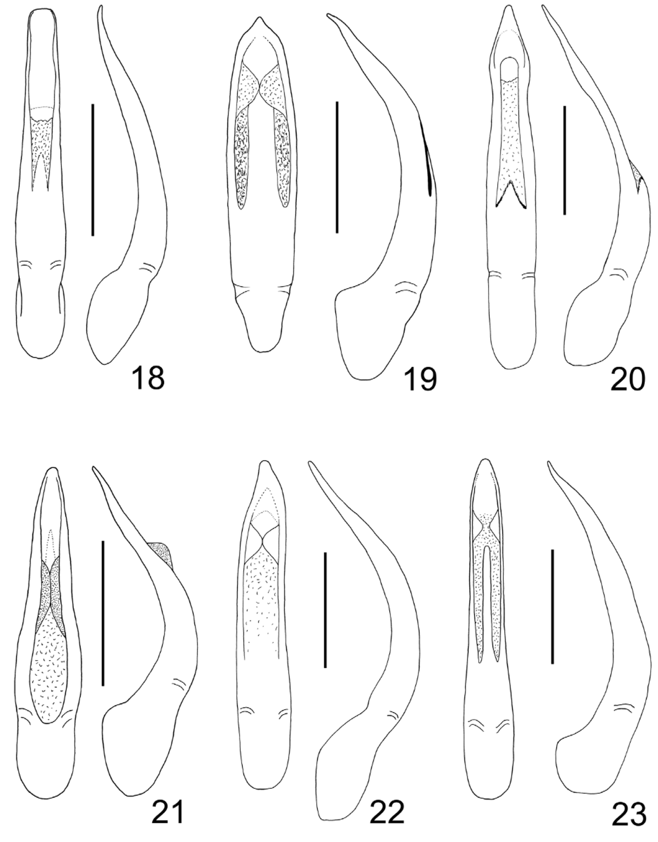
Median lobe of aedeagus asymmetrical, both apex and median part slightly deflected to left ( Fig. 22 View FIGURES 18 – 23 ).
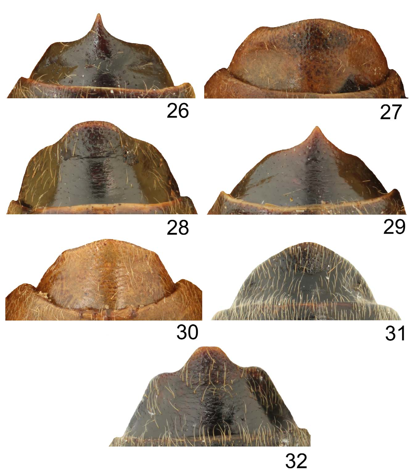
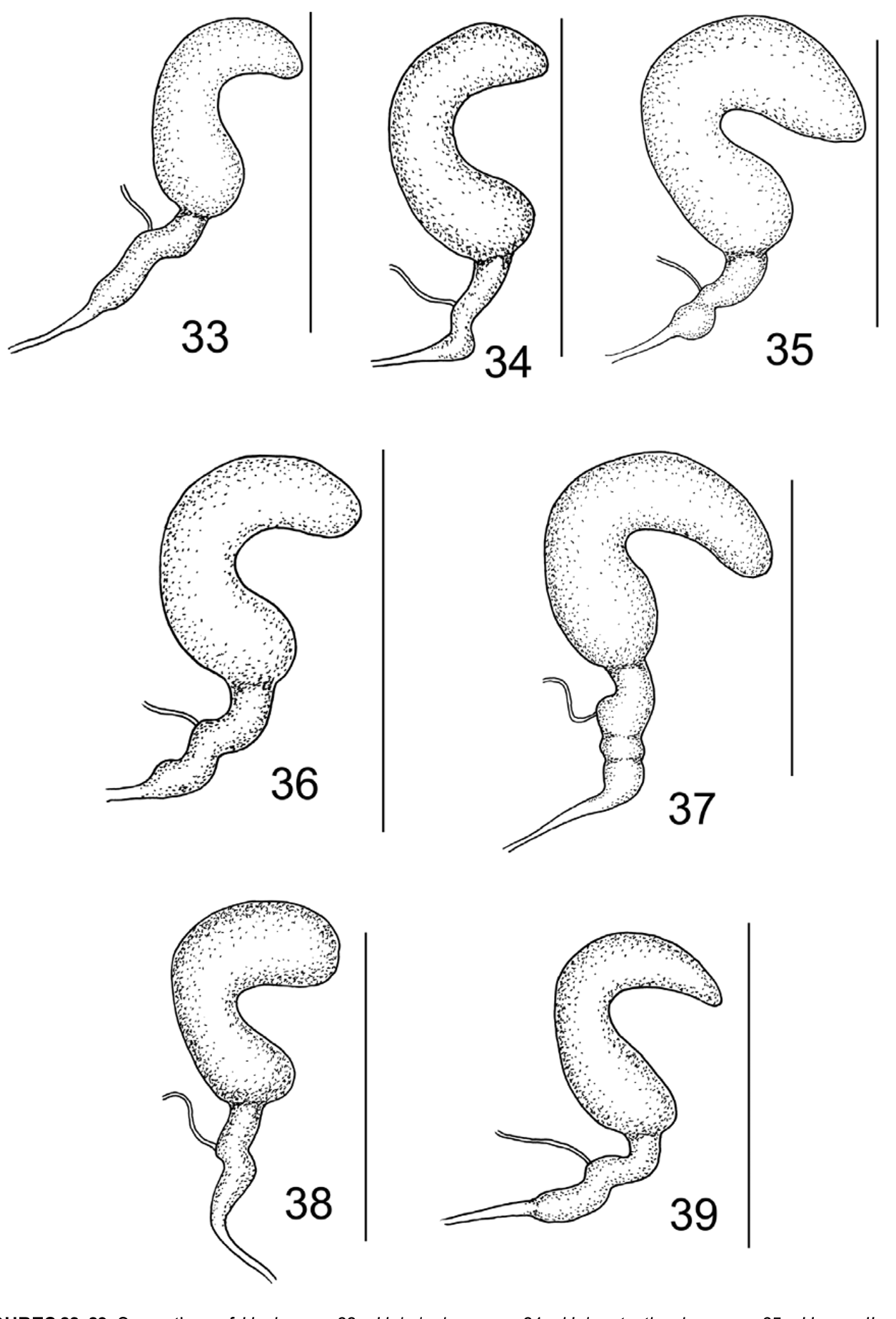
Female. Interocular space wide, 1.68 times as wide as transverse diameter of eye. Elytra more or less dull. All first tarsomeres shorter and thinner than in male. Last ventrite relatively longer, with almost evenly convex apex and distinct obtuse lateral angles ( Fig. 30 View FIGURES 26 – 32 ). Spermatheca: cornu strongly bent with distinctly enlarged basal third, proximal spermathecal duct bent in apical third with two constrictions in middle ( Fig. 37 View FIGURES 33 – 39 ).
Differential diagnosis. Hoplasoma mindanense resembles H. konstantinovi sp. nov. in having completely yellow ventral side. They can be separated by the structure of the median lobe of aedeagus which is assymetrical in H. mindanense (both apex and median part slightly deflected to the left) while the same is symmetrical in H. konstantinovi sp. nov. ( Figs 19, 22 View FIGURES 18 – 23 ). Abdominal appendages of both species are connected basally, but the same in H. konstantinovi sp. nov. are triangular in shape and longer, reaching the base of ventrite 5 ( Fig. 11 View FIGURES 10 – 17 ) while those in H. mindanense are nearly parallel sided and shorter, reaching slightly beyond the base of ventrite 4 ( Fig. 14 View FIGURES 10 – 17 ). The female differs in the shape of the last ventrite which is narrower, with broadly convex posterior margin in H. mindanense while the same in H. konstantinovi sp. nov. is broader, with sinuate posterior margin convex in the middle ( Figs 27, 30 View FIGURES 26 – 32 ).
Distribution. The Philippines (Mindanao).
Comments. As the gender of Hoplasoma was fixed as neuter ( Bezdĕk 2008), the ending of the name has been modified as “ mindanense ”.

No known copyright restrictions apply. See Agosti, D., Egloff, W., 2009. Taxonomic information exchange and copyright: the Plazi approach. BMC Research Notes 2009, 2:53 for further explanation.
|
Kingdom |
|
|
Phylum |
|
|
Class |
|
|
Order |
|
|
Family |
|
|
SubFamily |
Galerucinae |
|
Genus |