
Amazochroma pedroi, Rafael N. Carvalho & Adriano B. Kury, 2018
|
publication ID |
https://doi.org/ 10.5852/ejt.2018.393 |
|
publication LSID |
lsid:zoobank.org:pub:0D9D591F-0D5E-4078-B328-5831A7CD06E8 |
|
DOI |
https://doi.org/10.5281/zenodo.5959976 |
|
persistent identifier |
https://treatment.plazi.org/id/1E6958EB-B762-4244-B16D-A96EBB853365 |
|
taxon LSID |
lsid:zoobank.org:act:1E6958EB-B762-4244-B16D-A96EBB853365 |
|
treatment provided by |
Plazi |
|
scientific name |
Amazochroma pedroi |
| status |
gen. et sp. nov. |
Amazochroma pedroi View in CoL gen. et sp. nov.
urn:lsid:zoobank.org:act:1E6958EB-B762-4244-B16D-A96EBB853365
Figs 1 E–F View Fig. 1 , 7–9 View Fig. 7 View Fig. 8 View Fig. 9
Material examined
Holotype
BRAZIL: ♂, AC, Rio Branco, FEC , Nov. 2010, O.S. Torres leg. ( MNRJ 2352 View Materials ).
Paratypes
BRAZIL: 1 ♂, same collection data as for holotype ( MNRJ 9266 View Materials ); 1 ♂ ( MNRJ 7631 View Materials ); 1 ♂, AC, Senador Guiomard , Reserva Extrativista de Catuaba, 2002, E.F. Morato leg. ( IBSP 10234 View Materials ); 1 ♀, RO, Monte Negro , 22–24 Jul. 2007, P.I. Silva Jr. leg. ( MNRJ 9267 View Materials ); 1 ♂, 1 ♀, RO, Monte Negro, LC12,5 – BR421, Km 15 , 10.372525° S, 63.28502° W, 17 Dec. 2013, P.H. Martins et al. leg. ( UFMG 16959 View Materials ); GoogleMaps 1 ♀, 1 juv., RO, Monte Negro, LC25, Km 10 , 10.24694° S, 63.4046° W, 19 Dec. 2013, Martins, P.H. et al. leg. ( UFMG 16963 View Materials ); GoogleMaps 4 ♂♂, 2 ♀♀, 2 juv., RO, Monte Negro, BR421, Km 49 , 10.25981° S, 63.28936° W, 16 Dec. 2013, P.H. Martins leg. ( UFMG 16964 View Materials ); GoogleMaps 2 ♂♂, 1♀, 1 juv., RO, Monte Negro, BR421, Km 30 , 10.1164° S, 63.230861° W, 18 Dec. 2013, P.H. Martins et al. leg. ( UFMG 16968 View Materials ); GoogleMaps 1 ♂, 2 ♀♀, RO, Porto Velho , Apr. 1996, Team IBSP/SMNK leg. (IBSP 0936).
Etymology
The species name honors our friend, the arachnologist Pedro Henrique Martins, who collected most of the type series and provided fine pictures of this species.
Diagnosis
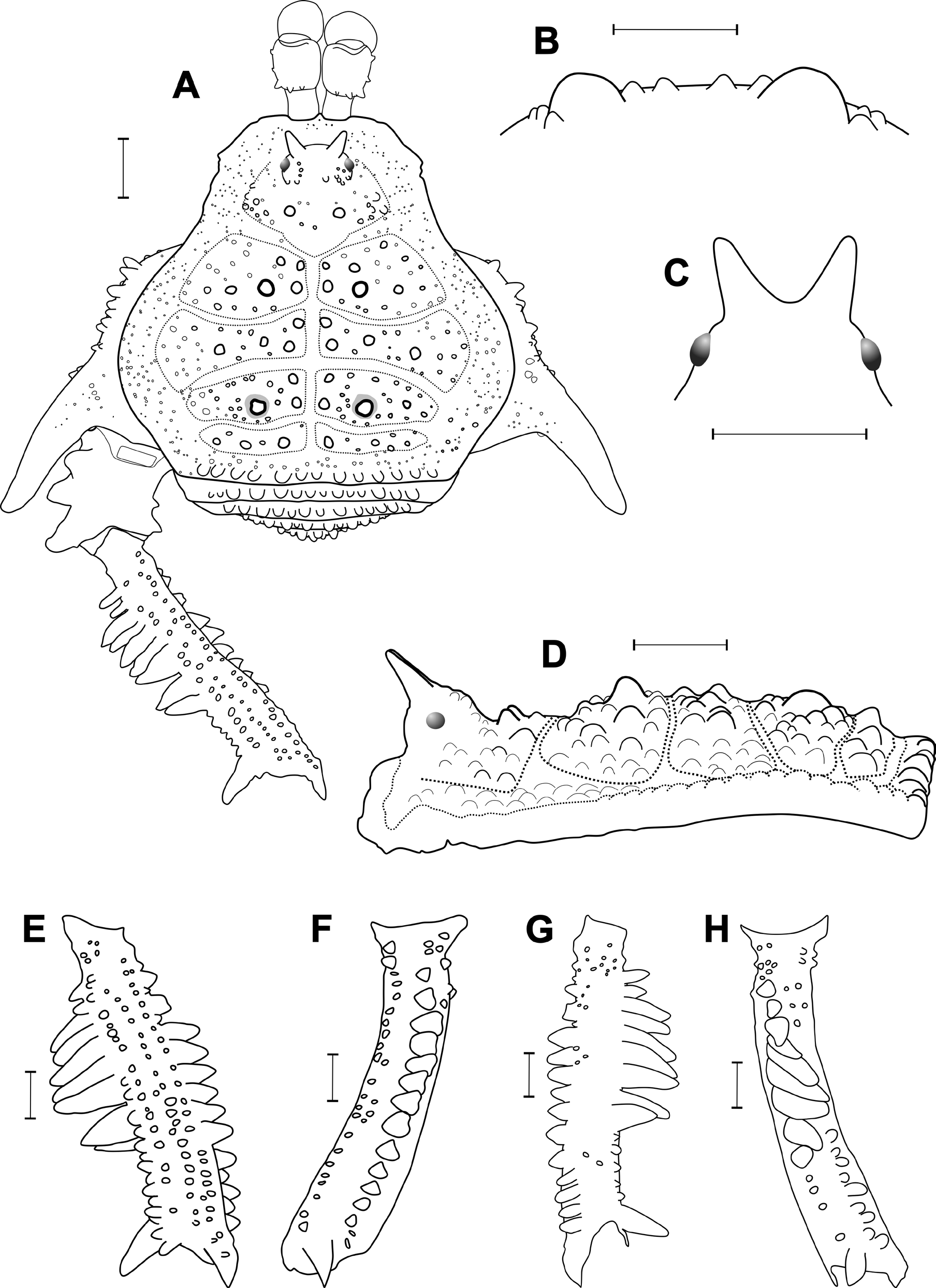
Carapace with a pair of paramedian larger tubercles, as large as found at paramedian of area I, darker, contrasting with background (absent in A. carvalhoi comb. nov.) ( Fig. 7A, D View Fig. 7 ); areas I–III with two higher tubercles next to the median groove (absent in A. carvalhoi comb. nov.) ( Fig. 7A View Fig. 7 ); area III with a pair of low, paramedian, rounded tubercles (lower and broader than the pair of high, paramedian, acuminated spines of A. carvalhoi comb. nov.) ( Fig. 7B, D View Fig. 7 ); Tr IV prolateral median with an apophysis (not occurring in A. carvalhoi comb. nov.) ( Fig. 7A View Fig. 7 ); Fe IV prolaterally and retrolaterally with high spines (instead of spines absent in A. carvalhoi comb. nov.) ( Fig. 7F, H View Fig. 7 ).
Description
Male (holotype)
CW 3.7, CL 2.4; AW 6.9, AL 3.9. Leg measurements in Table 5, tarsal counts in Table 4.
DORSUM. Dorsal scutum almost as long as wide, abdominal scutum with lateral margins strongly convex, widest at area II and highest at area III ( Fig. 7A, D View Fig. 7 ). Carapace with several tubercles on posterior region, with a pair of paramedian larger tubercles, darker, contrasting with background ( Fig. 7A, D View Fig. 7 ). Cheliceral sockets shallow, with a small apophysis in center. Ocularium elliptical, high, inclined frontwards, placed in middle of carapace, armed with a pair of divergent high spines fused at baseline and inclined frontwards ( Fig. 7A, C–D View Fig. 7 ). Mesotergum divided into four clearly defined areas. Areas I, III and IV divided into left and right halves by median groove. Area II anterior lateral border invading slightly space of area I and posterior lateral border gently invading space of area III. AS lateral borders with ordinary tubercles on full extent. All areas with several tubercles (darker than background). Area I with a pair of paramedian tubercles higher than the others and two medium tubercles (one anterior and one posterior) highlighted next to median groove. Area II with two medium tubercles (one anterior and one posterior) highlighted next to median groove and two medium tubercles highlighted abreast on horizontal medium. Area III with a pair of low, paramedian, rounded tubercles and two medium tubercles (one anterior and one posterior, about half size of paramedian) highlighted next to median groove ( Fig. 7A–B, D View Fig. 7 ). Area IV with six to eight minor tubercles plus horizontal row of three rounded, larger tubercles on each side. Posterior border of dorsal scutum and free tergites with a horizontal row of rounded tubercles.
VENTER. Cx I–III parallel to each other; each with ventral transverse rows of 6–10 setiferous tubercles (Cx I main row with higher and sharper tubercles). Cx IV much larger than the others, directed obliquely. Stigmatic area Y-shaped, clearly sunken relative to distal part of coxa IV. Intercoxal bridges well marked. Stigmata clearly visible. Free sternites and anal operculum each with one transverse row of small tubercles.
CHELICERA. Basichelicerite elongate, bulla well marked, with marginal setiferous tubercles – three ectal, three posterior, one mesal; hand not swollen ( Fig. 7A View Fig. 7 ).
PEDIPALPUS. Tr with three dorso-median rounded tubercles in a median elevation and two ventro-median setiferous tubercle (mesal highest in comparison to median). Fe with one meso-distal and one mesal, ventro-basal setiferous tubercles. Pa unarmed. Ti with two rows of setiferous tubercles; four (IiIi) ventromesal and four (IiIi) ventro-ectal, of which the two distal are geminated. Ta with two rows of setiferous tubercles; three (IIi) ventro-mesal and four (IiIi) ventro-ectal.
LEGS:. Tr I–III each with several ventral tubercles. Fe I–III slightly sinuous. Fe I and Ti I with prodorsal, prolateral, proventral, retroventral, retrolateral and retrodorsal rows of small tubercles. Fe II with a little retrodorsal distal tubercle, not forming a spur. Fe II and Ti II with prodorsal, proventral, retroventral and retrodorsal rows of small tubercles. Fe III substraight. Fe III and Ti III with prodorsal and retrodorsal rows of small tubercles and proventral and retroventral rows of acuminated tubercles. Fe III and Mt III with a well-developed retrodorsal, distal spur. Posterior border of Cx IV not reaching posterior border of dorsal scutum longitudinally. Cx IV with a prolateral apical caniniform apophysis, moderately elongate ( Fig. 7A View Fig. 7 ). Cx IV with prolateral, proventral, ventral and retrolateral rows of tubercles. Tr IV with two distal, retrolateral spines (ii). Tr IV median proventrally with one broad spearhead apophysis. Tr IV ventrally with several tubercles along its entire length. Tr IV prolaterally with three broad, conical apophyses. Fe IV substraight, curved from medial region toward retrolateral. Fe IV with dorsal, retrodorsal row of small tubercles ( Fig. 7E–F View Fig. 7 ). Fe IV with medial-distal, prodorsal row of rounded tubercles ( Fig. 7E, H View Fig. 7 ); Fe IV with proximal-medial, prolateral row of nine conical and substraight spines and a large, conical spine on distal region ( Fig. 7H View Fig. 7 ). Fe IV with seven medial, retrolateral higher acuminated spines followed by four smaller, acuminated tubercles on distal region ( Fig. 7F View Fig. 7 ); Fe IV with small distal, retrodorsal spur ( Fig. 7E–F View Fig. 7 ); Fe IV with proventral, ventral and retroventral diffuse tubercles. Pa IV covered by tubercles in dorsal view. Pa IV with proventral and retroventral, medial-distal row of three acuminate tubercles. Ti IV with prodorsal, prolateral, proventral, retroventral and retrodorsal rows of acuminate tubercles. Ti IV with proventral and retroventral, distal spur. Tarsal counts: 6(3)-6(3)/?- 8(3)/7-7/7-7.
PENIS. VP divided into two regions: distal part rectangular, proximal part trapezoidal ( Fig. 8A–C View Fig. 8 ). Ventral surface of VP entirely covered with microsetae of type 1 ( Fig. 8C View Fig. 8 , 9B View Fig. 9 ). All macrosetae inserted on lateral of VP: A1–A3, cylindrical, thick, on basal third of VP, A1 oriented dorsally, A2 oriented sidewards, A3 oriented ventrally ( Fig. 8A–C View Fig. 8 ); B1 inserted ventrally, below the line of A3 ( Fig. 8C View Fig. 8 ); C1–C3 slender, only moderately elongate, forming a row on the distal part of VP, C1–C2 close together on the distal portion, C3 on medial basal portion ( Figs 8A–C View Fig. 8 , 9B View Fig. 9 ); D1 medium, midway between C3 and A1 ( Figs 8A–C View Fig. 8 , 9B View Fig. 9 ); E1–E2 inserted ventrally, E1 between the height of C1–C2, E2 slightly below C3 ( Figs 8B View Fig. 8 , 9B View Fig. 9 ). Glans sac short, arising from middle bulge on podium, not extended as a dorsal process ( Figs 8A–B View Fig. 8 , 9A View Fig. 9 ). Stylus stout, cylindrical and S-shaped curved to dorsal ( Figs 8A–B View Fig. 8 , 9A–B View Fig. 9 ). Apex of stylus flattened dorsoventrally, covered by spines ventrally ( Fig. 10B, D–F View Fig. 10 ). Ventral process of stylus gently curved, with half longitudinal length of stylus ( Figs 8A–B View Fig. 8 , 9A, C View Fig. 9 ). Apex of ventral process of stylus forming a small flabellum with flaps deeply serrated ( Fig. 9A, C View Fig. 9 ).
COLOR (in vivo). Color background of scutum and coxae Deep Red (13), with paramedian tubercles of areas I and III Very Dark Red (17). Dry marks Very Pale Purplish Blue (202) distributed on Cx IV, free tergites I–III and uniformly all over dorsal scutum encircling the tubercles; also appearing faintly on basal parts of legs I–IV. Chelicerae, pedipalps (glossier than legs) and trochanters I to IV and base (proximal 15%) of femora III–IV Deep Orange (51). Femora and tibiae III and IV mostly (medial 70% of femur) Blackish Red (21). Distal 15% of femora III–IV and patellae III–IV Strong Orange (50). Tibiae III–IV repeating the color of femora, without lighter detail at base. Metatarsi and tarsi III–IV Brownish Orange (54). Legs I and II similar to posterior ones, but with colors much attenuated.
VARIATION. Besides the variation in tarsal counts, shown in Table 4, the minor (“beta”) males only show a difference in attenuated armature of Fe IV proventral and retrolateral rows of spines. Distribution and development of tubercles and apophyses in our sample fairly uniform.
Female (paratype, MNRJ 9267)
CW 4.2, CL 2.5; AW 7.4, AL 4.6. Cx IV with much weaker armature, main apophysis reduced to a simple spine. Fe IV thinner and less curved when compared to that of male. Fe IV with fewer spines on distal proventral axis and a retrolateral distal spur.
Distribution
BRAZIL, Acre (NT 0 166 – Southwest Amazon Moist Forests) and Rondônia (NT 0 135 – Madeira- Tapajós Moist Forests). ( Fig. 2 View Fig. 2 ).


No known copyright restrictions apply. See Agosti, D., Egloff, W., 2009. Taxonomic information exchange and copyright: the Plazi approach. BMC Research Notes 2009, 2:53 for further explanation.
|
Kingdom |
|
|
Phylum |
|
|
Class |
|
|
Order |
|
|
Family |
|
|
Genus |