
Eratigena inermis Bolzern, Angelo, Burckhardt, Daniel & Hänggi, Ambros, 2013
|
publication ID |
https://doi.org/ 10.1111/zoj.12040 |
|
publication LSID |
lsid:zoobank.org:pub:28796C66-FD49-4FA9-8D0F-21DD495AA88A |
|
DOI |
https://doi.org/10.5281/zenodo.6983321 |
|
persistent identifier |
https://treatment.plazi.org/id/BD701413-E210-B66D-5742-FE3EC28313AE |
|
treatment provided by |
Marcus |
|
scientific name |
Eratigena inermis |
| status |
comb. nov. |
ERATIGENA INERMIS ( SIMON, 1870) View in CoL COMB. NOV.
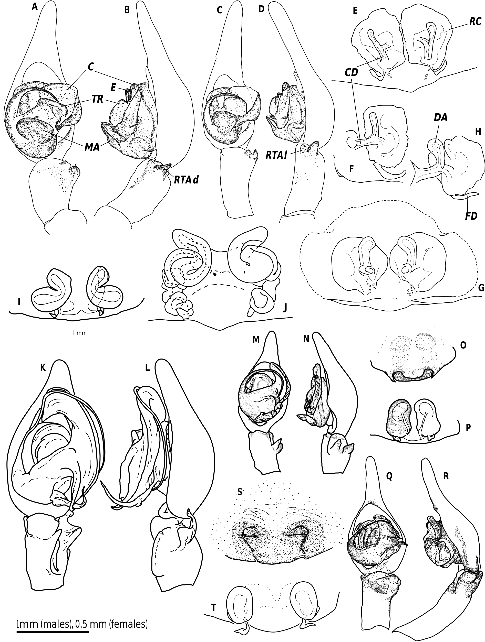
( FIGS 13C–D, K–L, R View Figure 13 , 14A–B, E–F View Figure 14 )
Tegenaria inermis Simon, 1870: 271–273 , pl. 1, figs 7, 11 (figures not useful for determination).
Types
Syntypes (several labels were present in the same tube, one from the type locality). Spain: León: Brañuelas; Navarra: Alsasua ; ‘Pyr. Raun Bonnes.’ (?), 1 ♂ ( MNHN, 1960, specimen selected and labelled as ‘neotype’ by R. De Blauwe). This specimen is here selected as lectotype in order to stabilize the nomenclature ( ICZN: art. 74.1; available online at http:// iczn.org/); remaining specimens, 4 ♂, 3 ♀, ( MNHN, 1960), paralectotypes .
Other material examined
France (6 ♂, 11 ♀) ; Portugal (7 ♀) ; Spain (13 ♂, 17 ♀) .
Diagnosis
Eratigena inermis differs from other congeneric species by having distinctly annulated legs (all other Eratigena species with other patterns), at least two femora with more than two dorsal spines (also observed in E. saeva comb. nov.; all other species with one or two spines), tibia I with prolateral spines (also observed in E. herculea , E. hispanica , and E. sicana ; all other species without prolateral spines), massive transversal ridge or bulge at the conductor of the male bulb ( Figs 13C–D View Figure 13 , 14A–B View Figure 14 , distinctly different to all other species), conductor dorsally with a small rounded bulge (as in several Tegenaria , but not in Eratigena species ), conspicuously large and strongly sclerotized MA, epigynal teeth absent, and long appendages at CD ( Fig. 14E–F View Figure 14 , as in E. herculea and E. hispanica ). It can be separated from the closely related E. vomeroi by having the basal part of the median apophysis more strongly sclerotized, the very special massive and prominent transversal ridge
at the conductor showing a distinct border line of sclerotization that is only indistinctly expressed in E. vomeroi , the long appendages anteriorly of the CD reaching at least to the top of the RC (or even beyond) in E. vomeroi but shorter in E. inermis ( Fig. 14E–H View Figure 14 ).
Description
Measurements: Male (N = 1): CL 4.75, CW 3.34, STL 2.11, STW 2.04, OL 5.10, OW 2.92. Leg I (7.4, 2.02, 7.39, 8.21, –), II (6.45, 1.83, 5.83, 6.75, 3.22), III (5.78, 1.64, 4.79, 6.88, 3.04), IV (6.51, 1.66, 6.11, 7.36, –). Pedipalp (2.31, 0.76, 0.92, 2.25), bulbL 1.15. Female (N = 3): CL 5.42–5.68, CW 3.73–3.91, STL 2.17–2.56, STW 2.17–2.43, OL 6.10–8.61, OW 3.70–5.64. Leg I (5.99–7.13, 1.87–2.12, 5.69–6.81, 5.85–7.48, 3.14– 3.61), II (5.35–6.39, 1.73–2.00, 4.64–5.86, 5.27–6.94, 2.81–3.14), III (4.9–6.13, 1.64–1.86, 4.02–5.10, 5.38– 7.09, 2.17–2.93), IV (6.14–7.51, 1.71–1.96, 5.36–6.67, 7.49–9.37, 3.07–3.66). Pedipalp (2.55–2.56, 0.97–0.98, 1.72–1.77, 2.64–2.89). EPL 0.65–0.73, EPW 0.94–1.19, ATL 0.29–0.42, ATW 0.45–0.58. Eyes: PME 0.22–0.26, PLE 0.22–0.28, AME 0.17–0.23, ALE 0.23–0.26. Eye distances: PME–PME 1 x PME, PME–AME 0.5–1 x PME, PME–PLE 0.5–1 x PME, PME–ALE 1–1.5 x PME, AME–AME 0.5–1 x AME, AME- ALE ≤ 0.5 x AME. CLY1 2.5–3 x AME, CLY2 1.5–2 x ALE.
Male palp: RTA with two branches, lateral branch bulge-like, dorsal branch a strongly sclerotized peak. Embolus length <1.25 x CB, originating at 8–10 o’clock position, distal tip at 2–4 o’clock position. Conductor drop-shaped, folded only at terminal half. Terminal end consists of two strongly sclerotized points and dorsally with a small rounded bulge. Transversal ridge of conductor massive and prominent with distinct margin between membranous and more sclerotized areas. Conductor membranously connected to tegulum. MA originating at 5–7 o’clock position, moderately protruding, much wider than long, pocket-like. Connection of MA to tegulum strongly sclerotized.
Epigyne and vulva: Epigyne medially with large bulge, anteriorly of which the CO are located in a transversal depression. Posterior sclerite absent. Epigynal teeth absent. Vulva consists of distinguishable CD, RC, and FD. CD short and straight with long appendages. RC irregularly formed and not constantly sclerotized. FD only represented by small, leaf-shaped appendages.
Other important characters: Cheliceral retromargin with six to seven teeth. Colulus rectangular with distal margin straight. PMS with one elevated minor ampullate gland spigot and three to four cylindrical gland spigots laterally. Tarsal trichobothria on cymbium and palp tarsus absent. Tarsal trichobothria eight to ten. Small teeth on paired claws of leg I 12–14. Leg spination: male palp (2–1–0–0, 2–0–0–0, 1–2p–0–0), female palp (2–0–0–0 or 2–1–0–0, 2–0–0, 2–2p–0–0), leg femora (3–3–3–0 or 3–3–4–0 or 3–4– 4–0, 3–3–3–0 or 3–3–4–0 or 3–5–4–0 or 4–6–5–0, 2–3–4–0 or 2–4–3–0 or 2–4–4–0 or 3–4–4–0, 2–2–2–0 or 2–3–2–0 or 2–3–3–0 or 2–4–2–0 or 3–2–2–0 or 3–3–3–0), patellae (all 2–0–0), tibiae [0–2–0–1+2p or 0–2–0–4p, 0–2–0–1+3p or 0–2–0–4p or 0–2–1–4p (dorsally on I & II two indistinct spines possible), 2–2– 2–1+3p or 2–2–2–3+1p or 2–2–2–4p, 2–2–2–1+3p or 2–2–2–2+2p or 2–2–2–3+1p or 2–2–2–4p], metatarsi (0–0–0–4p+1, 0–2–0–4p+1 or 0–2–1–4p+1, 0–4–4– 4p+1 or 0–5–4–4p+1, 0–5–4–4p+1).
Coloration: Two symmetrical longitudinal dark bands dorsally on carapace present (sometimes reduced to triangular dots). Distinct sternal pattern of median region and three symmetrical pale dots laterally, most distal pair fused to the median region ( Fig. 13R View Figure 13 ). Opisthosoma with three pale bands anteriorly, continuing to the back in chevrons. Legs annulated. ALS somewhat darkened, PLS both segments darkened.
Distribution
Reported from the northern part of France and from Spain and Portugal.
Comments
Further useful drawings for the determination of this species were provided by Simon (1937) and Brignoli (1978a).

| MNHN |
Museum National d'Histoire Naturelle |
No known copyright restrictions apply. See Agosti, D., Egloff, W., 2009. Taxonomic information exchange and copyright: the Plazi approach. BMC Research Notes 2009, 2:53 for further explanation.
|
Kingdom |
|
|
Phylum |
|
|
Class |
|
|
Order |
|
|
Family |
|
|
Genus |
Eratigena inermis
| Bolzern, Angelo, Burckhardt, Daniel & Hänggi, Ambros 2013 |
Tegenaria inermis
| Simon E 1870: 273 |