
Hymenicoides robertsi, Naruse, Tohru & Ng, Peter K. L., 2007
|
publication ID |
https://doi.org/ 10.5281/zenodo.179195 |
|
DOI |
https://doi.org/10.5281/zenodo.6251674 |
|
persistent identifier |
https://treatment.plazi.org/id/BB10DE28-6C3A-2C64-4485-FA50FBE12D90 |
|
treatment provided by |
Plazi |
|
scientific name |
Hymenicoides robertsi |
| status |
sp. nov. |
Hymenicoides robertsi View in CoL new species
( Figs. 1 View FIGURE 1 a, b, 3–6)
Material examined. Holotype: 1 male, 4.9 × 6.0 mm, ZRC 2007.0108, Kyaukdaw market on lower Kaladan River (tidal), Rakhine, Myanmar, coll. T. R. Roberts, 26 Mar. 2004.
Paratypes: 1 male, 5.0 × 6.2 mm, ZRC 2007.0109; 42 males, 3.0 × 3.3 – 5.2 × 6.2 mm, ZRC 2007.0110; 1 female, 4.8 × 5.6 mm, ZRC 2007.0111; 4 females, 3.9 × 5.5 mm – 4.8 × 5.6 mm, ZRC 2007.0112; 16 ovig., 4.1 × 4.9 mm – 4.5 × 5.4 mm, ZRC 2007.0113; 5 males, 4.2 × 5.0 – 5.0 × 5.8 mm, 2 ovig., 3.8 × 4.2, 3.8 × 4.3 mm, NHM 2007.600–606; 5 males, 4.0 × 4.8 – 4.9 × 5.7 mm, 2 ovig., 3.8 × 4.4 mm, 4.2 × 4.8 mm, MNHN- B30393. All paratypes were collected together with the holotype.
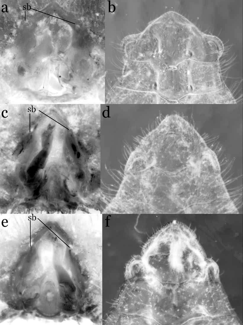
Description. Carapace ( Fig. 4 View FIGURE 4 a) oval, CW 1.13–1.24 times CL (mean 1.18, n = 9) CL; dorsal surface flat, surrounded by continuous rim, regions well demarcated by grooves, H-shaped gastric groove continuous with cervical groove, cervical groove branching anteriorly, branches confluent with anterolateral rim. Rostrum vestigial, triangular. Lateral margin of carapace lacking tooth or lobe, side wall of posterolateral region slightly expanded laterally, with longitudinal groove along posterolateral rim below. Conical tooth present between antennules, completely disconnected from vestigial rostrum; orbit indiscernible, without tooth on outer part of eye. Epistome long, placed anterior-dorsally to buccal cavern, ischium of third maxilliped partially covering posterior margin; posterior margin with trapezoidal convexity with median notch.
Eyes moderately developed, visible dorsally. Antennule with long coxa and basis; basis more than half length of coxa. Third maxilliped ( Fig. 4 View FIGURE 4 b) narrow, covering about one-quarter of buccal cavern; mid-length of ischium about three-quarters of merus, distal inner angle produced; palp long, propodus about half length of dactylus, dactylus as long as merus, tip of dactylus almost reaching proximal end of ischium in situ; exopod short, reaching about proximal two-thirds of merus, with distinct flagellum.
Male with relatively wide abdominal cavity ( Fig. 1 View FIGURE 1 a), sternal button narrow, high, just posterior to imaginary line joining inner ends of sutures between sternites 5 and 6. Female thoracic sternite 3 separated from sternite 4 by posteriorly convex rim, sterites 4–8 medially fused, vulva on imaginary line joining inner ends of sutures between sternites 5 and 6 on medial fused plate of thoracic sternum, vulva with longitudinally elliptical basal mount.
Chelipeds symmetrical, relatively stouter in males; male cheliped with short merus, ventral outer margin with subdistal tooth, inner margin of merus to carpus lined with long stiff setae, outer surface of carpus rounded, not strongly elevated, outer margin with subdistal tooth; chela ( Fig. 4 View FIGURE 4 c, d) with rounded palm, almost glabrous, outer surface inflated, with dorso-ventrally flattened tubercle on outer surface of proximal lower part of base of immovable finger, tubercle absent in females; fingers with ovoid gape when closed, tips slightly hoof-like; immovable finger with ca. 6 low teeth; teeth of movable finger similar to those of immovable finger.
Ambulatory legs ( Fig. 4 View FIGURE 4 a) slender, long, second longest, inner margin of propodi to dactyli fringed densely lined with long plumose setae; meri slightly longer than respective propodi, distal anterior angle not produced; dactyli ( Fig. 4 View FIGURE 4 e) terminating in sharp tooth, with subterminal inner tooth placed far from distal tooth.
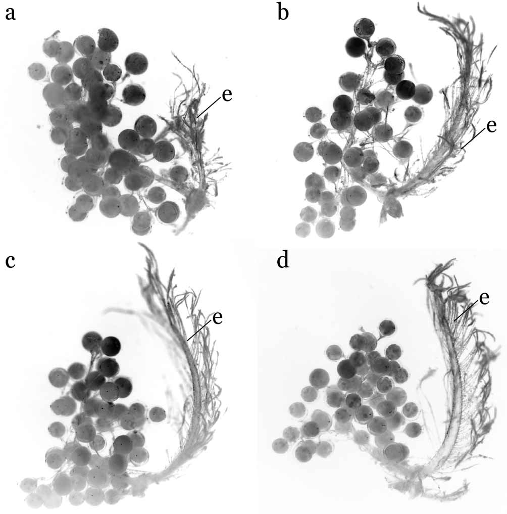
Male abdomen-pleotelson ( Fig. 5 View FIGURE 5 a) 6-segmented; first segment thick, hard, distal margin widely concave; second to fifth segments with distal margin concave medially, third segment widest; pleotelson trilobed, lateral lobes distinct, auriculate, inner surface of lateral lobes thickened externally, forming socket for sternal button on inner surface ( Fig. 1 View FIGURE 1 a, b). G1 ( Fig. 5 View FIGURE 5 c, d) stout, strongly bent on distal half, with longitudinal line of long, stiff setae on distal to medial inner angle of shaft; distal inner end with beak-like chitinous projection, distal outer angle swollen, covered by tiny granule, distal outer angle dorsally connected with thumb-like projection. G2 ( Fig. 5 View FIGURE 5 e) short, less than half length of G1. Female abdomen-pleotelson ( Fig. 5 View FIGURE 5 b) demarcated to 6 segments, boundary between second to fifth segments movable, more or less fixed in other boundaries; second to fifth segments with corneous wide ridge medially, that of pleotelson only on proximal half, pleotelson longest; pleopods ( Fig. 6 View FIGURE 6 ) on second to fifth segments, long, biramous from near base, developing from distal outer angle of inner surface of each segment.
Eggs spherical, small, diameter 0.27 – 0.30 mm [mean = 0.28, n = 15 from three females (3.9 × 4.4 mm, 4.1 × 4.7 mm, 4.2 × 5.0 mm)], attached on endopods of pleopods, exopods growing inwards, partially covering dome-shaped abdomen.
Habitat and distribution. All specimens of Hymenicoides robertsi were obtained from the Kyaukdaw market on the lower Kaladan River (with tidal influence), Rakhine, Myanmar. The specimens were found crawling over a large number of young Pisodonophis snake eels in a tray in the market (T. R. Roberts, pers. comm.) and had been apparently collected in the nearby area. The species is known only from Myanmar at present.
Etymology. We are pleased to name this species for the well known ichthyologist, Tyson R. Roberts, who very generously provided the specimens of this new species as well as those of H. carteri from Bangladesh for our study.
Remarks. Hymenicoides robertsi new species, is clearly different from H. carteri in its proportionately more slender ambulatory legs ( Fig. 3 View FIGURE 3 a; Kemp, 1917: Fig. 17). In addition, both species can be distinguished by the armature of the inner margin of the ambulatory dactyli. In H. robertsi , all the dactyli have only one subterminal tooth, which is placed some distance from the tip, while in H. carteri , the third to fifth dactyli have 3–11 small teeth (posterior legs with fewer teeth) and a subdistal large tooth which is placed closer to the tip. Another major difference is the single beak-like chitinous projection of the G1 of H. robertsi (biramous shorter projection in H. carteri ) ( Figs. 2 View FIGURE 2 a, b, 4a, e, 5c, d; Kemp, 1917: Figs. 17, 20).


| ZRC |
Zoological Reference Collection, National University of Singapore |
No known copyright restrictions apply. See Agosti, D., Egloff, W., 2009. Taxonomic information exchange and copyright: the Plazi approach. BMC Research Notes 2009, 2:53 for further explanation.