
Tyrannosaurus rex, Osborn, 1905, Osborn, 1905
|
publication ID |
https://doi.org/10.5281/zenodo.3479733 |
|
persistent identifier |
https://treatment.plazi.org/id/BA3D4333-FF98-5E04-FEB3-F6440A30FC08 |
|
treatment provided by |
Jeremy (2019-10-10 12:45:12, last updated 2019-10-10 16:59:18) |
|
scientific name |
Tyrannosaurus rex |
| status |
|
The 13 m long carnivorous dinosaur Tyrannosaurus rex is arguably the dinosaur best known by the general public ( Brochu, 2003). With a new dinosaur gallery scheduled to open in 2018, the national natural history museum of the Netherlands, Naturalis Biodiversity Center, in Leiden, set out to acquire an original skeleton of this dinosaur. A skeleton of Tyrannosaurus rex was discovered in Spring and excavated in the Fall of 2013, from a sandstone stream channel of the Hell Creek Formation near Jordan, Montana, USA. The excavation relied upon a close collaboration between Black Hills Institute and Naturalis ( Schulp et al., 2015). Reasonably complete skeletons of Tyrannosaurus are few and far between, and so far only two skeletons substantially more than 50% complete have been found ( Larson & Carpenter, 2008). At the moment of submission of the present paper (March 2016), the Naturalis T. rex skeleton is still being prepared, and is currently
scheduled to go on public display in Leiden in September 2016, where it is registered under collection number RGM 792.000.
The specimen comprises a well-preserved skull, partial cervical and dorsal vertebral series, an almost-complete rib-cage, scapula-coracoid, furcula, a complete pelvis, the right leg, and about half of the tail. Probably no other innovation has had a similarly profound impact on the study of fossil vertebrates in the last few decades than the increased accessibility and improvement in image quality of CT scanning (e.g., Leiggi & May, 2003). The nondestructive character of CT allows for features otherwise inaccessible, to be visualized, described, compared and analyzed ( Mallison, 2011; Abel et al., 2012). Medical CTscanners, with a bore suited to fit most humans, and, occasionally, veterinary CT scanners with a larger bore, now routinely allow for scanning of human-sized objects; however, the specifications of those imaging systems are optimized for objects less dense than fossils, and the X-ray performance is dimensioned for X-ray exposures “as low as reasonably achievable” (the ALARA safety principle). This is not necessarily the specification set required for successful imaging of higher density objects such as large, heavily permineralized fossils.
View MaterialsSuccessful scans of extremely large paleontological objects can, therefore, be challenging. Scanning the skull of one of the largest carnivores ever to have walked the earth is certainly beyond the capabilities of a regular medical CT system. The skull of the remarkably complete T. rex skeleton FMNH PR2081 , perhaps better known by its nickname “Sue” (now on display at the Field Museum in Chicago) was scanned -almost two decades agoat Rocketdyne Division of Boeing North America. This scan was completed in a Minatron 205 scanner, yielding 748 coronal slices, 2 mm in thickness ( Brochu, 2003). For many paleontological questions however, higher-resolution imaging than 2 mm voxel size is necessary. In this contribution, we elaborate on the technical challenges in scanning the skull of the Naturalis T. rex . A more detailed description of the morphology, partially based on the CT scan data discussed here, will be submitted for publication elsewhere.
2. Materials and Methods
System Setup
During the last few years the Fraunhofer Development Center X-Ray Technology (EZRT) focused on the research of CT imaging methods in the high energy regime to overcome the otherwise quite narrow scope and limitations of conventional X-ray systems regarding specimen size and material composition. This finally resulted in the development of a CT system capable of scanning very large objects of up to 5 m in at least one axis. The so called XXL-CT system was installed in Furth, Germany in 2013 ( Fuchs, 2016). To our knowledge, the XXL-CT is the first and largest publicly available CT system for the inspection of such objects. The XXL-CT features some unique characteristics when compared to conventional CT systems. Imaging of objects like complete cars or shipping containers require penetration lengths of more than 200 mm of steel or equivalent. To achieve enough penetration power, the system relies upon a linear accelerator (linac) with up to 9 MeV X-ray energy and a dose rate of up to 25 Gy/min at 1 m distance. Figure 1 shows the X-ray bremsstrahlung spectra for 6 and 9 MeV with 10 cm of Aluminum as prefilter. The data was acquired with Monte-Carlo simulation toolkit ROSI ( Giersch 2003). Both spectra look fairly similar with a mean energy of 1.8 and 2.5 MeV respectively.
When choosing the appropriate detection system, the still significantly higher X-ray energies compared to conventional systems have to be taken into account. At present, flat panel detectors are not a practical option when scaning large objects efficiently, due to both technological limitations and the X-ray attenuation characteristics. The dominant attenuation effect in the MeV range is Compton scattering (compare Berger 1998). Here a line detector provides a significantly better image quality which is mainly due to the nondetection of scattering in the plane orthogonal to the sensor. Certainly the most significant trade-off is an increase in scanning time. Another effect arising from Compton scattering is that the imaging properties are mainly determined by the density of the penetrated material and only to a small amount by the atomic number. This results in a better linearity for multi-material specimens as well as a better detectability of low-Z and high-Z elements. We chose a line detector array of about 4 meters in length and a pixel pitch of 0.4 mm. This results in nearly 10,000 pixels. A 10 mm thick scintillator of cadmium tungstate provides a reasonable quantum efficiency of 45% for a 9 MeV bremsstrahlung spectrum. Both the linac and the horizontally orientated detector are mounted on two towers of 8 m height (see figure 2 View Fig ). A three-meter diameter turntable is placed in front of the detector tower. The system allows for reconstruction volumes of 3.2 m (and more with limited angular range) in diameter and 5 m in height. The maximum specimen weight is 10,000 kg.
The CT scanning process with this system is done in a fan beam geometry. The source and detector are moving synchronously upwards during image acquisition. To compute a 3Drepresentation of the specimen a multitude of projections are acquired each from a different
viewing angle. This is achieved by rotating the turntable between each subsequent image by one small angular step.
Composition of the fossil
Excavating and removing large fossils from the field is generally done using a technique called ‘plaster jacketing' (e.g., Leiggi & May, 2005). Briefly summarized, a fossil is exposed by removing the surrounding matrix, and the exposed bones are impregnated with a consolidant. Subsequently, a trench is dug around the fossil, essentially leaving the fossil exposed on a ‘pedestal'. The bone surface is covered in aluminum foil or wet tissues to act as a barrier to the plaster jacket, which is subsequently applied. This plaster jacket is essentially a composite material, consisting of burlap and plaster of Paris. In larger blocks (such as the T. rex skull block) the jacket is reinforced with a wooden support structure, which also acts to provide grip and leverage in handling the block. The resulting plaster jacket, then, has become a composite of a wide variety of materials with a wide variety of X-ray absorbing properties: fossil bone, fossil tooth enamel, surrounding sandstone, the occasional concretionary growth of the mineral pyrite (FeS2; clearly visible in the scan), plaster, burlap, wood and metal screws to hold the wooden frame together. Figure 3 View Fig shows a photograph of the skull, within its overturned plaster jacket, being prepared at the excavation site.
Preparation of the skull for scanning
The CT scan geometry of the XXL-CT setup made it desirable to place the skull in an upright orientation, i.e. the skull on the turntable is rotated around its longitudinal axis. This yields two advantages over the natural orientation. First the maximum X-ray penetration length through the skull and surrounding sandstone matrix is significantly reduced (factor of 2) and more homogeneous over the 360° range (see figure 4) and second the smaller cross-section reduces blurring effects due to X-rays impinging the detector at an angle (signal spread over more than one pixel). Both effects highly influence the image quality both in terms of contrast as well as spatial resolution.
This provided an additional challenge in preparing the specimen for transport and scanning, as the way the fossil was crated had to ensure that the specimen was sufficiently tightly packed to allow for tipping 90° in the first place, while at the same time guarantee sufficient cushioning as to have the fossil survive transport (road and air transport, where turbulence cannot be ruled out), and on the other hand guaranteeing that the specimen did not move at all in the crate while scanning. This was achieved by first completing the plaster jacket so that the fossil was completely enclosed and the surface of the skull protected from movement within the jacket. Secondly the jacket had to be immobilized within the wooden crate. This was accomplished by adding more wooden supports to the plaster jacket and then screwing additional wooden supports between the jacket and the crate. To completely eliminate the possibility of movement, the remaining air space within the crate was then filled with 2# density polyurethane foam.
CT-Scan parameters
The skull, at the time of scanning, was still encased in the sandstone matrix and the plaster jacket and immobilized within the wooden crate. The crate containing the T. rex skull weighed in at about 700 kg. For scanning the crate was put in an upright orientation and hoisted onto the turntable with a ceiling crane. The CT scan was performed with the acquisition parameters summarized in table 1. Taking into account the system magnification of 1.2, the effective pixel size was 0.33 mm; however the CT reconstruction was performed at lower resolution with a binned voxel size of 1 mm to significantly improve the signal-to-noise ratio.
3. Results
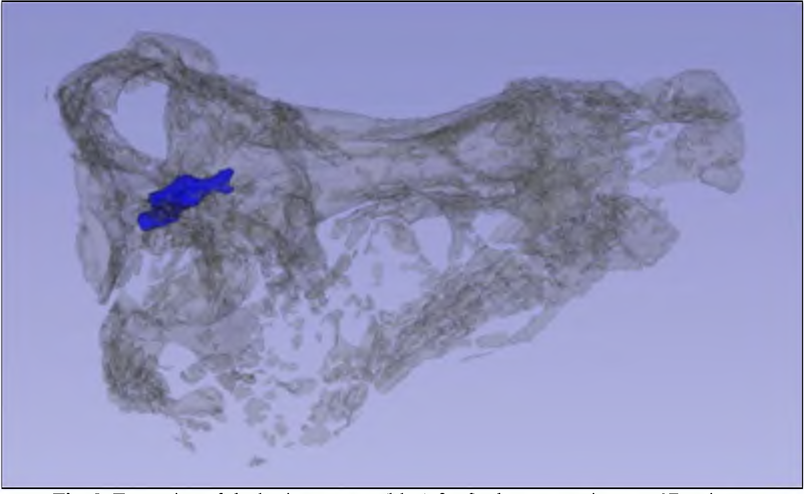
The CT reconstruction was performed on the whole dataset including the surrounding crate and support structure. Figure 5 View Fig shows one slice through the dataset. Generally, the bones (light grey) can be clearly distinguished from the sandstone matrix (dark grey) due to its significant difference in density. Inside the bone, one can discern occasional high-intensity spots which represent pyrite concretions. Other materials involved in packing and crating have generally much lower X-ray absorption, except for the screws used in the wooden support frame. As can be seen in the upright corner of figure 5 even the polyurethane foam is visible by using a different grey value scaling.
.
4. Discussion and Outlook
The scan has already allowed for much more efficient preparation work on the fossil, by showing which areas deserve particular attention. Many internal features are clearly visible, including pneumatic chambers and the portion of the skull that was originally occupied by the brain ( Witmer & Ridgely, 2009). Of particular interest are the morphology of the nerve channels, semicircular canals of the inner ear ( Witmer & Ridgely, 2009), location and form of the stapes, the possibility of respiratory turbinates ( Witmer, 1997; Brochu, 2003; Witmer & Ridgely, 2009), and the fact that this scan shows new fragile structures not visible with lower resolution CT scans - and which we suspect may have been obliterated by mechanical preparation of other specimens. We do hope that this scan, too, will contribute to a more informed reconstruction of soft tissues ( Witmer, 1997; Brochu, 2003).
Part of the magic of X-ray imaging techniques is that the invisible becomes visible. The CT of the T. rex skull allowed for a virtual endocast of the brain cavity to be made (compare Witmer & Ridgely, 2009). A 3D-print of the “brain” of the T. rex (see Figure 8 View Fig ) already played a role in lectures and (online) classes; the printed brain will also find a place in the upcoming exhibition at Naturalis.
The skull of the new T. rex shows multiple pathologies, some of which are captured in detail in the CT scan. Pathologies include a rather large bone infection which has removed bone tissue in the anterior part of the right maxilla (=the front end of the upper jaw); this element however was discovered separate from the main skull block, and therefore was not part of the scan. The same applies for the posterior mandibular unit (= the rear part of the lower jaw), which is graced by a series of healed puncture wounds; considering the size and spacing of the wounds, in all likelihood this individual was bitten by another T. rex - and lived for the wounds to heal. Other pathologies on the skull include scratch marks on the left side of the skull. At Naturalis we plan to present the CT data of the skull in an interactive display, where a laser line projected on a scaled, moveable 3D-print of the skull acts as a pointer to the CT-imagery shown beside the skull. Interactive “hot zones” allow to connect explanatory text and video to the features highlighted in the scan.
Abel, R. L., Laurini, C. R., & Richter, M. (2012). A palaeobiologist's guide to ' virtual' micro-CT preparation. Palaeontologia Electronica.
Berger M. J., Hubbell J. H., Seltzer S. M., Coursey J. S., and Zucker D. S. (1998) XCOM: Photon Cross Sections Database, NIST Standard Reference Database 8.
Brochu, C. A. (2003) Osteology of Tyrannosaurus rex: Insights from a nearly complete Skeleton and High-Resolution Computed Tomographic Analysis of the Skull. Journal of Vertebrate Paleontology, 22: sup 4, 1 - 138, DOI: 10.1080 / 02724634.2003.10010947
Fuchs, T. et al. (2016). High-energy 3 D X-ray computed tomography on very large objects, to be published.
Giersch, J. et al. (2003) ROSI an object-oriented and parallel-computing monte carlo simulation for X-ray imaging. Nuclear Instruments and Methods in Physics Research 509.
Larson, P. & K. Carpenter (Eds.), 2008. Tyrannosaurus rex: The Tyrant King. Indiana University Press, Bloomington, Indiana, 435 pp.
Leiggi, P. & P. May (Eds.), 2005. Vertebrate paleontological techniques, Vol. 1. Cambridge University Press, New York., 366 pp.
Mallison, H. (2011). Digitizing Methods for Paleontology: Applications, Benefits and Limitations. In Computational Paleontology (pp. 7 - 43). Berlin, Heidelberg: Springer Berlin Heidelberg. http: // doi. org / 10.1007 / 978 - 3 - 642 - 16271 - 8 _ 2
Schulp, A. S., D. Bastiaans, P. Kaskes, P. Manning & P. Larson (2015): A New, Mature and Pathologic specimen of Tyrannosaurus rex. Society of Vertebrate Paleontology Annual Meeting, Dallas.
Witmer, L. M. (1997). The evolution of the antorbital cavity of archosaurs: a study in softtissue reconstruction in the fossil record with an analysis of the function of pneumaticity. Society of Vertebrate Paleontology Memoir 3: 75 pp.
Witmer, L. M., & R. C. Ridgely (2009). New Insights Into the Brain, Braincase, and Ear Region of Tyrannosaurs (Dinosauria, Theropoda), with Implications for Sensory Organization and Behavior. The Anatomical Record: Advances in Integrative Anatomy and Evolutionary Biology, 292 (9), 1266 - 1296. http: // doi. org / 10.1002 / ar. 20983


Fig. 5. CT slice through the skull. The skull bones clearly stand out from the sandstone matrix and the supporting structures.
No known copyright restrictions apply. See Agosti, D., Egloff, W., 2009. Taxonomic information exchange and copyright: the Plazi approach. BMC Research Notes 2009, 2:53 for further explanation.
|
Kingdom |
|
|
Phylum |
|
|
Class |
|
|
Order |
|
|
Family |
|
|
Genus |
1 (by jeremy, 2019-10-10 12:45:12)
2 (by ExternalLinkService, 2019-10-10 16:59:18)
3 (by ExternalLinkService, 2019-10-11 18:16:38)
4 (by admin, 2019-10-11 21:26:55)
5 (by admin, 2019-10-11 21:36:10)
6 (by ExternalLinkService, 2019-10-11 21:41:34)
7 (by jeremy, 2020-02-06 14:58:49)
8 (by jeremy, 2020-09-11 07:46:14)
9 (by jeremy, 2020-09-11 07:50:35)
10 (by jeremy, 2021-03-04 19:06:08)
11 (by felipe, 2021-08-18 16:48:25)
12 (by ExternalLinkService, 2021-09-22 15:25:55)
13 (by ExternalLinkService, 2021-11-02 23:56:40)
14 (by jeremy, 2023-07-17 14:11:51)